UNIVERSITE DE GENEVE FACULTE DE MEDECINE Section de Médecine Clinique Département de Pédiatrie
Service d’Orthopédie Pédiatrique
Thèse préparée sous la direction du Professeur André Kaelin
Hémimélie tibiale : présentation de quatre cas
Thèse
Présentée à la Faculté de Médecine De l’Université de Genève
Pour obtenir le grade de Docteur en médecine par Romain DAYER de Hérémence (VS) Thèse n°10418 Genève 2005
Table des Matières Page
1. Résumé 5 2. Introduction 6 3. Présentation de cas 14 4. Discussion: Embryologie normale 29 Concept de champ du développement 31 Aspects génétiques 33 Principes thérapeutiques 355. Dissection commentée 38 6. Conclusion 44 7. Bibliographie 45
Hémimélie tibiale : présentation de quatre cas
RésuméL’hémimélie tibiale est une anomalie congénitale rare, survenant environ chez une naissance pour un million. Elle consiste en une aplasie ou hypoplasie du tibia selon le type d’hémimélie ; la fibula est normale. La classification la plus largement utilisée dans la littérature est la
classification Jones, basée sur la description radiologique.
Cette anomalie peut être unilatérale et isolée, bilatérale et isolée, et finalement uni- ou bilatérale et associées à d’autres malformations, ceci habituellement dans le contexte d’un syndrome d’hémimélie tibiale. Ce travail porte sur 4 patients présentant cette pathologie: les deux
premières patientes sont des jumelles monozygotiques, qui reflètent la variabilité d’expression « intragénotypique » du syndrome d’aplasie tibiale et d’ectrodactylie, les deux autres patients présentent des formes sporadiques. Les aspects embryologiques, génétiques et thérapeutiques de cette entité sont passés en revue. La dissection post-amputation des pièces opératoires issues du quatrième cas est présentée en ensuite.
Introduction en français
L’hémimélie tibiale est une anomalie congénitale rare,
survenant environ chez une naissance pour un million selon Brown. Elle consiste en une aplasie ou une hypoplasie du tibia selon le type
d’hémimélie ; la fibula est quant à elle normale. Cette anomalie a pour la première fois été décrite par Otto en 1841. En 1905, Myers rapporte le cas d’un garçon de deux ans avec une aplasie totale, opéré avec succès d’un repositionnement de la fibula et d’une arthrodèse de la cheville. Dans une revue de la littérature de cette époque, il avait retrouvé quarante-six cas d’hémimélie tibiale, parmi lesquels quarante-trois étaient des aplasies totales, chez 34 patients. La classification la plus largement utilisée dans la littérature est la classification de Jones (fig.1), qui est basée sur la description radiologique ;
- type I : tibia absent (divisé en Ia ; épiphyse fémorale inférieure hypoplasique, et Ib ; épiphyse fémorale inférieure normale)
- type II : tibia distal absent - type III : tibia proximal absent
Figure 1
Classification de Jones [Jones et al. 1978].
Type Description radiologique I a
- Tibia absent
- Epiphyse fémorale inférieure hypoplasique
I b
- Tibia absent
- Epiphyse fémorale inférieure normale II
- Tibia distal absent III
- Tibia proximal absent IV
Cette anomalie congénitale peut être unilatérale et isolée (c’est-à-dire sans malformation associée), bilatérale et isolée, et
finalement uni- ou bilatérale et associées à d’autres malformations, ceci habituellement dans le contexte d’un syndrome d’hémimélie tibiale, qui peut alors compliquer le problème du conseil génétique chez les familles présentant un enfant atteint. Un exemple d’un tel syndrome est le
syndrome d’hémimélie tibiale et des mains/pieds fendus, décrit par Majewski. Cette variabilité du contexte de l’hémimélie tibiale complique une prise en charge thérapeutique déjà difficile. Ce travail porte sur 4 patients (avec 6 membres atteints) présentant cette pathologie : les deux premières patientes sont des jumelles monozygotiques, qui reflètent la variabilité d’expression « intragénotypique » du syndrome d’aplasie tibiale et d’ectrodactylie, les deux autres patients présentent quant à eux des formes sporadiques. Les aspects embryologiques, génétiques et thérapeutiques de cette entité sont passés en revue. Les cas
sporadiques et isolés représentent la majorité des cas d’aplasie tibiale. Néanmoins, quatre syndromes d’hémimélie tibiale dominants ont été décrits, et ceux-ci sont très rares. Il est important de savoir différencier une atteinte sporadique d’un contexte syndromique, afin de pouvoir proposer un conseil génétique adéquat. L’attitude thérapeutique dépend quant à elle non seulement du type d’aplasie, mais aussi d’éventuelles malformations associées (surtout au niveau du pied sous-jacent), et
Ia, le traitement de choix est la désarticulation du genou, de préférence dans la première année de vie. Cette solution peut sembler mutilante, mais elle est préférable d’un point de vue fonctionnel, car elle permettra un appareillage précoce et retardera moins l’acquisition de la marche par rapport à une prise en charge conservatrice. Pour le type Ib, les
solutions envisageables sont soit la désarticulation du genou, soit une amputation selon Syme (après s’être assuré de la présence d’une ébauche cartilagineuse du tibia proximal), éventuellement associée à une synostose tibio-fibulaire ultérieure selon les auteurs, permettant ainsi de conserver un genou fonctionnel. Pour le type II, une amputation selon Syme et une synostose tibio-fibulaire sont parfois proposées. Une
alternative à l’amputation consiste à d’abord corriger le pied sous-jacent, à réaliser une synostose tibio-fibulaire, puis à aligner l’extrémité, pour enfin réaliser un allongement du membre selon Ilizarov. Le type III requiert une amputation de Syme ou de Chopart, étant donné que la partie proximale du tibia initialement cartilagineuse va se développer pour former un genou fonctionnel. Le type IV est généralement traité par reconstruction chirurgicale de la cheville, mais le problème d’inégalité de longueur des membres inférieures va persister, et nécessitera plusieurs traitements successifs par fixateur externe. Une solution plus agressive peut être une amputation selon Syme, et la prothèse sera alors adaptée à la croissance.
Enfin la dissection post-amputation des pièces opératoires issues du quatrième cas est présentée et comparée aux données anatomiques déjà rapportées dans la littérature. Elle démontre les
anomalies anatomiques osseuses et non-osseuses associées à l’aplasie tibiale, notamment au niveau musculaire, vasculaire et nerveux.
Tibial hemimelia: a report of four cases
IntroductionTibial hemimelia is a rare congenital anomaly which is typically characterized by an aplastic/hypoplasic tibia with a relatively intact fibula [Jones et al. 1978]. First described by Otto in 1841 [Otto 1841], it occurs approximately in one per 1,000,000 live births in the United States [Brown 1971]. In 1905, Myers reported the case of a two-year-old boy with total absence of the tibia successfully treated with transplantation of the fibula and ankle-joint arthrodesis. In a review of the literature at that time, he identified 46 cases of tibial defects; among these, 43 reported a total absence of this bone (in 34 patients) [Myers 1905]. The widely used classification in the literature was proposed by Jones et al [Jones et al. 1978] and is based on radiological description (fig.1):
• type I: tibia absent (further divided in Ia: hypoplastic lower femoral epiphysis; and Ib: normal lower femoral epiphysis)
• type II: distal tibia not seen • type III: proximal tibia not seen • type IV: tibio-fibular distal diastasis.
This congenital deficiency can be unilateral and isolated (without associated malformations), bilateral and isolated, or uni- or bilateral and associated with other malformations [Herbaux 2000], usually in the context of a tibial hemimelia syndrome. This raises the
issue of genetic counseling. An example of such a syndrome is the tibial hemimelia split-hand/foot syndrome [Majewski et al. 1996]. Moreover, this variability of context of tibial aplasia complicates even more an already difficult therapeutic management.
We report here on four cases of tibial aplasia: two were observed in monozygotic twins and reflect the “intragenotypic” expression variability of the syndrome of tibial aplasia and ectrodactyly; the two others represent sporadic cases. We review also the embryologic, genetic and therapeutic considerations related to these deformities. Finally, we discuss the postamputation dissection of the left leg, and left and right feet of case 4 in comparison with previous anatomical findings on tibial aplasia.
Figure 1
Distribution of types of tibial deficiencies at presentation according to the classification of Jones et al [Jones et al. 1978].
Type Radiological description I a
- Tibia not seen
- Hypoplastic lower femoral epiphysis I b
- Tibia not seen
- Normal lower femoral epiphysis II
- Distal tibia not seen III
- Proximal tibia not seen IV
Case reports
Cases 1 and 2 are monozygotic female twins. DNA testing confirms the monozygosity. Pregnancy and delivery were unremarkable. Parents are non consanguineous. There were no other sibs. Family history was positive for malformations (fig. 2). Paternal grandfather had syndactyly of the third and fourth fingers of the right hand, and right first toe duplication. He also had a paternal female cousin with bilateral foot syndactyly and his paternal grandmother was known for having one split hand. On the maternal side, a great-grandmother presented hip dysplasia, and her sister had unilateral phocomelia (hand->elbow). Hip dysplasia recurred in one of her male children.
Figure 2
Genealogical tree of cases 1 and 2.
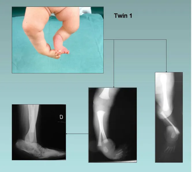
Case 1
The girl was first seen at the age of two months and presented with malformation of the lower limbs and left hand (fig. 3). Left leg: tibial aplasia type Ia with a left femur 10% shorter than the right, absent patella; left foot: club foot, totally absent first ray. Right leg: tibial aplasia type IV; right foot: clubfoot with a hypoplastic internal ray. Left hand: inter-metacarpal split with a partially absent third ray, a supernumerary metacarpus and a partial syndactyly of the fourth commissure. Neuropsychological development was normal.
At one year, a left knee desarticulation and a right foot reposition with astragalectomy was performed using two transcalcaneo-fibular pins and a horizontal pin to fix rotation. During the same surgical procedure, hand correction was carried out with excision of the third and supernumerary metacarpus, placing together the second and fourth metacarpus, cutaneous plastics of the second commissure, and deepening of the fourth commissure. A prosthesis was fitted to her left limb. Seven months later, she could walk alone with the prosthesis and the left hand showed a good functionality. The prosthesis was regularly adapted to her growth. However, progressive internal rotation of the right tibia, equinus and adductus of the underlying foot required two further operations at three years of age (fibular osteotomy and soft tissue release) and at six years of age (fibular osteotomy and calcaneal osteotomy using a plaque fixed exteriorly on calcaneus). At the last clinical follow-up, the patient was an active and dynamic young girl of fourteen years, schooled and practicing swimming.
Figure 3 Case 1.
Upper left: clinical presentation at the age of two months. Lower right: radiographs of right and left limbs at two months.
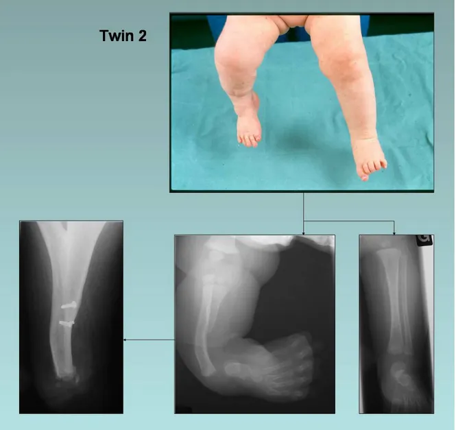
Case 2
This monozygotic twin sister of case 1 was seen for the first time with her sib at the age of two months (fig. 4) and presented with malformations of the right lower limb and left upper limb. Radiographs revealed right tibial aplasia type II. She presented also a right club foot, a left hand with an inter-metacarpal split, partial aplasia of the third ray and a supernumerary metacarpus. In other respects, neuropsychological development was within normal limits.
She was first operated at the age of four months. A soft tissue release on the medial side of the right foot, an astragalectomy and a fibulo-calcaneal synostosis (using a trans-fibulo-calcaneal pin) were carried out with the intent to align her distal lower limb. Nevertheless, her right foot kept a supple tendency to varus and adductus. An orthesis was adapted to fix it in a position to permit normal gait acquisition. At the age of one year she could walk with this orthesis. During the same operative session, tibialisation of the right fibula and hand surgery (excision of the third and the supernumerary metacarpus, internal collateral ligament of index MCP plastics, and cutaneous plastics of the second new commissure) were performed. Four months later, a lower limb length inequality (the right one being 1.5 cm shorter than the left) was observed, and a functional left hand. Due to the persistence of right foot adductus and varus, a new soft tissue release was carried out at the age of three
and length inequality was now measured at 5 cm (compensated by a shoe lift). However, the right foot showed a good functionality and was not painful. Over time, leg discrepancy increased, right knee instability appeared, and the right foot accentuated its varus and adductus with internal rotation and equinus. At 11 years, the patient experienced a non-traumatic external patellar dislocation. A knee orthesis was prescribed to protect the right knee above the foot. This very active young girl requested a right foot amputation because it prevented the good fitting of her right lower limb-lengthening prosthesis. At fourteen years, a Boyd amputation was performed on the right side. However, due to secondary detachment of the calcaneal fragment, impaired wound healing and persistence of a painful stump, it had to be converted to a Syme amputation. Postoperative evolution was good and permitted good prosthesis fitting with excellent functional results.
Figure 4 Case 2.
Upper right: clinical presentation at the age of two months. Lower right: radiographs of right and left limbs at two months.
Case 3
This boy was first seen at the age of three weeks (fig. 5). Delivery had to be induced three weeks before term because of preeclampsia. Parents were not consanguineous. Family history was negative for congenital abnormalities. There were no other sibs. Radiographs revealed a right tibial aplasia type II with a short right fibula (right fibula, 6 cm; left, 7 cm). Clinically, he presented a right short leg and a right clubfoot, but apparently no other malformation. Neuropsychological development was normal.
At the age of nine months he underwent a surgical correction of the foot using two trans-calcaneo-astragalo-fibular pins to fix the rotation and a trans-fibulo-astragalo-cuboido-first metacarpus pin to correct adduction. At one year of age, he began to walk with an orthesis. At the age of two years, distal translation of fibula was performed using an Orthofix external fixator. Two pins were placed on proximal and distal tibial fragment, and a third pin was placed on the distal fibula. A first extemporaneous distraction of 1 cm was performed and followed by progressive distraction carried out during one month to obtain a 3 cm total translation of the fibula. At that time, removal of the external fixator was carried out and a tibio-fibular synostosis was performed using two screws and two cerclages (fig. 6). Two months later, synostosis showed signs of clinical and radiological consolidation and permitted removal of the cast. At four years of age, he presented a 5 cm shortening of the
right leg. An arthrodesis of the talus and the lower end of the fibula [De Sanctis et al. 1990] using two trans-plantar Kirchner pines and one pin to fix rotation was carried out to facilitate future lengthening. Ten weeks later, radiological signs of consolidation of the arthrodesis permitted removal the cast. He rapidly began to walk with an orthesis and compensated sole without pain and functional impairment. At six years of age, he showed a 6 cm shortening of the right leg which was totally corrected using an Ilizarov external fixator treatment during four and half months. At eight years of age, he began skiing. At eleven, clinical follow-up revealed a 7 cm shortening of the right leg. A new Ilizarov external treatment was started and symmetrical leg length was obtained.
Figure 5 Case 3.
Left: clinical presentation at three weeks old.
Right: radiograph at first day of life showing tibial aplasia type II and short fibula.
Figure 6
Case 4
This girl was first seen at the age of four years and had been brought to Geneva from Guinea with humanitarian aid. She presented bilateral tibial aplasia type Ia on the left side (fig. 7), and type II on the right side (fig. 8) with bilateral club feet, but without any other malformation. She could walk on her knees. Apparently, first-degree relatives presented no congenital abnormalities. We had no information about pregnancy and delivery. Neuropsychological development seemed not to be delayed.
Due to the short time allocated for her stay by the humanitarian program and her age, but with the aim to obtain the best functional result, the two limbs were operated during the same surgical procedure: Boyd amputation with tibio-peroneal synostosis on the right side, and knee desarticulation on the left side (fig. 9). Postoperative evolution was complicated by incomplete right tibio-peroneal fusion which needed re-operation 19 days later. Prostheses were fitted one and half months later on the left, and two and half months later on the right. The gait was satisfactory at that time.
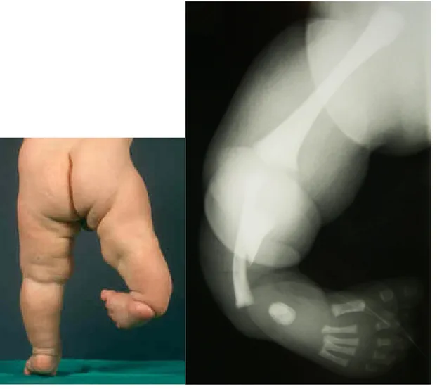
Figure 7
Case 4: clinical presentation and radiographic examination of the left leg (tibial aplasia type Ia).
Figure 8
Case 4: clinical presentation and radiographic examination of the right leg (tibial aplasia type II).
Figure 9
Case 4: clinical result and radiographic examination of the right leg three months after surgery.
Discussion Embryology
Limb morphogenesis takes place from the fourth to the eighth week; lower limb development lags slightly (a few days) behind the
development of the upper limbs [Clavert 2000, Larsen 2001]. The first morphogenetic movement which can be described, adduction, takes place in the hip [Clavert 2000]. By the end of the fourth week, the lower limbs buds appear at the level of L3 to L5 and are formed by a
mesenchymal core of mesoderm covered by an ectodermal cap. An ectoderm apical ridge, formed by the thickening of the ectoderm, promotes the outgrowth of the limb bud in a proximodistal manner [Larsen 2001]. During the fifth week, mesenchyme of the limb buds undergoes condensations to form mesenchymal bone models. Then, during the sixth week, chondrification within these mesenchymal bones produces cartilaginous precursors. All the structural elements of the upper and lower limbs are distinct by the end of the eighth week. Bone models are developed in a proximodistal sequence, and patterning in the developing limb is regulated by homeobox-containing (Hox) genes
[Moore 2003].
Ossification of these hyaline cartilage bone models begins in the long bone in the eighth to twelfth weeks. It initially takes place in the diaphysis of the bones from primary centres of ossification. By the twelfth week, these primary centres have appeared in nearly all bones of the
limbs. Digital rays appear on the foot plates during the seventh week (one week after the digital rays of the hand plate). A selective
programmed cell death sculpts out these rays to free the fingers and toes. The first secondary ossification centres to appear are those at the knee (centres for the distal end of the femur and the proximal end of the tibia). They appear during the last month of intrauterine life (34 to 38 weeks after fertilization) and are usually present at birth. Most secondary centres of ossification appear nevertheless after birth; epiphysis is the part of the bone formed by this ossification. Thus, the bones (as blood vessels) of the limbs originate from the lateral plate mesoderm of the limb buds, while limb muscles arise from somatic mesoderm that invades the limb bud [Moore 2003, Larsen 2001].
The concept of developmental field and of developmental field defects
As some specific malformations of the lower limbs have been demonstrated to show causal heterogeneity (for example, occurrence of fibular aplasia in sporadic cases, but also in autosomal dominant, autosomal recessive and sporadic syndrome [Lewin and Opitz 1986]), some authors have proposed the concept of developmental field defects, i.e., “dysmorphogenetically reactive units”: “a set of embryonic primordia that reacted identically to different dysmorphogenetic causes” [Opitz 1985]. These primordia must also constitute a morphogenetically reactive unit during the normal ontogeny, hence a developmental field [Lewin and Opitz 1986, Opitz 1985].
Two distinct developmental fields have been described in the lower limbs after careful clinical observations of specific malformations. Most of the time, the fibular field includes the pubic portion of the pelvis, proximal femur, patella, anterior cruciate ligament and, secondarily, all the nerves, muscles, arteries and veins intimately associated with fibular development, as well as the lateral and/or axial foot rays (i.e., fibular foot rays). Of note, it never includes the hallux and is almost never associated with polydactyly [Lewin and Opitz 1986]. Talus, cuboid, and variable degrees of talocalcaneal fusion which might also involve the cuboid (if present) and other tarsal bones have also been included in defects of this field [O’Rahilly 1951]. Less commonly, the ischion may be
involved. Furthermore, the rare cases of fibuloulnar dimelia reported allowed to confirm that in upper and lower limbs, respectively, ulna and fibula and radius and tibia are homologous structures. Concordant ulnar and fibular (and radial and tibial) defects are present if both upper and lower limbs are involved [Lewin and Opitz 1986].
Given that tibial aplasia is much rarer than fibular aplasia, the most common of the four hemimelias, the tibial field defect is not so clearly described in the literature. It appears to involve the distal half of the femur (thus causing distal femoral defects such as femoral duplication or bifurcation associated with tibial defects), tibia, and hallux, or preaxial toes with preaxial polydactyly. Occasionally, it is associated homologously with hand ectrodactyly, similar to tibial aplasia ectrodactyly or the Gollop-Wolfgang complex (tibial aplasia, ectrodactyly and femoral bifurcation) [Lewin and Opitz 1986, Richieri-Costa et al 1987a]. These two developmental field defects have been confirmed by clinical data in other reports [Khoury et al 1999, Pavone et al 1989, Sorge et al 1995].
Genetics
Tibial defects are most often unilateral and sporadic [Majewski et al. 1985]. There are four recognized autosomal dominant tibial hemimelia syndromes: 1) the tibial hemimelia-foot polydactyly-triphalangeal thumbs syndrome (Werner syndrome), 2) the tibial hemimelia diplopodia syndrome, 3) the tibial hemimelia-split-hand foot syndrome, and 4) the tibial hemimelia-micromelia-trigonobrachycephaly syndrome [Richieri-Costa et al. 1987b]. Tibial hemimelia-split-hand foot syndrome is a very rare malformation with a highly variable phenotype, which ranges from isolated minor malformations like anonychia of some toes to isolated syndactyly of fingers, to split-hand/foot with or without tibial aplasia (uni- or bilateral) [Majewski et al. 1985, Richieri-Costa et al. 1987b]. In most cases, there is an autosomal dominant inheritance with variable expressivity and reduced penetrance, but some examples suggest an autosomal recessive inheritance and sporadic cases have been described [Majewski et al. 1985, Richieri-Costa et al. 1987b]. Case 1 and 2 seem to present this syndrome. To our knowledge these are the first monozygotic twins with tibial hemimelia split-hand foot syndrome described in the literature. They further demonstrate the highly variable phenotypic manifestations of this syndrome, despite an identical genotype. Given reduced penetrance and phenotypic variability, genetic counseling is difficult in such families. Risk to the offspring of unaffected person with one affected (or gene assumed carrier by familial history)
parent is maximally 8,6% at a penetrance of 60%. It decreases with higher or lower penetrance [Aylsworth and Kirkman 1979]. Risk to a sib of an isolated patient is maximally 12,5% at a penetrance of 50% [Frota-Pessoa et al. 1976]. Finally, recurrence risk to offspring of healthy sibs of affected may be up to 25% [Majewski et al. 1996]. Because of these risks, prenatal diagnosis should be recommended in these situations. Prenatal diagnoses of unilateral tibial aplasia [Dreyfus et al. 1996] and of tibial hemimelia syndrome [Ramirez et al. 1994] by real-time ultrasound have been reported. In those two cases, pregnancy was electively terminated.
Case 3 illustrates an isolated unilateral tibial aplasia, without family history of even minor malformation. This points to a teratologic incident or a de novo mutation in the affected patient. Risk of recurrence is probably very low, but must take in account the possibility of germline mosaicism in one of the parents. It must be kept in mind that the search for minor anomalies in the family of affected patient is essential for accurate genetic counseling [Richieri-Costa et al. 1987b].
Case 4 is an example of a bilateral isolated case (without other malformation) with healthy first degree relatives. This could rule out a tibial hemimelia syndrome, but we cannot affirm this without a precise and complete familial history.
Therapeutics
Different, and sometimes contradictory, guidelines for treatment have been proposed. It seems that some conservative procedures initially proposed have now to be rejected. For type Ia, the treatment of choice is knee disarticulation, preferably during the first year of life [Loder and Herring 1987] before the cerebral image takes place.
Several authors [Epps et al. 1989, Epps et al. 1991, Fernandez-Palazzi et al. 1998, Kalamchi and Dawe 1985, Loder and Herring 1987, Schoenecker et al. 1989] have reported the inefficiency of the Brown operation (centralisation of fibula, followed by Syme amputation [Brown 1965]) for treatment of type Ia aplasia. This procedure was followed in most cases by flexion contracture of the knee and requiring one or more secondary procedures such as knee disarticulation, posterior release, extension osteotomy, femorofibular arthrodesis, and biceps to quadriceps transfer. Furthermore, it delayed early rehabilitation and fitting with a prosthesis.
For type Ib, the therapeutic choice is more difficult. According to Fernandez-Palazzi, types Ia and Ib require knee disarticulation [Fernandez-Palazzi et al. 1998]. However, Schoenecker recommends to verify the presence of tibia by arthrography, direct surgical exploration of knee joint, or ultrasonograhy [Schoenecker et al. 1989]. If a cartilaginous anlage of the proximal tibia is observed, knee disarticulation should be deferred and a prosthesis fitted after a Syme amputation. Tibiofibular
synostosis may be possibly performed later and may permit to preserve a functional knee.
Management of type II deficiencies must include an attempt to preserve a functional knee as these patients have a predicted satisfactory function as below-the-knee amputees [Schoenecker et al. 1989]. For this reason, Syme amputation and tibiofibular synostosis are proposed [Fernandez-Palazzi et al. 1998, Schoenecker et al. 1989]. However, de Sanctis [de Sanctis et al. 1990] presented an alternative to amputation in type II tibial aplasia consisting of four steps: early foot correction; tibiofibular synostosis; alignment of the extremity; and finally leg lengthening by Ilizarov`s technique. Results reported on three cases are encouraging. Furthermore, Carranza-Bencano [Carranza-Bencano and Gonzales-Rodriguez 1999] reported the case of a 15-year-old girl with type II unilateral tibial agenesia, a 13.5 cm shortening of the right limb, tibiofibular synostosis, cavus, adductus and varus of the right foot who had rejected amputation. Lower limb inequality and foot deformity were treated by means of an external fixator, and she could walk freely. These cases, although certainly not sufficient in terms of number, suggest a more conservative therapeutic choice for type II aplasia.
The atittude seems more univocal for patients with a type III malformation. Syme or Chopart amputation is recommended [Fernandez-Palazzi et al. 1998, Schoenecker et al. 1989] since the
initially cartilaginous proximal part of the tibia will develop to form a functional knee.
Reconstructive surgery of the ankle (closure of the diastasis) is possible for a type IV deformity, but the additional problem of lower limb length inequality will persist [Schoenecker et al. 1989]. It can be corrected nevertheless with successive external fixator treatments. Another more aggressive option proposed for type IV is Syme amputation [Fernandez-Palazzi et al. 1998, Schoenecker et al. 1989] with the fitted prosthesis lengthened according to growth.
Finally, associated malformations, particularly those concerning the underlying foot (equinovarus deformation, hypoplasia of the internal ray, partial duplication of the foot, rear foot synostosis [Bronfen et al. 1994, Diamond 1979]), have to be taken into account in the management of tibial congenital deficiencies for a better functional result.
Dissection
We present the postoperative dissection of the left leg, and left and right feet of case 4, and compare our observations with previous anatomical findings on tibial aplasia.
Left leg
Bone: there was no fibrous band replacing the tibia (this fibrous band is frequently described in the literature [Dankmeijer 1935, Evans and Smith 1926, Freund 1936, Hootnick et al. 1983, 1987, M`laren 1888-1889, Rodriguez-Baeza et al. 1997], but it appears not to be always present [Selke and Bogusch 1989]).
Muscles: muscles normally originating from the tibia had their proximal insertion on the fibula as described by Selke [Selke and Bogusch 1989] (in the case of a fibrous band replacing the tibia, muscles normally originating from the tibia have their origin on this fibrous band [Hootnick et al. 1983, 1987], or on a cartilaginous structure found at the distal articular surface of the knee [Rodriguez-Baeza et al. 1997]). All the muscles of the leg were present except for the tibialis posterior. The same anomaly has been reported by Rodriguez-Baeza in the case of tibial aplasia with ectrodactyly and suggests that the development of the skeletal elements have an important role in the differentiation of the muscles notably, but also of tendons, arteries and nerves.
the flexor hallucis brevis and flexor digitorum brevis. Similar findings have also been reported [Selke and Bogusch 1989] and, according to Selke, indicate that there are connections between tendons of muscles from different blastemas. No plantaris tendon was identified, but this is also frequently reported [Bergman et al. 1984, Hootnick et al. 1983, 1987].
Nerves: all the nerves were present and normally distributed, except for the tibial nerve, which was absent; this has not been described by other authors [Hootnick et al. 1983, 1987, Rodriguez-Baeza et al. 1997]. An anomaly described in the literature is the failure of the superficial peroneal nerve to enter the dorsum of the foot [Hootnick et al. 1987], but this is not always the rule [Hootnick et al.1983, Rodriguez-Baeza et al. 1997].
Arteries: the anterior tibial and dorsalis pedis arteries were absent. Reduction or absence of these two arteries and an incomplete plantar arch have also been noted [Ben-Menachem and Butler 1974, Edelson and Husseini 1984, Greider et al. 1982, Hootnick et al. 1980, 1982, 1983, 1984, 1987, Levinsohn et al. 1991]. Hootnick speculated that reduction in blood flow during development could put the limb at risk of teratogenic damage, and that vascular anomalies and congenital tibial aplasia are etiologically related [Hootnick et al. 1987].
The inferior cartilaginous surface of the fibula was anteriorly oriented and articulate with the posterior surface of the talus (fig. 10). There was an important movement restriction at this level, and we found a fibrous tissue connecting the two cartilaginous surfaces. Histological analysis revealed fibrous cartilage. This could be another example of a tibiofibular syndesmosis, as described by Selke [Selke and Bogusch 1989], rather than the normal diarthrosis.
Left foot
All the bones were present (fig. 11), including the sesamoids, but we noted marked hypoplasia of the cuboid and discrete hypoplasia of the median and intermediary cuneiforms. An anterior talo-calcaneal fusion was observed; tarsal coalition in tibial aplasia is often reported [Hootnick et al. 1983, 1987, Selke and Bogusch 1989, Diamond 1979]. For Diamond [Diamond 1979], massive tarsal coalition appeared to be an obligatory part of what he called the tibial hemimelic syndrome. Detailed soft tissue dissection of the two feet was not performed because of inadequate tissue conservation due to the use of an incorrect conservation solution.
Figure 10
Left fibula and foot (note the anterior orientation of the inferior articular surface of the fibula).
Figure 11
Right foot
Skeletal elements were the same as the left foot, i.e., all present and normally distributed (fig. 12). Hypoplasia of the cuboid and of the median and intermediary cuneiforms were also present. For the same reason as for the left foot, we could not perform extra-skeletal dissection.
Figure 12
Conclusion
Tibial hemimelia, although a rare congenital anomaly, is a well-described entity in pediatric orthopedics. Knowledge of the different aspects of this pathology is essential for an adequate global management. First, correct diagnosis, classification, and evaluation of the associated malformations will permit to orient appropriate therapy, with priority always given to the functional result. Furthermore, surgeons must keep in mind the various anatomical anomalies associated with this condition. In this perspective, certain pre-operative examinations may help to prove the presence or absence of specific anatomical structures, for example, arteriography, to evaluate the permeability of the anterior tibial artery. Also of importance, a careful assessment of family history and an attentive physical examination of the patient with reporting of every minor anomaly (such as anonychia of some toes) will help to define the need for genetic counseling which remains difficult in the case of tibial hemimelia syndrome, given the reduced penetrance. The dissection presented here shows that the anatomy of the leg and foot in tibial aplasia is variable; some anomalies are frequent, but not always present. Of note, not only are the bones affected, but also the soft tissues such as muscles, vascular elements, and nerves.
Bibliography
1. Aylsworth AS, Kirkman HN. Genetic counseling for autosomal
dominant disorders with incomplete penetrance. Birth Defects Orig Artic
Ser 1979;15:25-38.
2. Ben-Menachem Y, Butler JE. Arteriography of the foot in congenital deformities. J Bone Joint Surg Am 1974;56:1625-30.
3. Bergman RA, Thompson SA, Afifi AK. Catalog of human variation. Catalog of human variation. Baltimore: Urban & Schwarzenberg, 1984:59-60.
4. Bronfen C, Rigault P, Padovani JP, Touzet P, Finidori G, Chaumien JP. [Foot deformities in longitudinal ectromelia of the lower limbs]. Int
Orthop 1994;18:139-49.
5. Brown FW. Construction of a knee joint in congenital total absence of the tibia (paraxial hemimelia tibia): a preliminary report. J Bone Joint
Surg Am 1965;47:695-704.
6. Brown FW. The Brown operation for total hemimelia tibia. In: Aitken GT, ed. Selected lower-limb anomalies: surgical and prosthetics
management. Washington, DC: National Academy of Sciences, 1971:21-8.
7. Carranza-Bencano A, Gonzalez-Rodriguez E. Unilateral tibial hemimelia with leg length inequality and varus foot: external fixator treatment. Foot Ankle Int 1999;20:392-6.
8. Clavert JM. Embryologie normale et pathologique des membres inférieurs, essai de classification des malformations. In: Carlioz H, Clavert JM, eds. Malformations congénitales des membres inférieurs. Paris: Elsevier, 2000:3-15.
9. Dankmeijer J. Congenital absence of the tibia. Anat Rec 1935;62:179-94.
10. De Sanctis N, Razzano E, Scognamiglio R, Rega AN. Tibial agenesis: a new rationale in management of type II--report of three cases with long-term follow-up. J Pediatr Orthop 1990;10:198-201. 11. Diamond LS. Tarsal coalition in paraxial hemimelia of lower extremity. Orthopaedic Review 1979;8:91-9.
12. Dreyfus M, Baldauf JJ, Rigaut E, Clavert JM, Gasser B, Ritter J. Prenatal diagnosis of unilateral tibial hemimelia. Ultrasound Obstet
Gynecol 1996;7:205-7.
13. Edelson JG, Husseini N. The pulseless club foot. J Bone Joint Surg
Br 1984;66:700-2.
14. Epps CH, Jr., Schneider PL. Treatment of hemimelias of the lower extremity. Long-term results. J Bone Joint Surg Am 1989;71:273-7. 15. Epps CH, Jr., Tooms RE, Edholm CD, Kruger LM, Bryant DD, 3rd. Failure of centralization of the fibula for congenital longitudinal deficiency of the tibia. J Bone Joint Surg Am 1991;73:858-67.
17. Fernandez-Palazzi F, Bendahan J, Rivas S. Congenital deficiency of the tibia: a report on 22 cases. J Pediatr Orthop B 1998;7:298-302.
18. Freund E. Congenital defects of femur, fibula and tibia. Arch Surg 1936;33:349-91.
19. Frota-Pessoa O, Otto PA, Plaza JR. The variation of recurrence risks with penetrance for isolated cases of autosomal dominant conditions. J
Hered 1976;67:256.
20. Greider TD, Siff SJ, Gerson P, Donovan MM. Arteriography in club foot. J Bone Joint Surg Am 1982;64:837-40.
21. Herbaux B. Hypoplasies du rayon tibial. In: Carlioz H, Clavert JM, eds. Malformations congénitales des membres inférieures. Paris: Elsevier, 2000:49-60.
22. Hootnick DR, Levinsohn EM, Crider RJ, Packard DS, Jr. Congenital arterial malformations associated with clubfoot. A report of two cases.
Clin Orthop 1982:160-3.
23. Hootnick DR, Levinsohn EM, Randall PA, Packard DS, Jr. Vascular dysgenesis associated with skeletal dysplasia of the lower limb. J Bone
Joint Surg Am 1980;62:1123-9.
24. Hootnick DR, Packard DS, Jr., Levinsohn EM. Congenital tibial aplasia with preaxial polydactyly: soft tissue anatomy as a clue to teratogenesis. Teratology 1983;27:169-79.
25. Hootnick DR, Packard DS, Jr., Levinsohn EM, Cady RB. Soft tissue anomalies in a patient with congenital tibial aplasia and talo-calcaneal
synchrondrosis. Teratology 1987;36:153-62.
26. Hootnick DR, Packard DS, Jr., Levinsohn EM, Lebowitz MR, Lubicky JP. The anatomy of a congenitally short limb with clubfoot and
ectrodactyly. Teratology 1984;29:155-64.
27. Jones D, Barnes J, Lloyd-Roberts GC. Congenital aplasia and
dysplasia of the tibia with intact fibula. Classification and management. J
Bone Joint Surg Br 1978;60:31-9.
28. Kalamchi A, Dawe RV. Congenital deficiency of the tibia. J Bone
Joint Surg Br 1985;67:581-4.
29. Khoury NJ, Haddad MC, Hourani MH. Tibial and fibular developmental fields defects. Eur Radiol 1999;9:1879-81.
30. Larsen WJ. Development of the limbs. In: Larsen WJ, ed. Human embryology. Philadelphia, PE: Churchill Linvingstone, 2001:315-47. 31. Levinsohn EM, Hootnick DR, Packard DS, Jr. Consistent arterial abnormalities associated with a variety of congenital malformations of the human lower limb. Invest Radiol 1991;26:364-73.
32. Lewin SO, Opitz JM. Fibular a/hypoplasia: review and documentation of the fibular developmental field. Am J Med Genet Suppl 1986;2:215-38. 33. Loder RT, Herring JA. Fibular transfer for congenital absence of the tibia: a reassessment. J Pediatr Orthop 1987;7:8-13.
34. Majewski E, Goecke T, Meinecke P. Ectrodactyly and absence (hypoplasia) of the tibia: are there dominant and recessive types? Am J
35. Majewski F, Kuster W, ter Haar B, Goecke T. Aplasia of tibia with split-hand/split-foot deformity. Report of six families with 35 cases and considerations about variability and penetrance. Hum Genet
1985;70:136-47.
36. M'Laren JS. A case of congenital absence of the tibia. J AnatPhysiol 1888-1889;23:598-605.
37. Moore KL. The skeletal system. In: Saunders, ed. The developing human: clinically oriented embryology, 2003:381-99.
38. Myers TH. Congenital absence of tibia: Transplantation of head of the fibula: Arthrodesis at the ankle-joint. Am J Othop Surg 1905;3:72-85. 39. Opitz JM. The developmental field concept. Am J Med Genet
1985;21:1-11.
40. O'Rahilly R. Morphological patterns in limb deficiencies and duplications. Am J Anat 1951;89:135-93.
41. Otto AW Monstorum sexeentorum descripto anatomica sumptibus. Breslau: Ferdinandi Hirt; 1841.
42. Pavone L, Viljoen D, Ardito S, Rizzo R, Neri G, Longo G, Beighton P. Two rare developmental defects of the lower limbs with confirmation of the Lewin and Opitz hypothesis on the fibular and tibial developmental fields. Am J Med Genet 1989;33:161-4.
43. Ramirez M, Hecht JT, Taylor S, Wilkins I. Tibial hemimelia syndrome: prenatal diagnosis by real-time ultrasound. Prenat Diagn 1994;14:167-71.
44. Richieri-Costa A, Brunoni D, Laredo Filho J, Kasinski S. Tibial aplasia-ectrodactyly as variant expression of the Gollop-Wolfgang complex: report of a Brazilian family. Am J Med Genet 1987;28:971-80. 45. Richieri-Costa A, Ferrareto I, Masiero D, da Silva CR. Tibial
hemimelia: report on 37 new cases, clinical and genetic considerations.
Am J Med Genet 1987;27:867-84.
46. Rodriguez-Baeza A, Minguella J, Soldado F, Ortega M, Escola J. Congenital tibial aplasia with preaxial polydactyly. A case report. Acta
Anat (Basel) 1997;160:51-61.
47. Schoenecker PL, Capelli AM, Millar EA, Sheen MR, Haher T, Aiona MD, Meyer LC. Congenital longitudinal deficiency of the tibia. J Bone
Joint Surg Am 1989;71:278-87.
48. Selke B, Bogusch G. [Changes in bones of the foot and development of inserting tendons of the lower leg musculature in tibial aplasia]. Acta
Anat (Basel) 1989;136:69-75.
49. Sorge G, Ardito S, Genuardi M, Pavone V, Rizzo R, Conti G, Neri G, Katz BE, Opitz JM. Proximal femoral focal deficiency (PFFD) and fibular A/hypoplasia (FA/H): a model of a developmental field defect. Am J Med