Révision taxonomique de la famille des Harrimaniidae (Hemichordata: Enteropneusta) incluant les descriptions de sept espèces de la côte Est du Pacifique
par Carine Deland
Département de sciences biologiques Faculté des Arts et des Sciences
Mémoire présenté à la Faculté des études supérieures en vue de l’obtention du grade de M.Sc.
en sciences biologiques
30 avril 2010
Ce mémoire intitulé :
Révision taxonomique de la famille des Harrimaniidae (Hemichordata: Enteropneusta) incluant les descriptions de sept espèces de la côte Est du Pacifique
Présenté par : Carine Deland
A été évalué par un jury composé des personnes suivantes :
Pierre Brunel président-rapporteur Christopher Cameron directeur de recherche Michel Anctil Membre du jury
Résumé
Cette étude comparative est une révision de la famille des Harrimaniidae basée sur les caractères morphologiques d'espèces connues et nouvelles provenant des collections de William E. Ritter, Theodore H. Bullock et Kandula P. Rao rassemblées au cours du 20e siècle. Les descriptions présentées ici portent le total des genres de cinq à neuf par l'ajout de Horstia n. gen., Mesoglossus n. gen., Ritteria n. gen et Saxipendium, un genre auparavant attribué à la famille monospécifique des Saxipendidae. Le nombre d'espèces est porté à 34 par la description de cinq nouvelles espèces du Pacifique oriental: Horstia kincaidi, Mesoglossus intermedius, Mesoglossus macginitiei,
Protoglossus mackiei et Ritteria ambigua. La description d'une sixième espèce, Stereobalanus willeyi Ritter et Davis, 1904 (nomen nudum) est présentée ici pour la
première fois, ainsi qu'une description abrégée de Saxipendium coronatum. Quatre espèces précédemment attribuées au genre Saccoglossus sont transférées au genre
Mesoglossus: M. bournei, M. caraibicus, M. gurneyi, et M. pygmaeus et Saccoglossus borealis est transféré au genre Harrimania. Une hypothèse phylogénétique sur la famille
des Harrimaniidae est émise, présentant l'évolution possible des caractères morphologiques au sein du groupe. Finalement, des notes sur la distribution géographique étendue mais discontinue de plusieurs espèces suggère que les entéropneustes auraient pu avoir une distribution ancienne continue et plus grande qui aurait été fragmentée par la suite.
Mots-clés: Taxonomie, révision systématique, morphologie, Enteropneusta,
Summary
This comparative study is a revision of the family Harrimaniidae based on morphological characters of described and undescribed species from the collections of William E. Ritter, Theodore H. Bullock and Kandula P. Rao, gathered in the 20th century. The new descriptions bring the total number of genera to nine by the addition of
Horstia n. gen., Mesoglossus n. gen., Ritteria n. gen and Saxipendium, a genus
previously assigned to the monospecific family Saxipendidae The number of species is increased to 34, resulting from the description of five new species from the eastern Pacific: Horstia kincaidi, Mesoglossus intermedius, Mesoglossus macginitiei,
Protoglossus mackiei and Ritteria ambigua. The description of a sixth species, Stereobalanus willeyi Ritter et Davis, 1904 (nomen nudum) is presented here for the first
time and a brief description of Saxipendium coronatum is also presented. Four species previously assigned to the genus Saccoglossus are transfered to the genus Mesoglossus:
M. bournei, M. caraibicus, M. gurneyi, and M. pygmaeus, while Saccoglossus borealis
is transfered to the genus Harrimania. A phylogenetic hypothesis on the Harrimaniidae is postulated presenting the possible evolution of morphological characters within the group. Finally, notes on the wide but spotty distribution of several species suggest that the Enteropneusta may have once had a wider distribution that has since become fragmented.
Key words: Taxonomy, systematic revision, morphology, Enteropneusta, Hemichordata,
Table des matières
Résumé--- Summary---Table des matières---Listes des tableaux---Liste des figures---Liste des sigles et des abréviations---
1. Introduction---
1.1 Biologie--- 1.2 Anatomie--- 1.3 Systématique--- 1.4 Données historiques inédites--- 1.5 Contribution des différents auteurs---
2. L'ancêtre deutérostomien et la génétique moderne--- 3. Objectifs du travail--- 4. Article: A taxonomic revision of the family Harrimaniidae (Hemichordata:
Enteropneusta) with descriptions of seven species from the Eastern Pacific--- 29
Accord des coauteurs--- Abstract--- Introduction--- The specimen collection--- Diagnostic morphological characters--- Material and Methods--- Results---
Systematic diagnoses of family and gerena--- Species descriptions---
Stereobalanus willeyi sp. nov.--- Protoglossus mackiei sp. nov--- Saxipendium coronatum Woodwick & Sesenbaugh, 1985--- Mesoglossus intermedius gen. & sp. nov.--- Mesoglossus macginitiei gen. & sp. nov.--- Ritteria ambigua gen. & sp. nov.--- Horstia kincaidi gen. & sp. nov.---
Discussion--- Zoogeography--- Phylogeny--- Acknowledgments--- Appendix---References--- 5. Discussion---
5.1 Validité des caractères taxonomiques--- 5.2 Phylogénétique--- 5.3 Zoogéographie--- 5.4 Suite du projet--- 5.5 Perspectives--- iii iv v vi vii x 1 2 3 4 6 7 9 12 14 15 16 17 18 19 20 22 22 30 30 34 37 41 44 48 52 56 57 59 61 62 66 71 72 73 74 75 75
Liste des tableaux
Table I Comparison of external and internal characters of the species of the family Harrimaniidae, excluding members of the genus Saccoglossus, which will be discussed in a subsequent paper. Characters from previously described species were either obtained from the literature, or, in the case of Harrimania maculosa and Saxipendium coronatum, from the literature and the holotypes. Question marks indicate when a character state is unknown.---62
Table II A dichotomous key to the enteropneust families Harrimaniidae, Spengelidae, Ptychoderidae and Torquaratoridae and to the genera of family Harrimaniidae.---64
Liste des figures
Figure 1 Schéma d'un entéropneuste généralisé, modifié de Cameron, 2005. a, anus; b, bouche; ch, caecum hépatique; cms, canal du mésocoele; cnd, corde nerveuse dorsale; co, coeur; cp, coelome périhémal; cpb, cavité péri-buccale; fb, fente branchiale; g, glomérule; go, gonade; md, mésentère dorsal; ms, mésocoele; mt, métacoele; opc, organe pré-oral cilié; pb, pore branchial; pp, pore du proboscis; ps, péritoine viscéral; pt, protocoele; qp, queue post-anale; s, stomocorde; sbr, sac branchial; sca, sinus cardiaque; se, septum; sqp, squelette du proboscis; vsd, vaisseau sanguin dorsal; vsv, vaisseau sanguin ventral.---4
Figure 2.1 Light micrographs of transverse sections of the proboscis of (A) Saccoglosus pusillus, showing the arrangement of the proboscis longitudinal musculature in concentric rings; (B)
Protoglossus mackiei n. sp., showing the arrangement of the proboscis longitudinal
musculature in radial plates; (C) Mesoglossus macginitiei n. gen. and sp., showing the diffuse arrangement of the proboscis longitudinal musculature. plm, proboscis longitudinal muscles. Scale bars = 500 !m.---23
Figure 2.2 Drawings of live animals and photographs of fixed specimens of the family Harrimaniidae.
(A) Drawing of the dorsal side of Horstia kincaidi n. gen. and sp.. (B) Drawing of the lateral side of Mesoglossus intermedius n. gen and sp.. (C) Drawing of the dorsal side of
Mesoglossus intermedius. (D) Drawing of the dorsal side of Stereobalanus willeyi n. sp.. (E)
Drawing of the lateral side of Stereobalanus willeyi. (F) Photograph of the dorsal side of
Horstia kincaidi. (G) Photograph of the lateral side of Ritteria ambigua n. gen. and sp.. (H)
Photograph of the dorsal side of Mesoglossus intermedius. (I) Photograph of the dorsal side of Stereobalanus willeyi. (J) Photograph of the lateral side of Stereobalanus willey. bp, branchial pore; c, collar; gr, genital ridge; p, proboscis; t, trunk. Scale bars = 2 mm.---29
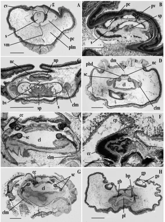
Figure 2.3 Light micrographs of transverse sections of Stereobalanus willeyi n. sp.. (A) Proboscis with
heart-kidney complex. (B) Posterior part of the proboscis showing the skeleton. (C) Proboscis neck. (D) Anterior region of the collar. (E) Anterior pharyngeal region of the trunk. (F) Genital region of the trunk. cl, collar lumen; cv, cardiac vesicle; g, glomerulus; gb, gill bar; go, gonad; nc, nerve cord; np, neuropore; pbr, parabranchial ridge; pc, proboscis coelom; pl, pharynx lumen; plm, proboscis longitudinal muscles; pv, proboscis vesicle; s, stomochord; sb, skeletal body; sc, skeletal cornua; sl, stomochord lumen; vc, ventral caecum; vm, ventral mesentery. Scale bars (A, D, E and F) = 1000 !m; (B) = 500 !m; (C) = 750 !m.---33
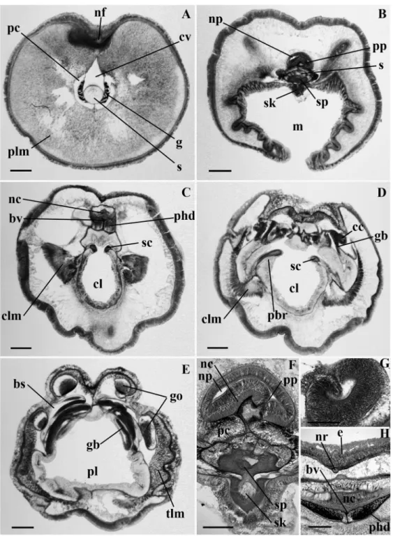
Figure 2.4 Light micrographs of transverse sections of Protoglossus mackiei n. sp.. (A) Proboscis with heart-kidney complex. (A inset) Proboscis complex. (B) Proboscis neck. (C) Proboscis neck showing the neuropore. (D) Collar, (inset) collar nerve cord. (E) Anterior pharyngeal region of the trunk, (inset) collar canal. (F) Pharyngeal region of the trunk. bp, branchial pore; bs, branchial sac; bv, blood vessel; cc, collar canal; clm, collar longitudinal muscles; cm, circular muscle layer; cv, cardiac vesicle; dm, dorsal mesentery; g, glomerulus; gb, gill bar; nc, nerve cord; ncl, nerve cord lacunae; nf, nerve fiber layer; np, neuropore; pbr, parabuccal ridge; pc, proboscis coelom; pl, pharynx lumen; plm, proboscis longitudinal muscles; phd, perihaemal diverticulum; pp, proboscis pore; pv, proboscis vesicle; s, stomochord, sc, skeletal cornua; sk, skeletal keel; sp, skeletal plate; tlm, trunk longitudinal muscles; vm, ventral mesentery. Scale bars (A, E, G and I) = 500 !m; (B, C, D and H) = 100 !m; (F) = 300 !m.---36
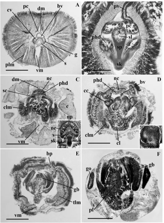
Figure 2.5 Light micrographs of transverse sections of Saxipendium coronatum. (A) Proboscis with heart-kidney complex. (B) Anterior region of the proboscis neck. (C) Posterior region of the proboscis neck showing the neuropore and skeleton plate. (D) Anterior region of the collar. (E) Posterior region of the collar showing the collar pores. (F) Collar canal and pore. (G) Posterior region of the collar showing the curved skeletal cornua. (H) Anterior region of the trunk. bp, branchial pore; bv, blood vessel; cc, collar canal; cl, collar lumen; clm, collar longitudinal muscles; cp, collar pore; cv, cardiac vesicle; dm, dorsal mesentery; g, glomerulus; gb, gill bar; go, gonad; gp, gonopre; nc, nerve cord; np, neuropore; pc, proboscis coelom; phd, periheamal diverticulum; pl, pharynx lumen; plm, proboscis longitudinal muscles; pv, proboscis vesicle; s, stomochord; sc, skeletal cornua; sp, skeletal plate; vm, ventral mesentery. Scale bars (A, C, D, G and H) = 1000 !m; (B and E) = 500 !m; (F) = 200 !m.---40
Figure 2.6 Light micrographs of transverse sections of Mesoglossus intermedius n. gen. and sp.. (A)
Proboscis with heart-kidney complex. (B) Proboscis neck. (C) Proboscis neck showing the neuropore. (D) Collar. (E) Posterior region of the collar, (inset) collar canal. (F) Anterior pharyngeal region of the trunk. bp, branchial pore; bv, blood vessel; cc, collar canal; clm, collar longitudinal muscles; cv, cardiac vesicle; dm, dorsal mesentery; g, glomerulus; gb, gill bar; nc, nerve cord; np, neuropore; pc; proboscis coelom; phd, perihaemal diverticulum; pl, pharynx lumen; plm, proboscis longitudinal muscles; pp, proboscis pore; s, stomochord; sc, skeletal cornua; sk, skeletal keel; sp, skeletal plate; tlm, trunk longitudinal muscles; vm, ventral mesentery. Scale bars (A, D, E and G) = 500 !m; (B) = 100 !m; (C and F) = 200 !m.---43
Figure 2.7 Light micrographs of transverse sections of Mesoglossus macginitiei n. gen. and sp.. (A)
Proboscis with heart-kidney comolex. (B) Proboscis coelomic cavity showing amoeboid-like cells. (C) Proboscis neck. (D) Anterior region of the collar showing the neuropore. (E) Anterior region of the collar showing the dorsal nerve cord crest, (inset) dorsal nerve cord crest. (F) Posterior region of the collar showing the peribuccal diverticula. (G) Anterior pharyngeal region of the trunk, (inset) collar canal. ac, amoeboid-like cells; bv, blood vessel; cc, collar canal; cl, collar lumen; clm, collar longitudinal muscles; cv, cardiac vesicle; dm, dorsal mesentery; g, glomerulus; gb, gill bar; go, gonad; nc, nerve cord; ncc, nerve cord crest; np, neuropore; pbd, peribuccal diverticula; pc, proboscis coelom; phd, perihaemal diverticulum; pl, pharynx lumen; plm, proboscis longitudinal muscles; proboscis vesicle; s, stomochord; sc, skeletal cornua; sk, skeletal keel; sp, skeletal plate; tlm, trunk longitudinal muscles; vm, ventral mesentery. Scale bars (A, D, G and H) = 500 !m; (B) = 50 !m; (C and I) = 200 !m; (F) = 300 !m.---47
Figure 2.8 Light micrographs of transverse sections of Ritteria ambigua. (A) Proboscis with
heart-kidney complex. (B) Junction of the proboscis and collar. (C) Anterior region of the collar. (D) Anterior pharyngeal region of the trunk. (E) Genital region of the trunk. (F) Proboscis skeleton, neuropore and proboscis pore. (G) collar canal. (H) collar nerve cord showing the nerve root. bs, branchial sac; bv; blood vessel; cc, collar canal; cl, collar lumen; clm, collar longitudinal muscles; cv, cardiac vesicle; e, epithelia; g, glomerulus; gb, gill bar; go, gonad; m, mouth; nc, nerve cord; nf, nerve fiber layer; np, neuropore; nr, nerve root; pbr, parabuccal ridge; pc, proboscis coelom; phd, perihaemal diverticulum; plm, proboscis longitudinal muscles; pp, proboscis pore; s, stomochord; sc, skeletal cornua; sk, skeletal keel; sp, skeletal plate; tlm, trunk longitudinal muscles. Scale bars (A, B, C, D, E) = 500 !m; (F, G, H) = 200 !m.---51
Figure 2.9 Light micrographs of transverse sections of Horstia kincaidi n. gen. and sp.. (A) Proboscis
with heart-kidney complex. (B) Proboscis neck. (C) Collar, (inset) collar nerve cord and stomochord (D) Anterior pharyngeal region of the trunk, (inset) collar canal. (E) Pharyngeal region of the trunk. (F) Genital region of the trunk. bp, branchial pore; bv, blood vessel; cc, collar canal; cl, collar lumen; clm, collar longitudinal muscles; cv, cardiac vesicle; dm, dorsal mesentery; g, glomerulus; gb, gill bar; go, gonad; nc, nerve cord; np, neuropore; pc, proboscis coelom; phd, perihaemal diverticulum; pl, pharynx lumen; plm, proboscis longitudinal muscles; pv, proboscis vesicle; s, stomochord; sc, skeletal plate; sk, skeletal keel; sl, stomochord lumen; sp, skeletal plate; tlm, trunk longitudinal muscles; vm, ventral mesentery. Scale bars (A, C, E, and G) = 500 !m; (B) = 200 !m; (D and F) = 100 !m; (H) = 300 !m.---55
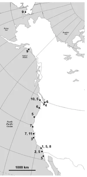
Figure 2.10 A map of the geographic distribution of harrimaniid enteropneusts on the west coast of North America. (1) Stereobalanus willeyi n. sp. (Newport Bay, CA (Ritter, 1900)); (2)
Stereobalanus canadensis (San Diego, CA (Bullock, personnal communication)); (3) Ritteria ambigua n. gen. and sp. (Monterey, CA); (4) Horstia kincaidi n. gen. and sp.
(Whidbey Island, WA); (5) Saccoglossus pusillus (Barkley Sound, BC, Cape Arago, OR, (Smith et al., 2003) San Pedro, San Diego, La Jolla, Anaheim, Newport Bay, CA, Ensenada, Mexico,); (6) Saccoglossus bromophenolosus (Padilla Bay, Willapa Bay, WA (Smith et al., 2003)); (7) Mesoglossus intermedius n. gen. and sp. (Shelter Cove, Moss Beach, CA); (8)
Mesoglossus macginitiei n. gen. and sp. (Newport Bay, CA); (9) Harrimania maculosa
(Prince William Sound, Kodiak, AK (Ritter, 1900), Providence Bay, Siberia); (10)
Harrimania planktophilus (Barkley Sound, BC (Cameron, 2002a)); (11) Protoglossus mackiei n. sp. (Moss Beach, CA).---58
Figure 2.11 A phylogenetic hypothesis for the family Harrimaniidae that was constructed using the tree of Cannon et al (2009) as a backbone, to which were added the new genera Horstia, Ritteria and Mesoglossus. (A) Proboscis longitudinal musculature in radial bunbles; (B) Direct development, absence of branchial synapticules, hepatic caeca, genital wings, trunk lateral septa, parabranchial ridges and circular muscles in the trunk; (C) Four tubular genital ridges restricted to the branchial region, reduced proboscis canals and pores; (D) No currently known characters appear to be unique to this group; (E) Dorsal and ventral proboscis septa; (F) Parabuccal ridges; (G) Prominent dorsal proboscis groove, proboscis short and conical, anterior edge of the collar is ruffled, anterior and posterior neuropores, parabuccal ridges extend to the posterior part of the collar, dorsal and ventral septa in the proboscis and collar; (H) Collar broader than long, no peribuccal diverticula, proboscis conical, a little longer than broad; (I) No genital ridges, gill pores on an elevated ridge, proboscis round and as long as broad, trunk very narrow; (J) Loss of radial proboscis longitudinal musculature; (K) Proboscis longitudinal musculature is diffuse; (L) Live in deep sea, coronate proboscis skeleton; (M) Absence of proboscis neck, two pairs of genital ridges, proboscis round, proboscis and collar broader than long, proboscis pore opens into the neuropore; (N) Collar pores, cardiac vesicle attached to dorsal the wall of the proboscis, externally visible gonopores, gill pores not visible externally; (G) Proboscis at least twice as long as broad, diffuse proboscis longitudinal musculature; (P) Proboscis at least twice as long as broad, proboscis longitudinal musculature in concentric rings.---60
Liste des sigles et des abréviations
a anus
ac amoeboid-like cells
ADNr Acide désoxyribonucléique ribosomal
b bouche bp branchial pore bs branchial sac bv blood vessel c collar cc collar canal ch caecum hépatique cl collar lumen
clm collar longitudinal muscles
cm circular muscle layer
cms canal du mésocoele
cnd corde nerveuse dorsale
co coeur cp coelome périhémal cpb cavité péri-buccale cv cardiac vesicle dm dorsal mesentery e epithelia fb fente branchiale g glomérule / glomerulus gb gill bar go gonade / gonad gr genital ridge m mouth md mésentère dorsal ms mésocoele mt métacoele nc nerve cord
ncc nerve cord crest
ncl nerve cord lacunae
nf nerve fiber layer
np neuropore
nr nerve root
opc organe pré-oral cilié
p proboscis pb pore branchial pbd peribuccal diverticula pbr parabuccal ridge pc proboscis coelom pl pharynx lumen
plm proboscis longitudinal muscles
phd periheamal diverticuluum
pp pore du proboscis / proboscis pore
ps péritoine viscéral pt protocoele pv proboscis vesicle qp queue post-anale s stomocorde / stomochord sb skeletal body
sbr sac branchial
sc skeletal cornua
sca sinus cardiaque
se septum sk skeletal keel sl stomochord lumen sp skeletal plate sqp squelette du proboscis t trunk
tlm trunk longitudinal muscles
vc ventral caecum
vm ventral mesentery
vsd vaisseau sanguin dorsal
1.1 BIOLOGIE
Les entéropneustes sont des vers benthiques exclusivement marins qui vivent, pour la plupart, dans les sédiments en zone peu profonde ou associés aux roches et aux crampons des algues laminaires. Certaines espèces ont également été trouvées en mer profonde sur les plaines abyssales (Holland, 2005, 2009) et près des sources hydrothermales (Woodwick et Sensenbaugh 1985). Ils sont présents partout sur le globe, mais la plupart des espèces ont été trouvées dans les eaux tropicales et sub-tropicales (Hyman, 1959). On sait très peu de choses en ce qui a trait à leur écologie et leur biologie, la plupart des espèces étant rares et peu étudiées.
Certaines espèces sont tubicoles et construisent des terriers en forme de ''U'' qui peuvent être repérés en zone intertidale par des tas d'excréments spiralés laissés à l'une des deux extrémités du terrier. Ces espèces présentent deux types de mode d'alimentation. Les espèces du genre Saccoglossus balaient la surface des sédiments à l'extérieur d'une des embouchures du terrier à l'aide de leur long proboscis, tandis que les espèces du genre Balanoglossus construisent plutôt une embouchure en forme d'entonnoir dans laquelle ils font tomber les sédiments, leur permettant ainsi de garder leur proboscis à l'intérieur du terrier (Hyman, 1959).
Certaines espèces ne construisent pas de terrier permanent et vivent simplement enfouies dans les sédiments, sous les roches ou parmi les racines des plantes, mais on ne sait que très peu de choses sur les espèces ayant ce mode de vie. Les entéropneustes s'alimentent de matière organique contenue dans les sédiments en transportant les particules de nourriture vers la bouche le long du proboscis à l'aide de cils et de mucus (Barrington 1940, Knight-Jones 1953, Thomas 1972). Quelques espèces, comme
Harrimania planktophilus, s'alimentent aussi par filtration à travers les fentes
pharyngiennes, à la manière des céphalocordés (Cameron, 2002).
Leur reproduction est de type sexué, mais certaines espèces comme
extrémité postérieure qui se regénère par la suite pour donner deux individus complets (Petersen & Ditadi, 1971). Les espèces de la famille des Harrimaniidae ont un développement de type direct, tandis que les espèces des autres familles produisent une larve tornaria planctonique semblable à la larve de certains échinodermes.
Les entéropneustes sécrètent plusieurs types de composés halogénés tels que les bromophénols et bromoindoles qui leur confèrent une odeur médicinale distinctive et qui ont, entre autres, pour fonction de repousser certains prédateurs et parasites (Higa et
al. 1980).
1.2 ANATOMIE
La taille des entéropneustes varie de quelques dizaines de millimètres pour la plupart, à plus de 1,50 m pour Balanoglossus gigas (Spengel, 1893). Ils ont une symétrie bilatérale simple et un corps trimérique (voir figure 1 pour le schéma d'un entéropneuste). Chacune des trois régions corporelles contient une cavité coelomique séparée des autres par des septa; le protocoele au niveau du proboscis, le mésocoele au niveau du collet et le métacoele au niveau du tronc. Ces cavités coelomiques présentent des diverticules et des pores communiquant avec l'extérieur du corps. Le ou les pore(s) du protocoele se situe(nt) au niveau du pédoncule du proboscis, ceux du mésocoele sont sous forme de canaux qui s'ouvrent au niveau de la première paire de branchies et le métacoele est percé de pores oesophagiens. Une caractéristique distinctive des entéropneustes est le diverticule de la cavité buccale qui se prolonge antérieurement à l'intérieur du proboscis. Cette structure se nomme la stomocorde et a souvent été comparée à la notocorde des céphalocordés de par sa structure similaire. La bouche est ventrale, à la jonction du proboscis et du collet. L'anus est terminal.
Les entéropneustes possèdent des branchies et des fentes pharyngiennes dorso-latérales supportées par un squelette de collagène. Une autre pièce squelettique aussi faite de collagène sert de support au proboscis. Les gonades sont situées en deux ou
quatre rangées dorso-latérales qui peuvent être proéminentes ou non, selon les espèces.
Le système circulatoire est composé des vaisseaux sanguins principaux, l’un ventral, l’autre dorsal, ainsi que d'un organe unique, le coeur-rein. Cet organe est composé d'un péricarde, d'un vaisseau sanguin et de glomérules. Les pulsations du coeur sont produites par des fibres musculaires insérées sur la stomocorde, qui sert de support à ce complexe.
Le système nerveux est diffus. On dénote toutefois un renflement de fibres nerveuses le long de la ligne médiane dorsale, qui peut parfois prendre l'aspect d'un tube plus ou moins complet, semblable à celui des céphalocordés. Les entéropneustes possèdent une musculature très fine, les muscles les plus distinctifs se trouvant dans le proboscis.
Figure1. Schéma d'un entéropneuste généralisé, modifié de Cameron, 2005. a, anus; b, bouche; ch, caecum hépatique; cms, canal du mésocoele; cnd, corde nerveuse dorsale; co, coeur; cp, coelome périhémal; cpb, cavité péri-buccale; fb, fente branchiale; g, glomérule; go, gonade; md, mésentère dorsal; ms, mésocoele; mt, métacoele; opc, organe pré-oral cilié; pb, pore branchial; pp, pore du proboscis; ps, péritoine viscéral; pt, protocoele; qp, queue post-anale; s, stomocorde; sbr, sac branchial; sca, sinus cardiaque; se, septum; sqp, squelette du proboscis; vsd, vaisseau sanguin dorsal; vsv, vaisseau sanguin ventral.
1.3 SYSTÉMATIQUE
faisant partie du phylum des hémicordés (Hemichordata): Enteropneusta, Pterobranchia, Planktosphaeroidea, Graptolithina (éteint). Jusqu'à présent, 78 espèces appartenant à 14 genres et 5 familles ont été identifiées à travers le monde, incluant 17 espèces en Amérique du Nord. Les cinq familles actuellement reconnues sont les Harrimaniidae Spengel, 1901, les Ptychoderidae Spengel, 1893, les Spengelidae Willey, 1897, les Saxipendiidae Woodwick & Sensenbaugh, 1985 et les Torquaratoridae Holland et al., 2005. La famille des Harrimaniidae compte présentement les 5 genres suivants:
Saccoglossus Schimkewitsch, 1892, Harrimania Spengel, 1893, Stereobalanus Spengel,
1901, Xenopleura Gilchrist, 1925 et Protoglossus van der Horst, 1939.
Nos connaissances actuelles sur les entéropneustes ne reflètent toutefois pas la diversité réelle du groupe. En raison de leur mode de vie endobenthique, les entéropneustes font partie de la faune marine qui passe souvent inaperçue et leur grande fragilité fait en sorte qu’on prélève rarement des spécimens intacts. Leur étude est donc beaucoup plus ardue que pour la majorité des autres invertébrés marins. De ce fait, le nombre réel d'espèces et leur distribution demeurent inconnus. Sur les côtes nord-américaines, de nombreuses populations n’ont jamais été étudiées.
Les entéropneustes sont un groupe relativement homogène d'animaux qui possèdent très peu de caractères uniques permettant de distinguer les différents genres et espèces. Il existe en plus une grande confusion dans la nomenclature des caractères utilisés pour la classification des espèces, certains termes provenant de descriptions anciennes souvent librement traduites de l'allemand, d’après les travaux de Spengel (1893, 1901). Comme plusieurs espèces ont été décrites à partir de spécimens uniques, de nombreux états de caractères ne sont pas connus pour l'ensemble des espèces et rendent la classification d'autant plus incertaine. Chacun des auteurs ayant basé sa description sur des suites de caractères anatomiques différents, accordant plus d'importance à certains et ignorant d'autres, nous nous retrouvons en présence de descriptions taxonomiques difficilement comparables les unes aux autres. De surcroît, comme plusieurs de ces espèces n'ont été trouvées qu'une seule fois et que les spécimens les plus communs sont souvent rares et fragiles, il est difficile de vérifier à nouveau la
présence des caractères ignorés.
Quelques ouvrages généraux sont toutefois disponibles sur la biologie et l'anatomie du groupe, dont les plus importants sont ceux de Hyman (The Invertebrates, 1959), Dawydoff (Traité de zoologie, 1948) et Horst (Bronn’s Klassen und Ordnungen
des Tierreichs, 1939). Le traité taxonomique le plus complet étant celui de Horst (1939),
écrit en allemand et aujourd'hui malheureusement désuet, un nouvel ouvrage taxonomique sur les enteropneustes s'avère nécessaire pour une meilleure compréhension du groupe, que ce soit simplement pour l'uniformisation de la nomenclature des caractères. C’est seulement en examinant de nombreux spécimens qu’on pourra déterminer quels sont les caractères taxonomiques stables et utiles. Ayant accès à la collection de Ritter et Bullock, nous espérons être en mesure de clarifier, du moins en partie, la validité des caractères au sein de la famille.
1.4 DONNÉES HISTORIQUES INÉDITES
En participant à l'expédition Harriman de 1899 en Alaska, William E. Ritter avait accumulé une collection considérable de spécimens d'entéropneustes provenant de la côte est du Pacifique et avait commencé, au début du siècle dernier, la rédaction d'un travail taxonomique sur ces nouvelles espèces. Les descriptions de deux espèces furent publiées séparément (Ritter 1900, Ritter & Davis 1904), mais l'ouvrage complet ne fut jamais terminé. En 1937, T. H. Bullock héritait du projet et ajoutait de nouveaux spécimens à la collection. Il confia plus tard le projet à K. P. Rao, puis à C. Burdon-Jones, mais malgré leur contribution la monographie ne vit jamais le jour. En 2004, Bullock confia finalement le manuscrit à Christopher Cameron en souhaitant qu'il puisse être complété.
Le présent mémoire est donc la suite d'un projet de longue haleine, bien connu des zoologistes de la côte ouest et parfois cité par les auteurs d'ouvrages importants sur la famille (Hyman, 1959 et Ricketts et al., 1985). Dans le présent travail, six espèces de
cette collection appartenant à la famille Harrimaniidae sont décrites et ajoutées aux quatre espèces de la famille présentement connues pour la côte ouest de l'Amérique du Nord, soit Saccoglossus pusillus (Ritter & Davis, 1904), Saccoglossus bromophenolosus (King, Giray, & Kornfield, 1994), Harrimania maculosa (Ritter, 1900) et Harrimania
planktophilus (Cameron, 2002). Bien que la majorité des espèces ici traitées soient peu
répandues ou représentées par un seul spécimen, plusieurs d'entre elles semblent communes localement. Il est d'ailleurs fort probable que certaines populations soient déjà connues des zoologistes et qu'on puisse rencontrer ces espèces le long des côtes, sans que l'on sache les identifier.
C'est pour cette raison qu'une description des spécimens découverts jusqu'à présent s'avère plus que nécessaire, puisque l'on sait désormais que les quatre espèces de la famille auparavant décrites pour cette côte ne sont pas représentatives de la richesse spécifique de cette région.
1.5 CONTRIBUTION DES DIFFÉRENTS AUTEURS
Les spécimens de la collection furent prélevés par de nombreux zoologistes au cours du 20e siècle et les coupes histologiques furent effectuées par plusieurs techniciens dans le laboratoire de T. H. Bullock. Les descriptions originales de Mesoglossus
intermedius gen. et sp. nov. et de Horstia kincaidi gen. et sp. nov. furent élaborées par
Ritter. Rao effectua une première révision des descriptions de Ritter et rédigea les descriptions originales de M. macginitiei gen. et sp. nov. et de Ritteria ambigua gen. et sp. nov.. Finalement, j'ai préparé moi-même la description de Protoglossus mackiei sp. nov. par l'observation de spécimens de la collection de Bullock.
De plus, j'ai effectué la révision et l'uniformisation de chacune des descriptions, j'ai ajouté de nouveaux caractères pour chacune des espèces, et j'ai ajouté des planches et figures illustrant l'anatomie des spécimens. J'ai rédigé une nouvelle description de l'espèce
Saxipendium coronatum à l'aide de la documentation et des coupes histologiques de
l'holotype, en utilisant la même suite de caractères que celle que j’ai utilisée pour les autres espèces décrites dans ce travail. J'ai aussi illustré les détails de l'anatomie de cette espèce à l'aide d'une nouvelle planche photographique. Finalement, j'ai construit un arbre phylogénétique, une table de caractères, ainsi qu'une clé d'identification à partir de données obtenues de la documentation pour les espèces pour lesquelles nous n'avions pas accès aux holotypes et à partir des coupes histologiques pour les espèces décrites dans ce travail.
L'origine de l'intérêt porté à la classe des enteropneustes est liée aux théories évolutionnaires et provient de la suggestion faite par Bateson (1885) selon laquelle ces animaux seraient des cordés et représenteraient le parent le plus primitif des vertébrés. Le nom Hemichordata fut d'ailleurs proposé par Bateson pour refléter le fait que les hémicordés partagent certaines caractéristiques morphologiques avec les cordés, telles les fentes pharyngiennes, un tube neural dorsal et la stomocorde, une structure ressemblant à la notocorde. Toutefois, l'hypothèse de l'inclusion des hémicordés au sein des cordés fut très contestée, entre autres par les auteurs de trois ouvrages zoologiques importants portant sur cette classe, soit Spengel (1893), Horst (1939) et Dawydoff (1948).
Une étape clé ayant récemment ravivé l'intérêt porté aux entéropneustes provient de l'étude moléculaire de Turbeville et al. (1994) qui confirma la monophylie des Ambulacraires (Hemichordata + Echinodermata) et leur position en tant que groupe frère des cordés. Depuis, plusieurs autres études moléculaires et morphologiques ont confirmé la position phylogénétique du groupe (Cameron et al. 2000, Swalla et al. 2000, Winchell
et al. 2002, Cameron 2005, Zeng & Swalla 2005, Bourlat et al. 2006). Plusieurs des
résultats de ces analyses phylogénétiques suggèrent la possibilité que l'ancêtre deutérostomien aurait été un ver semblable aux entéropneustes actuels, ayant des fentes pharyngiennes supportées par un squelette de collagène acellulaire (Cameron et al. 2000, Swalla et al. 2000, Zeng & Swalla 2005). Ces résultats font en sorte que les entéropneustes jouissent à nouveau d'une certaine popularité dans les domaines de la phylogénétique et de l'évolution. Néanmoins, plusieurs questions demeurent sans réponse au sujet de la nature des caractères deutérostomiens ancestraux. La réponse à ces questions réside, entre autres, dans la distribution des caractères homologues à l'intérieur des divers groupes d'animaux deutérostomiens, et plus particulièrement au sein des entéropneustes (Ruppert, 2005). Comme les entéropneustes sont plus près des échinodermes qu'ils ne le sont des cordés, tout caractère homologue partagé par les deux groupes pourrait avoir été un attribut du dernier ancêtre commun. En fait, au moins deux caractères anatomiques des entéropneustes seraient homologues de ceux des cordés: les fentes branchiales et la queue post-anale (ventrale chez ce groupe, mais considérée
homologue par l'expression des gènes Hox) (Lowe et al., 2003). Les entéropneustes recèlent peut-être même davantage d'homologies, mais malgré le rôle central qu'ils occupent dans les hypothèses sur l'évolution des deutérostomiens, leur anatomie n'a été que peu étudiée depuis les travaux de Horst (1939), Bullock (1945), Rao (1954) et plus récemment Cameron (2005). En raison de ce manque d'information, on sait d'ailleurs très peu de choses sur l'évolution des caractères morphologiques au sein même du groupe et par conséquent, sur la systématique des divers genres et familles.
Les études moléculaires et morphologiques récentes ayant tenté de clarifier les relations au sein des entéropneustes ont mené à l'élaboration d'hypothèses contradictoires concernant la position des familles et même en ce qui a trait aux classes du phylum Hemichordata. Les données provenant de l'analyse morphologique (Cameron, 2005) indiquent que les entéropneustes sont un groupe monophylétique, plaçant les ptérobranches à la base des hémicordés. Elles démontrent aussi la monophylie des familles Spengelidae et Ptychoderidae, mais ne présentent aucune résolution quant aux autres familles dont les membres ont une anatomie plus simple. Une analyse de l'ADNr 28s (Winchell et al., 2002) démontre la monophylie des entéropneustes en plaçant les ptérobranches comme groupe frère des entéropneustes, mais ne clarifie pas la systématique des familles en raison d'un jeu de données trop limité. Finalement, diverses études basées sur les séquences d'ADNr 18s démontrent que les entéropneustes sont un groupe paraphylétique et placent les ptérobranches en tant que groupe frère de la famille Harrimaniidae (Cameron et al. 2000, Bourlat et al. 2003, Cannon et al. 2009).
Si la dernière hypothèse s'avère exacte et que les ptérobranches ont évolué à partir de vers entéropneustes, l'étude de la morphologie de ces derniers devient d'autant plus importante pour la découverte des caractères deutérostomiens ancestraux, puisque les entéropneustes posséderaient un plan corporel moins dérivé que celui des ptérobranches.
L'objectif premier de ce mémoire est de faire la description anatomique de spécimens appartenant à de nouvelles espèces de la famille des Harrimaniidae provenant de populations de la côte ouest de l'Amérique du Nord, dans une perspective de classification taxonomique. À l'aide d'une collection de coupes histologiques de spécimens amassée au cours du siècle dernier, d'abord par W. E. Ritter et T. H. Bullock, puis par K. P. Rao, nous avons pu ajouter cinq nouvelles espèces et trois nouveaux genres à la famille Harrimaniidae, en plus de faire la description d'une espèce découverte par Ritter, qui n'avait pas encore été véritablement décrite. Nous avons aussi tranféré l'espèce Saxipendium coronatum dans cette famille et en avons fait une nouvelle description.
Ayant eu l'opportunité de travailler sur ce qui est probablement la collection d'entéropneustes la plus importante au monde quant au nombre de spécimens et d'espèces, l'objectif second de ce projet est de réviser la taxonomie de la famille Harrimaniidae en vérifiant d'abord, à l'aide des spécimens disponibles, les caractères morphologiques déjà décrits dans la documentation, et ensuite d'établir des listes de caractères pertinents pour la classification.
4. Article: A taxonomic revision of the family Harrimaniidae
(Hemichordata: Enteropneusta) with descriptions of seven species from
Accord des coauteurs
Étudiant : Carine Deland
Programme : M.Sc. en sciences biologiques (2-235-1-0)
Description de l’article
Liste des auteurs : Carine Deland
Christopher B. Cameron Kandula P. Rao (décédé) Theodore H. Bullock (décédé) William E. Ritter (décédé)
Titre: A taxonomic revision of the family Harrimaniidae (Hemichordata:
Enteropneusta) with descriptions of seven species from the Eastern Pacific
L’article a été soumis à la revue Zootaxa le 26 juin 2009 et publié le 24 mars 2010
Déclaration de tous les coauteurs autres que l’étudiant
À titre de coauteur de l’article identifié ci-dessus, je suis d’accord pour que Carine Deland inclue cet article dans son mémoire de maîtrise qui a pour titre Révision
taxonomique de la famille des Harrimaniidae (Hemichordata: Enteropneusta) incluant les descriptions de sept espèces de la côte Est du Pacifique
Christopher B. Cameron
ABSTRACT
The family Harrimaniidae of the enteropneust hemichordates is revised on the basis of morphological characters. The number of harrimaniid genera is increased to nine by the addition of Horstia n. gen., Mesoglossus n. gen., Ritteria n. gen and
Saxipendium, a genus previously assigned to the monospecific family Saxipendidae. The
number of species is increased to thirty-four, resulting from the description of five new species from the eastern Pacific: Horstia kincaidi, Mesoglossus intermedius,
Mesoglossus macginitiei, Protoglossus mackiei and Ritteria ambigua. A description is
supplied for a sixth harrimaniid species, Stereobalanus willeyi (Ritter and Davis, 1904), which previously had the status of a nomen nudum. Four harrimaniids previously assigned to the genus Saccoglossus are transfered to the genus Mesoglossus: M. bournei,
M. caraibicus, M. gurneyi, and M. pygmaeus, while Saccoglossus borealis is reassigned
to the genus Harrimania. Notes on habitat and zoogeography are included for the seven foregoing species, a table of diagnostic characters for old and new species and a dichotomous key for the enteropneust families and harrimaniid genera are provided. Finally, a phylogenetic hypothesis to the Harrimaniidae is postulated with discussion on the evolution of the group.
INTRODUCTION
The family Harrimaniidae is one of the five families of enteropneusts. Of the 79 enteropneust species known to date, 28 are classified in this family, which consists at the time of writing of the following five genera: Harrimania, Protoglossus, Saccoglossus,
Stereobalanus and Xenopleura. Members of this family include common and widely
distributed North American species such as Saccoglossus kowalevskii (Agassiz, 1873) and Saccoglossus pusillus (Ritter, 1902). Found in all latitudes, from the intertidal zone to the deep sea, enteropneusts inhabit sand or mud, occasionally under rocks or among seaweed holdfasts. Typical habitats are clean coral sand flats exposed at low tides and black mud under clean sea water. Some species may be located by a coiled casting of sand thrown up in a cone at one end of the burrow. But most species are encountered only by chance digging in the right place.
Our work is based in part on an unpublished manuscript and specimen collection initiated a century ago by William E. Ritter and later continued by Theodore H. Bullock and Kandula P. Rao. Our present study incorporates a part of this unpublished material to revise and update the family Harrimaniidae, to which we add five new species and three new genera. In addition, a description is provided for Stereobalanus willeyi, heretofore a nomen nudum. Moreover, an expanded description is provided for
Saxipnedium coronatum (Woodwick and Sensenbaugh, 1985) and these findings
coupled with recent molecular phylogenetic results (Cannon et al., 2009) indicate that the monospecific genus Saxipendium should be transferred to the Harimaniidae. From the older unpublished material plus our own contributions, we synthesize a new generic classification within the family.
Our morphological descriptions of haramaniid genera and species is incorporated into a dichotomous key as well as a table listing the characteristic features of each species of the family. It is hoped that our textural material along with the photographic plates and generic relationships will make the work more accessible to non-specialists.
THE SPECIMEN COLLECTION
As an outgrowth of his extensive studies on tunicates, Ritter took up the subject of the Enteropneusta in the last decade of the nineteenth century and published several accounts dealing with natural history, embryology and taxonomy (Ritter, 1900; Ritter, 1902; Ritter, 1908; Ritter & Davis, 1904). Over a period of years he accumulated a considerable body of material, representing seven new species from the west coast of the United States and Alaska. He obtained many of his specimens during the Harriman Alaska Expedition of 1899, the results of which appeared in a series of volumes (C. Hart Merriam, ed., Harriman Alaska Expedition, 13 volumes. New York: Doubleday, Page and Company, Washington, D.C.: Smithsonian Institution, 1901-1914). The then recent summary by Spengel (1893) of the enteropneusts of the world listed no species from this coast so that Ritter, with a proportionately considerable new fauna relative to the forty or so species known for the entire group at that time projected a monograph, and put into draft descriptions and figures of all these forms. Descriptions of two of the species were later published separately (Ritter, 1900; Ritter & Davis, 1904), but the full monograph was never completed.
Shortly before he died in 1944, Professor Ritter gave his enteropneust slides and manuscript to Theodore H. Bullock, who had recently finished a doctoral dissertation on the neuroanatomy of the group (Bullock, 1940, 1944, 1945). Bullock subsequently undertook to update and complete the paper, with the aid of Kandula Pampapathi Rao who had previously studied the group extensively (Rao, 1952, 1953, 1954, 1955, 1957, 1962). They then found new material in the Albatross collection through the kindness of C.A. Kofoid and S.F. Light, both of the Department of Zoology, University of California, Berkeley and in the collections at the Scripps Institution of Oceanography, through the courtesy of Percy Barnhart. In the ensuing years Bullock found a number of additional new eastern Pacific species, as well as receiving valuable specimens from others.
C. Burdon-Jones inherited the task of completing the monograph in the early seventies but had not made any significant contribution to the work. In 2003 Bullock, then well into his retirement years, urged that Burdon-Jones, then at a later age and in poor health, return what material he had to California so that the whole collection could be deposited at the Smithsonian Museum in Washington. Bullock feeling a deep obligation to see the monograph complete then contacted Cameron, who had developed a graduate thesis on the group, including the description of a new species (Cameron, 2002a), to complete the work. Following the death of Bullock in 2005, Cameron decided that it would be most practical to publish the new material as several smaller papers instead of as a comprehensive monograph.
DIAGNOSTIC MORPHOLOGICAL CHARACTERS
Species (and even genera and families) of enteropneust hemichordates differ mainly in their unique combinations of morphological features and presence or absence of soft parts, traced in serial sections. Not many features are by themselves diagnostic. These features are only to a limited degree established, from examination of many individuals of abundant species, to be consistent characters. Even less is known of variation that might be due to seasonal, ontogenetic, local environmental factors and the like. In general, only single or very few specimens are examined for all relevant characters, both because of the rarity of finds and because of the formidable labor of serial sectioning and interpreting sizable, sandy, coiled worms. The paucity of hard parts and the distortions of soft parts in fixation add to the problems.
The net result is almost certain to be that some of the characters used are not good species characters and we do not place undue weight on the validity of the taxa named. Nevertheless the characters have a certain modicum of reliability to judge from the few species repeatedly examined and are sanctified by usage. Until better characters, e.g. molecular and other biochemical signatures are validated, we perforce deal with these.
We have faced the same dilemma as previous authors, namely, whether to risk confusion by ignoring morphological differences that have not been well shown to be significant or to risk confusion by paying attention to them, in short whether to lump or split. Our decision, after some years of dealing with this group in various respects, is that the lesser evil is to split. Especially when such a high percentage of the published species are represented by unique specimens or part of a specimen, errors uncovered in the future will be easier to correct by throwing names into synonymy than the reverse. This has already happened in the widespread and abundant species of Ptychodera, of which only two are now recognized. We have been particularly conscious of the possibility that this may be called for in multispecific genera like Saccoglossus and
Mesoglossus.
In the present state of understanding there is no justification for collapsing harrimaniid species into synonymy, even with the substantial number of sectioned specimens we were able to examine. The resulting bulk of material and number of species at hand is quite likely to be considerably in excess of any assembly of material that has been available to any previous author. This certainly does not mean that we have arrived at greater insight into evaluation of characters or regard our taxa as more likely to last. It does correlate with a serious concern, a long hesitancy to perpetrate new genera and species and a decision to do so only after fairly protracted efforts.
MATERIAL AND METHODS
The specimens used in this study are part of T. H. Bullock's enteropneust slides collection deposited at the Smithsonian Institution National Museum of Natural History, Washington, DC. Most of the material in this collection has been fixed in Bouin’s solution, while some of it was fixed in formol-acetic-alcohol and all the material has been archived by transfer to 80% alcohol with 10% glycerin. Sections were cut either
in paraffin or in low viscosity nitrocellulose and mounted on glass slides. Heidenhain’s iron alum haematoxylin, Masson’s trichrome or Mallory’s triple stain were used for staining the sections. Specimens were viewed and photographed with a Q Imaging Retiga-2000R digital camera mounted on an Olympus BX51 compound microscope and on an Olympus SZX16 stereomicroscope for lower magnifications.
Being soft and fragile enteropneusts are usually taken as pieces. These are nevertheless valuable. Whatever enteropneust material is collected should be carried back to the laboratory in separate containers since the chances of their suffering damage, if mixed with hard shells or hard skinned marine organisms, is very great. They should be preserved soon after collection. Good preservation is most desirable as all serious study depends upon serial sectioning and histologic staining. It is quite desirable to leave them in a tray of clean sea water for a few hours to allow evacuation of the sand from the gut; frequent removal of the sand and the mucous sheath with its sand grains may avoid reingestion. Fixation without coiling is aided if each worm is lifted into the air by a match stick under the middle of the specimen and killed by dropping fixative solution over it for a few minutes. It may then be lowered into the fixative. A general histologic fixative such as Bouin’s, Hedenhain’s Susa fluid, formol-acetic-alcohol or 10% formalin is satisfactory. Changing the fluid twice in the first 24 hours and again after several days is more than desirable. Labels should give not only details of the geographic locality, date of collections and name and address of collector, but also of the nature of substratum, color of the body parts in life and method of preservation. A sketch or photograph with measurements in life showing length and diameter of different regions is helpful.
RESULTS
SYSTEMATIC DIAGNOSES OF FAMILY AND GENERA
CLASS ENTEROPNEUSTA Gegenbaur, 1870
FAMILY HARRIMANIIDAE Spengel, 1901
Balanoglossidae Willey, 1899
Enteropneusta characterized by the absence of circular muscle fibers in the trunk. In those cases where the development has been studied, the typical tornaria larva is absent and the development is direct. In addition to these two unique features may be added the following characters: absence of lateral septa, absence of vermiform process of the stomochord, absence of hepatic caeca in the trunk and absence of synapticulae joining the primary and secondary gill bars (for a drawing of a generalized enteropneust, see Fig. 1 from Cameron, 2005). The dorsal nerve roots in the collar mesentery and the intestinal pores may be present or absent. The skeletal cornua extend at least to the middle of the collar.
Of the five known genera included in this family, Harrimania, Saccoglossus,
Protoglossus, Stereobalanus and Xenopleura, the first two occur in the Eastern Pacific.
In addition, as a result of the present work, Protoglossus and Stereobalanus genera can now be added to this list, along with the three new genera described here (Ritteria,
Horstia and Mesoglossus) and Saxipendium, which is transferred to the family
Figure 2.1 Light micrographs of transverse sections of the proboscis of (A) Saccoglosus pusillus, showing the arrangement of the proboscis longitudinal musculature in concentric rings; (B) Protoglossus mackiei n. sp., showing the arrangement of the proboscis longitudinal musculature in radial plates; (C) Mesoglossus
macginitiei n. gen. and sp., showing the diffuse arrangement of the proboscis longitudinal musculature.
plm, proboscis longitudinal muscles. Scale bars = 500 !m.
Genus Saccoglossus Schimkewitsch, 1892
Dolichoglossus Spengel, 1893Type species: Saccoglossus kowalevskii (Agassiz, 1873)
The proboscis is usually long; a middorsal longitudinal groove may be present. The collar is usually very short compared to the proboscis. Dorsal inter-branchial genital ridges and dorsal gonads are absent, but lateral extra-branchial genital ridges may be present. Intestinal pores are often present. Perihaemal cavities are always present. Peribuccal cavities are usually present, but not always. More important than all the above characters, the genus is characterized by the arrangement of the longitudinal muscle fibers of the proboscis in several concentric rings (Fig. 2.1 A). Many species favor quiet flats not too far from the mouth of a bay, in muddy sand; live in "permanent" tubes and throw up low conical mounds of quasi-spiral castings from the anus.
The genus as revised above would now include the following species: S. apatensis (Thomas, 1956), S. aulakoeis (Thomas, 1968), S. bromophenolosus (King, Giray & Kornfield, 1994), S. horsti (Brambell & Goodhart, 1941), S. hwangtauensis (Tchang & Koo, 1935), S. inhacensis (Kapelus, 1936), S. kowalevskii (Agassiz, 1873), S.
1895), S. pusillus (Ritter, 1902), S. ruber (Tattersall, 1905), S. sulcatus (Spengel, 1893) and five new species collected from the Eastern Pacific which will be described in a forthcoming article on the genus.
Genus Harrimania Spengel, 1893
Type species: Harrimania kupfferi (von Willemoes-Suhm, 1871)
The proboscis is conical and a little longer than broad; the collar is broader than long. The longitudinal muscles of the proboscis are arranged in radial plates (Fig. 2.1 B). The genus is often characterized by two proboscis pores. Intestinal pores and peribuccal cavities are absent. In H. maculosa (Ritter, 1900) and H. planktophilus (Cameron, 2002a), the cornua are very long and extend into the trunk. They also form parabuccal ridges on either side of the buccal cavity in the collar. Both dorsal and lateral gonads can be present and occur as simple sacs arranged in long rows. The dorsal gonads, when present, are confined to the branchial region.
Three species from this genus are currently described, two of which are from the Eastern Pacific region: H. maculosa and H. planktophilus. From the description given above, we should now reassign Saccoglossus borealis (Okuda & Yamada, 1955) to this genus, for its great resemblance to H. planktophilus in its possession of a radial proboscis musculature, a broad collar, a left proboscis pore, lateral gonads, parabuccal ridges and the absence of peribuccal cavities.
Genus Stereobalanus Spengel, 1901
Balanoglossus Spengel, 1893Type species: Stereobalanus canadensis (Spengel, 1893)
The genus is characterized by broad dorsolateral and ventrolateral genital ridges, with the broad gill opening situated in between. The gill tongues are externally visible.
The longitudinal musculature of the proboscis is arranged in radial plates. Abdominal pores may or may not be present. Perihaemal cavities are present but the peribuccal coelomic cavities are absent. The genus possesses two proboscis pores, but they are greatly reduced. Contrarily to the report of Spengel (1893), collar ducts are present in this genus (Reinhard, 1942).
Two species, namely, S. canadensis and S. willeyi are included in this genus and both are represented on the west coast of North America.
Genus Xenopleura Gilchrist, 1925
Type species: Xenopleura vivipara Gilchrist, 1925
The genus is characterized by the presence of medullary folds in the trunk extending posteriorly on the dorsal side into low pleurae. The proboscis is not elongate, and its longitudinal muscles are scattered (Fig. 2.1 C). The stomochord is without a vermiform process, and is continuous through the buccal cavity as two dorso-lateral folds. There is one proboscis pore. The posterior margin of the collar is fused with the trunk and no nerve roots are present in the collar nerve cord. The branchial skeleton lacks synapticula, and internal hepatic caeca are present in the trunk. This genus, only described from one specimen, could be viviparous.
Genus Protoglossus van der Horst, 1939
Balanocephalus Caullery & Mesnil, 1900; Protobalanus Caullery & Mesnil, 1904
Types species: Protoglossus koehleri (Caullery & Mesnil, 1900)
This genus is characterized by a short conical proboscis with a deep posterior dorsal groove and a conspicuous horseshoe-shaped pre-oral ciliary organ. The longitudinal musculature of the proboscis is radial. Its paired dorsal gonads are not prominent. It is also considered as having the simplest arrangement of body cavities of
all enteropneusts in not possessing peribuccal cavities and having rudimentary or no perihaemal cavities (Burdon-Jones, 1956). A left proboscis pore is present as are dorsal and ventral mesenteries in the proboscis and collar. The cornua of the skeleton extend to the posterior margin of the collar forming parabuccal ridges on each side of the buccal cavity.
Genus Saxipendium Woodwick & Sensenbaugh, 1985
Type species: Saxipendium coronatum Woodwick & Sensenbaugh, 1985
This genus, previously assigned to its own family is here transferred to the family Harrimaniidae. It possesses the general characters of the family and can be distinguished from the other genera of the family by the following characters: diffuse longitudinal proboscis mucles, coronate body of the proboscis skeleton, absence of skeleton keel, long recurved skeletal cornua, collar canals with pores opening to the outside of the body, dorso-lateral genital ridges with the gonopores externally visible and the possible presence of testicular antra.
Genus Mesoglossus gen. nov.
Type species: Mesoglossus bournei (Menon, 1904)
Saccoglossus bournei Menon, 1904
The genus has the following features distinguishing it from other genera of Harrimaniidae. The proboscis, about twice as long as wide, has no conspicuous dorsal groove. Its longitudinal musculature is arranged diffusely, not in concentric rings or radial bundles. A proboscis pore is present usually on the left side. Perihaemal cavities are usually present. Collar canals are present. A ventral mesentery is present in the proboscis and both dorsal and ventral mesenteries are present in the collar. Dorsal gonads are absent, only the lateral gonads are present. Peribuccal spaces may be present or absent.
It is evident from the details given above that the closest relations of this species are four species previously assigned to the genus Saccoglossus but here transferred to the genus – namely, Mesoglossus: M. bournei, M. caraibicus (van der Horst, 1924), M.
gurneyi (Robinson, 1927), and M. pygmaeus (Hinrichs & Jacobi, 1938). Although all
these species and M. intermedius resemble species of Saccoglossus a great deal, they are distinguished from the latter by the fact that the longitudinal muscle fibers of the proboscis are uniformly distributed without being arranged into concentric rings as in
Saccoglossus (or into radial groups as in Harrimania and Stereobalanus). Mesoglossus
is too distinct to be confused with Stereobalanus. Hence all the forms which have previously been put under Saccoglossus but which exhibit no regular arrangement of the proboscis longitudinal muscle fibers into concentric rings are here transferred to the genus Mesoglossus, with M. bournei as the type species.
Remarks: The description given above is primarily based on Ritter’s account in his manuscript of circa 1900 and a re-examination of his own slides and of material collected by others. At the beginning of the twentieth century, no Saccoglossus species had been described with a diffuse distribution of longitudinal fibers in the proboscis, except for Benham's description of S. otagoensis (Benham, 1895). But it is now known that in S. otagoensis the longitudinal muscle fibers in the proboscis are actually arranged in 3 or 4 concentric rings (van der Horst, 1939). Thus S. otagoensis cannot be considered as belonging to Mesoglossus, which means that M. bournei is the earliest described species under the genus and should be considered as its type species.
Genus Ritteria gen. nov.
Type species: Ritteria ambigua gen. & sp. nov.
Created for the single species R. ambigua, the genus conforms to the diagnosis of the family and may be defined tentatively by the following selection of characters of the type species. The proboscis is short and its longitudinal muscles are dispersed diffusely, not concentrically or radially. There is a single, left proboscis pore. The proboscis neck
is severely reduced and the stomochord lumen broken into lacunae. There is neither dorsal nor ventral septum in the collar and no peribuccal cavities. Dorsal gonads are present and, with the lateral gonads forming two pairs of genital ridges, leaving the branchial pores recessed in the groove between them.
The genus is named in honor of Professor W.E. Ritter, but for whose interest and initiative our knowledge of the enteropneusts would have been very much poorer. He never saw the material on which this name is based so that we are not deterred by his insistent modesty.
Genus Horstia gen. nov.
Type species: Horstia kincaidi gen. & sp. nov.
Having the characters of the family, this genus is distinguished from others of the Harrimaniidae by the following combination of features. The proboscis is very short and round, hardly as long as broad. Its longitudinal muscles are arranged in radial plates. A proboscis pore is present on the right or the left. An anterior neuropore is present and there are large lacunae in the collar nerve cord. Peribuccal cavities are absent. The gonads do not form ridges; the cross section of the trunk is regular and nearly circular with the gill pores unusually prominent, even elevated a bit above the general surface level. There are no dorsal gonads. The gonads are conspicuous as modules projecting prominently from the surface. Thus Horstia differs in at least three or four characters from the nearest genera (Mesoglossus, Saccoglossus, Harrimania and Ritteria) and currently comprises only one species (H. kincaidi), which would otherwise have to be forced into one of these genera, doing violence to what we believe are defining generic features. Horstia is of course only tentatively defined by the foregoing selection of characters of its single species; further discoveries may reveal some of generic rank in the specific description below.
Figure 2.2 Drawings of live animals and photographs of fixed specimens of the family Harrimaniidae. (A) Drawing of the dorsal side of Horstia kincaidi n. gen. and sp.. (B) Drawing of the lateral side of
Mesoglossus intermedius n. gen and sp.. (C) Drawing of the dorsal side of Mesoglossus intermedius. (D)
Drawing of the dorsal side of Stereobalanus willeyi n. sp.. (E) Drawing of the lateral side of Stereobalanus
willeyi. (F) Photograph of the dorsal side of Horstia kincaidi. (G) Photograph of the lateral side of Ritteria ambigua n. gen. and sp.. (H) Photograph of the dorsal side of Mesoglossus intermedius. (I) Photograph of
the dorsal side of Stereobalanus willeyi. (J) Photograph of the lateral side of Stereobalanus willey. bp, branchial pore; c, collar; gr, genital ridge; p, proboscis; t, trunk. Scale bars = 2 mm.
SPECIES DESCRIPTIONS
Stereobalanus willeyi sp. nov.
Stereobalanus willeyi Ritter & Davis, 1904: nomen nodum
Three anterior fragments were collected around 1900 by Ritter by dredging extremely soft, sandy mud at a depth of about 80 meters in San Pedro Channel, off the coast near Newport, Orange County, California. Accession no. NMNH 71441 is the holotype and Bullock 129 is the paratype.
External features (Fig. 2.2 D, E, I, J): The proboscis is conical, cylindrical in
section and about 1 cm long. The collar is less than half the length of the proboscis and rather broader than long, with an expanded rim at the anterior edge. The four genital ridges are distinct and column-like and confined to the branchial region. The branchio-genital region is slightly longer than the proboscis. A prominent ventral keel is seen in the abdominal region. The proboscis is cream color, the collar the same but somewhat lighter. Gonads in the female are bright yellow. The pharyngeal region, excepting the gonads, is nearly the same color as the collar; the abdomen is nearly black; gills as seen from dorsal side when gonads are pressed asunder, appear black.
Internal features (Fig. 2.3): The circular muscle layer of the proboscis is well
developed and somewhat thicker than the nerve fiber layer of the ectoderm. Longitudinal muscle fibers are in well defined radial plates (Fig. 2.3 A), particularly toward the base of the proboscis. The stomochord is in the form of a tube without a ventral caecum and with a thick and even wall (Fig. 2.3 B). The stomochordal neck is completely obliterated and no trace of it is found in the neck. The stomochordal sheath is extremely thick. The glomerulus is very limited in development and much scattered, arising as two irregular masses from the dorsal side of the stomochordal sheath (Fig. 2.3 A). The proboscis cavity is obliterated by musculature and glomerulus posteriorly. The cardiac vesicle is rudimentary and has no bifid tip. Proboscis vesicles with canals and pores, one on each side, are reduced to a mere trace. There certainly appears to be some communication