O
pen
A
rchive
T
oulouse
A
rchive
O
uverte
(OATAO)
OATAO is an open access repository that collects the work of some Toulouse
researchers and makes it freely available over the web where possible.
This is
an author's
version published in:
https://oatao.univ-toulouse.fr/23068
Official URL : https://doi.org/10.1016/j.otsr.2017.07.011
To cite this version :
Any correspondence concerning this service should be sent to the repository administrator:
[email protected]
Ehlinger, Matthieu and D’Ambrosio, Alfred and Vie, Pascal and Leclercq, Sylvain and
Bonnomet, François and Bonnevialle, Paul and Lustig, Sébastien and Parratte, Sébastien
and Colmar, Michel and Argenson, Jean-Noël Total knee arthroplasty after opening– versus
closing-wedge high tibial osteotomy. A 135-case series with minimum 5-year follow-up. (2017)
Orthopaedics & Traumatology: Surgery & Research, 103 (7). 1035-1039. ISSN 1877-0568
OATAO
Total
knee arthroplasty after opening– versus closing-wedge high
tibial
osteotomy. A 135-case series with minimum 5-year follow-up
M. Ehlinger
a,∗,
A. D’Ambrosio
a,
P. Vie
b,
S. Leclerc
c,
F. Bonnomet
a,
P. Bonnevialle
d,
S. Lustig
e,
S. Parratte
f,
M. Colmar
g,
J.-N. Argenson
f,
and the French Society of Orthopedic
Surgery, Traumatology (SoFCOT)
ha Service de chirurgie orthopédique et de traumatologie, hôpital de Hautepierre, 1, avenue Molière, 67098 Strasbourg cedex, France b Clinique du Cèdre, 950, rue de la Haie, 76230 Bois-Guillaume, France
c CHP St-Martin, 18, rue des Roquemonts, 14000 Caen, France
d Département d’orthopédie et de traumatologie, hôpital P.P.-Riquet, place Baylac, 31052 Toulouse cedex, France
e Département de chirurgie orthopédique, centre Albert-Trillat, hôpital de la Croix-Rousse, 103, boulevard de la Croix-Rousse, France
f Département de chirurgie orthopédique, hôpital Sainte-Marguerite, hôpital universitaire de Marseille, 270, boulevard Sainte-Marguerite, 13009 Marseille,
France
g Hôpital privé des Côtes-d’Armor, 12, rue Franc¸ ois-Jacob, 22198 Plerin, France h 56, rue Boissonade, 75014 Paris cedex, France
Keywords:
Total knee arthroplasty
Closing-wedge high tibial osteotomy Opening-wedge high tibial osteotomy Outcome
Complications
a b s t r a c t
Introduction: High tibial osteotomy (HTO) is effective in treating isolated medial osteoarthritis of the knee, but subsequent deterioration is inevitable, and total knee arthroplasty (TKA) is then an option. The present study sought to compare TKA following medial opening-wedge HTO (OW-HTO) versus lateral closing-wedge HTO (CW-HTO) in terms of intraoperative data and clinical results. The study hypothesis was that there is no significant difference in clinical results or complications in TKA following OW-HTO or CW-HTO.
Material and method: A retrospective multicenter (9 centers) study was conducted for the French Society of Orthopedic Surgery and Traumatology (SoFCOT), including 135 TKAs following HTO (58 OW and 77 CW) at a minimum 5 years’ follow-up. Mean interval between HTO and TKA was 134 months and was longer in case of CW-HTO (P < 0.0001). Mean age at TKA was 65.4 years and older in case of CW-HTO (P = 0.021). Tibial slope was greater in case of OW-HTO (P = 0.024). Prior to TKA, 55.7% of patients could walk without canes, 98.4% found stairs difficult or impossible and only 19.1% could manage a walking
distance greater than 1000 m. Mean flexion was 110◦; 54.2% of patients showed frontal knee stability
and 87.8% sagittal stability; 60.1% had a mechanical axis in varus, without difference according to OW-or CW-HTO.
Results: Hardware was almost systematically removed (in 98.5% of cases): in the same step for OW-HTO (P = 0.018) or often in 2 steps for CW-HTO. The primary approach was generally re-used (54.2%), but less frequently in the CW-HTO group (P = 0.0004). Lateral or medial ligament release was not associated in respectively 78.2% and 79.7% of cases. The TKA implant was usually without stem (87.2%) and was fitted using a conventional technique (74.4%). At a mean 87 months’ follow-up, 78.5% of patients could walk without canes, stairs were still difficult or impossible for 67%, and 74.1% could now walk further
than 1000 m; mean flexion was 110.5◦. Overall, 91.5% of patients showed frontal knee stability and 98.2%
sagittal stability, without difference according to OW- or CW-HTO. There were 15 complications within 3 months, more often in the OW-HTO group (12.3%) although not significantly, and with no difference in severity. Late complications comprised loosening (5.5%) and infection (3.6%) and were more frequent in the CW-HTO group (12%) (P < 0.05).
Discussion: The study hypothesis was partially confirmed. The only technical differences concerned hard-ware removal, often performed in two steps in case of CW-HTO, and TKA approach, which differed from the primary approach in case of CW-HTO. Clinical results were comparable between OW- and CW-HTO, but late complications were more frequent in the CW-HTO group.
Level of evidence: III; comparative retrospective study.
∗ Corresponding author.
1. Introduction
High tibial osteotomy (HTO), by opening wedge (OW-HTO) or closing wedge (CW-HTO), is effective in treating medial osteoarthritis of the knee in young subjects[1], without difference between the two types of osteotomy[2]. Results are satisfactory, but longer-term deterioration is unavoidable[3,4]. Khoshbin et al.
[5]reported 67% 10-year survival in 2671 HTOs, with risk factors comprising age > 46 years, female gender, comorbidity and history of arthroscopy.
Surgical revision is thus necessary, usually in the form of total knee arthroplasty (TKA). Insall et al.[6]reported a 23% rate of TKA at 10 years after HTO.
TKA is reputed to be difficult following HTO, requiring man-agement of the previous scar, extra-articular deformity, possible residual material, patellar height and ligament balance. Systematic literature reviews, meta-analyses and large-scale registries either find no difference between primary TKA and TKA following HTO or else report higher complications rates following HTO[7–16]. There have been few studies comparing TKA following OW-HTO or fol-lowing CW-HTO. Han et al.[7], in a systematic literature review, reported no difference.
The present study sought to compare TKA following medial OW-HTO versus lateral CW-OW-HTO in terms of intraoperative data, clinical results and complications. The study hypothesis was that there is no significant difference in clinical results or complications in TKA following OW-HTO or CW-HTO.
2. Patients and method
2.1. Series
A retrospective multicenter study was conducted for the French Society of Orthopedic Surgery and Traumatology (SoFCOT) as part of the November 2016 symposium, including 135 TKAs follow-ing HTO performed over a 5-year period from January 1, 2005 to December 31, 2009. Nine centers participated.
Inclusion criteria comprised: TKA after OW-HTO or after CW-HTO (whatever the model of TKA), with at least 5 years’ follow-up after TKA.
Exclusion criteria comprised: • femoral osteotomy;
• revision of osteotomy by unicompartmental implant; • revision of osteotomy;
• tibial medial closing-wedge; • tibial lateral opening-wedge.
Table 1
Description of the series at the time of TKA.
Full series TKA after OW-HTO TKA after CW-HTO P
(OW/CW) n 135 58 77 Female 73 (54%) 32 (55.2%) 41 (53.2%) NS Age (years) 65.4 63.2 67 P = 0.021 Weight (kg) 85.5 84 86.7 NS Unassisted walking 75/131 (55.7%) 35/58 (60.3%) 40/77 (51.9%) NS Stairs impossible 40/130 (30.7%) 17/57 (29.8%) 23/73 (31.5%) NS Stairs difficult 88/130 (67.7%) 39/57 (68.4%) 49/73 (67.1%) NS Walking distance > 1000 m 25/131 (19.1%) 12/58 (20.7%) 13/73 (17.8%) NS
Not leaving the house 12/131 (9.2%) 8/58 (13.8%) 4/73 (5.5%) NS
Mean flexion (◦) 110◦(70 to140. med 110) 110◦(75 to 140. med 110) 108◦(70 to140. med 110) NS
Mean extension (◦) −5◦(0 to−30. med 0) −4◦(0 to−20. med 0) −6◦(0 to−30. med 0) NS
Frontal stability (< 5◦) 71/131 (54.2%) 34/58 (58.6%) 37/73 (50.7%) NS
Sagittal stability (< 5◦) 115/131 (87.8%) 50/58 (86.2%) 65/73 (89%) NS
Radiologic frontal axis (< 180◦) 77/128 (60.1%) 35/57 (61.4%) 42/71 (59.1%) NS
Mean tibial slope (◦) 3.6◦(−10 to 20. med 0) 4.4◦(−8 to 15. med 5) 2.7◦(−10 to 20. med 3) P = 0.024
OW-HTO: opening-wedge high tibial osteotomy; CW-HTO: closing-wedge high tibial osteotomy; TKA: total knee arthroplasty; NS: non-significant.
The series comprised 135 patients (54% female): 58 OW-HTOs (43%) and 77 CW-HTOs (57%). Mean age was 65.4 years (range, 31–87; median, 64) and mean weight 85 kgs (range, 44–185; median, 82). Before the TKA, 55.7% could walk without canes; 98.4% found stairs difficult or impossible; 80.9% could walk only less than 1000 m. Mean flexion was 110◦; the knee was stable frontally in 54.2% of cases, and sagittally in 87.8%. The mechanical axis was in varus in 60.1% of cases (Table 1).
The two groups differed in as much as age was greater in the CW group (P = 0.021) and tibial slope greater in the OW group (P = 0.024) (Table 1).
2.2. Method
Clinical (flexion, stability, autonomy, walking distance, stairs, walking aids) and radiological data (HKA angle) were assessed at last follow-up. Pain was self-scored as “none”, “occasional” (once a month), “moderate” (once a week) or “severe” (every day). Results were considered “good” if autonomy (walking and stairs) and pain improved.
Laxity was assessed clinically according to the examiner’s judg-ment. The mechanical axis was considered to be in varus if HKA < 180◦. Loosening was defined by progressive radiolucency. Complications were noted: earlier than 3 months and at last follow-up.
Kaplan–Meier survival curves were drawn up for each HTO group, considering TKA revision as endpoint.
2.3. Statistical analysis
Statistical analysis was descriptive (range, mean, median). Qual-itative variables were compared on Fisher’s test and quantQual-itative variables on Wilcoxon test for matched pairs. The significance threshold was set at P < 0.05. Analysis used SAS software.
3. Results
3.1. Intraoperative data
Mean HTO-to-TKA interval was 134 months and was longer in the CW group (P < 0.0001) (Table 2).
TKA was in all cases posterior-stabilized without further con-straint, but the model was left up to the surgeon.
The groups did not differ (Table 2) in terms of conventional or navigation-assisted technique, use of an extension stem or fre-quency of lateral or medial release.
Table 2
Intraoperative data.
Full series TKA after OW-HTO TKA after CW-HTO P
(OW/CW)
n 135 58 77
Mean HTO-TKA interval (months) 134 (8–393. med 117) 104 (8–332. med 99) 155 (12–393. med 140) < 0.0001
Hardware removal 129/131 (98.5%) 55/56 (98.2%) 74/75 (98.7%) NS
1-step ablation 62/131 (47.3%) 32/56 (57.1%) 30/75 (40%) P = 0.018
Primary incision re-used 71/131 (54.2%) 41/57 (71.9%) 30/74 (40.5%) P = 0.0004
No lateral ligament release 104/133 (78.2%) 43/58 (74.1%) 61/75 (81.3%) NS
No medial ligament release 106/133 (79.7%) 46/58 (79.3%) 60/75 (80%) NS
TKA with stem 17/133 (12.8%) 8/58 (13.8%) 9/75 (12%) NS
Conventional TKA 99/133 (74.4%) 45/58 (77.6%) 54/75 (72%) NS
TKA with navigation 34/133 (25.5%) 13/58 (22.4%) 21/75 (28%) NS
OW-HTO: opening-wedge high tibial osteotomy; CW-HTO: closing-wedge high tibial osteotomy; TKA: total knee arthroplasty; NS: non-significant. Table 3
Comparison between pre-TKA and last follow-up clinical data for the full series.
Pre-TKA Last follow-up P
n 135 135 No or occasional pain 5/91 (5.5%) 88/118 (74.6%) < 0.0001 Unassisted walking 75/131 (55.7%) 91/116 (78.5%) < 0.0001 Stairs impossible 40/130 (30.7%) 10/115 (8.7%) < 0.001 Stairs difficult 88/130 (67.7%) 67/115 (58.2%) < 0.0001 Walking distance > 1000 m 25/131 (19.1%) 86/116 (74.1%) < 0.0001
Not leaving the house 12/131 (9.2%) 2/116 (1.7%) < 0.001
Mean flexion (◦) 110◦(70 to140, med 110) 110.5◦(80 to130, med 110) NS
Mean extension (◦) −5◦(0 to−30, med 0) −0.8◦(−20 to 0, med 0) < 0.0001
Frontal stability (< 5◦) 71/131 (54.2%) 108/118 (91.5%) < 0.0001
Sagittal stability (< 5◦) 115/131 (87.8%) 115/117 (98.2%) < 0.005
NS: non-significant; pre-TKA: pre-total knee arthroplasty. Table 4
Outcomes: comparison of TKA after opening wedge osteotomie or closing wedge osteotomy.
TKA after OW-HTO TKA after -CW-HTO P
(OW/CW) n 58 77 No or occasional pain 42/56 (75%) 46/62 (74.2%) NS Unassisted walking 44/54 (81.5%) 47/62 (75.8%) NS Stairs impossible 2/55 (3.6%) 8/60 (13.3%) NS Stairs difficult 33/55 (60%) 34/60 (56.7%) NS Walking distance > 1000 m 41/55 (74.5%) 45/61 (73.8%) NS
Not leaving the house 1/55 (1.8%) 1/61 (1.6%) NS
Mean flexion (◦) 111◦(80 to 130, med 110) 110◦(65 to 140, med 110) NS
Mean extension (◦) 0◦(−10 to 0, med 0) −1◦(−20 to 0, med 0) NS
Frontal stability (< 5◦) 50/55 (90.9%) 58/63 (92%) NS
Sagittal stability (< 5◦) 53/54 (98.1%) 62/63 (98.4%) NS
Radiologic varus (< 180◦) 31/53 (58.5%) 36/61 (59%) NS
OW-HTO: opening-wedge high tibial osteotomy; CW-HTO: closing-wedge high tibial osteotomy; TKA: total knee arthroplasty; NS: non-significant.
The primary incision was more frequently re-used in the OW group (P = 0.0004).
Hardware was removed in 98.5% of cases and in the same sur-gical step in 47.3%. One-step removal was more frequent in case of OW-HTO (P = 0.018).
3.2. Clinical and radiological results
Table 3shows overall results. At a mean 86.7 months, post-TKA
(range, 2–136 months: 88.9 months [4–134] in OW-HTO and 84.9 months [2–136] in CW-HTO), there was significant improvement in pain, unassisted walking, stairs, walking distance, leaving the house, extension and stability.
There were no clinical or radiological differences between groups (Table 4).
3.3. Complications
There were 15 early complications (Table 5). Although they were more frequent in the OW group (12.3% versus 8.3%), the difference was not significant (P > 0.05).
Table 5
Early complications (< 3 months).
Complication n OW-HTO CW-HTO
Skin necrosis 5 2 3
Common peroneal nerve injury
1 – 1
Posterior tibial nerve injury 1 1 –
Rotational polyethylene dislocation
1 – 1
Infection 3 2 1
Joint stiffness 4 3 1
OW-HTO: opening-wedge high tibial osteotomy; CW-HTO: closing-wedge high tib-ial osteotomy.
In the 111 files documenting late complications during follow-up, there were 11 cases of revision surgery: 6 for loosening (5.5%) at a mean 59 months, 4 for infection (3.6%) at a mean 34 months and 1 for a “technical problem”. Late complications were more frequent in the CW-HTO group (12% versus 6%; P < 0.05).
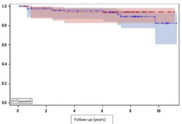
Fig. 1. Survival curve. Blue: survival of total knee arthroplasty after closing-wedge high tibial osteotomy. Red: survival of total knee arthroplasty after opening-wedge high tibial osteotomy, 95% confidence intervals.
3.4. Survival curves
Ten-year survival was 94% (95% CI: [83–98]) in the OW-HTO group and 82% (95% CI: [61–93]) in the CW-HTO group, without significant difference (P = 0.338) (Fig. 1).
4. Discussion
Surgical habits for TKA after HTO were unchanged despite this context of HTO revision, with few stems used and mostly conven-tional surgery. In the CW group, hardware removal was mainly performed as a separate step and the primary approach was not mainly reutilized for TKA. Long-term clinical outcome was good and comparable between groups. Late complications, however, were more frequent in the CW group (12% versus 6%). The study hypoth-esis was thus partially confirmed.
The present results are comparable to those in the literature (systematic reviews, meta-analyses, comparative series and large-scale registries[7–16]) for TKA following HTO and primary TKA, with improved autonomy and conserved range of motion. Analyz-ing the literature in the light of levels of evidence, however, refines these conclusions.
Level-IV retrospective series confirm clinical improvement, but with a relatively high rate of complications (6–15%), revision surgery and malalignment[7–11], as in the present study. Gupta et al.[11]reported a 50-point improvement in IKS score and 32◦ improvement in flexion, but with a major complications rate of 14%. In contrast, at 15 years’ follow-up, the Norwegian registry[8]found no difference in survival between primary TKA and TKA following HTO, although the proportions of reasons for revision differed: in primary TKA failure comprised 28% loosening or wear, 21% infec-tion and 6% pain and in TKA following HTO 35% loosening or wear, 12% infection and 11% pain.
In level-III case-control studies comparing primary TKA versus TKA following HTO, results are less clear-cut and more
controver-sial[12–16]. Clinical results were comparable in 2 reports[13,15]
and better in primary TKA in 2 others[14,16]. Complications rates were comparable in 4 series[12–14,16], with identical revision rates in only 1 report[16]and good alignment in 3[13–15]. The most striking report was by Efe et al.[14], with nearly 10◦ gain in flexion, a 12-point improvement in IKS score and a 50% lower revision rate in primary TKA.
Finally, Meding et al.[17]published an original study with 15 years’ follow-up, comparing primary TKA and TKA following HTO in the same patient; in the 39 patients, there were no clinical or
radiological differences, with 100% 15-year survival in primary TKA and 97% in TKA following HTO.
The most frequent complications in the present post-HTO-TKA series were those commonly reported:
• skin complications; • infection;
• stiffness.
There were no differences according TKA after OW-HTO or TKA after CW-HTO in our study, in agreement with the literature
[7,18–20]. BastosFihlo et al.[18], comparing 117 TKAs after
CW-and 24 after OW-HTO, found identical gain in range of motion CW-and functional scores and comparable alignment correction, although with more frequent complementary intraoperative procedures in the CW group:
• 1 medial release;
• 1 anterior tibial tuberosity osteotomy; • 1 quadriceps snip;
• more frequent posterolateral release[18].
Generally, all authors underline that TKA is more demand-ing after HTO, with more frequent complementary procedures
[7,11,18–22]. This is attributed to architectural changes in the prox-imal tibia due to HTO: altered tibial slope, epiphyseal enlargement, metaphyseal malunion, possible lateral impingement by the tib-ial base in CW-HTO, and change in patellar height[2,7,15,23,24], as well as vascular risk in CW-HTO[25]. The recent introduction of personalized cutting-guides in OW-HTO has reduced architec-tural alteration in tibial slope, improving reproducibility[26]. The present study did not confirm these technical findings, but did high-light the fact that hardware removal tended to be performed in a separate step in the CW group and with a new incision for TKA. This was not reported by Han et al.[7]or Preston et al.[19], who found no difference according to the approach used for revision TKA.
Despite these frequently highlighted technical issues, the lit-erature shows that a gliding prosthesis without adding a stem is usually enough to stabilize the knee[11,21,27–31]. The rationale for posterostabilization is that there is frequent posterior cruciate ligament failure, especially in case of severe bone defect. Pos-terostabilization improves knee stability and clinical outcome. The present study confirmed these findings. Navigation may be useful
[15]but was not used in most of the present cases. The inclusion period was too early for the use of personalized cutting guides to be analyzed.
The study involved certain limitations. The retrospective mul-ticenter design could have incurred a risk of missing data and variation in techniques and practices, but in fact this did not hap-pen. The multicenter design, on the other hand, enabled a large series to be included, indeed one of the largest in the literature for TKA after HTO. The minimum 5-year follow-up was sufficient to meet objectives and test the study hypothesis. The lack of functional scoring and precise radiologic assessment was another limitation; but comparative study on the same set of technical and clinical criteria limited bias.
5. Conclusion
The present series of TKA following HTO confirmed that clinical outcome is good, with no differences between CW- and OW-HTO, except for more frequent late complications with CW-HTO. In case of CW- compared to OW-HTO: 0.4 0.2 0.0 ~ 10 1 Follow-up (years)
• hardware removal is more frequently performed in two steps; • the TKA approach is more often not the primary HTO approach.
Disclosure of interest
ME: consultant for Depuy-Synthes, Lepine, NewClip. AD declares that he has no competing interest. PV: consultant for Medacta.
SLe: consultant for Ceraver. FB: consultant for Amplitudeet Serf.
PB: consultant for Stryker and Depuy-Synthes.
SLu: consultant for Medactaand Smith&Nephew, institutional research support for Amplitude and Tornier.
SP: consultant for Zimmer-Biomet, Arthrex, Newclip, Graftys, Adler Ortho.
MC: consultant for Ceraver. JNA: consultant for Zimmer-Biomet.
Acknowledgments
The authors thank all the symposium members and their teams: P. Bizot (Paris), J.L. Briard (Rouen), O. Cantin (Lyon), G. Dereudre (Rouen), A. Grandjean (Paris), M. Ollivier (Marseille), G. Pirovano (Marseille) and S. Putman (Lille).
Special thanks to Pr. Duhamel’s biostatistics department in Lille.
References
[1]Gstottner M, Pedross F, Liedensteiner M, Bach C. Long-term outcome after high tibial osteotomy. Arch Orthop Trauma surg 2008;128:111–5.
[2]Sun H, Zhou L, Li F, Duan J. Comparison between closing-wedge and opening-wedge high tibial osteotomy in patients with medial knee osteoarthritis: a systematic review and meta-analysis. J Knee Surg 2017;30: 158–65.
[3]Papachristou G, Plessas S, Sourlas J, Levidiotis C, Chronopoulos E, Papachristou C. Deterioration of long-term results following high tibial osteotomy in patients under 60 years of age. Int Orthop 2006;30:403–8.
[4]Virolainen P, Aro HT. High tibial osteotomy for the treatment of osteoarthritis of the knee: a review of the literature and a meta-analysis of follow-up studies. Arch Orthop Trauma Surg 2004;124:258–61.
[5]Khoshbin A, Sheth U, Ogilvie-Harris D, MahomedN, Jenkinson R, Gandhi R, et al. The effect of patient, provider and surgical factors on survivorship of high tib-ial osteotomy to total knee arthroplasty: a population based study. Knee Surg Sports Traumatol Arthrosc 2017;25:887–94.
[6]Insall JN, Joseph DM, Msika C. High tibial osteotomy for varusgonarthrosis. A long-term follow-up study. J Bone Joint Surg 1984;66A:1040–8.
[7]Han JH, Yang JH, Bhandare NN, Suh DW, Lee JS, Chang YS, et al. Total knee arthroplasty after failed high tibial osteotomy: a systematic review of open versus closed wedge osteotomy. Knee Surg Sports Traumatol Arthrosc 2016;24:2567–77.
[8]Badawy M, Fenstad AM, InderkvamK, Havelin LI, Furnes O. The risk of revision in total knee arthroplasty is not affected by previous high tibial osteotomy. Acta Orthop 2015;86:734–9.
[9]Niinimaki T, Eskelinen A, Ohtone P, Phuto AP, Mann BS, Leppilahti J. Total kneearthroplasty after high tibial osteotomy: a registry-based case-control study of 1036 knees. Arch Orthop Trauma Surg 2014;134: 73–7.
[10]vanRaaij TM, Reijman M, Furlan AD, Verhaar JA. Total knee arthroplasty after high tibial osteotomy. A systematic review. BMC Musculoskelet Disord 2009;10:88.
[11]Gupta H, Dahiya V, Vasdev A, Rajgopel A. Outcomes of total knee arthroplasty following high tibial osteotomy. Indian J Orthop 2013;47:469–73.
[12]Haslam P, Armstrong M, Geutjens G, Wilton TJ. Total knee arthroplasty after failed high tibial osteotomy long-term follow-up of matched groups. J Arthro-plasty 2007;22:245–50.
[13]Kazakos KJ, Chatzipapas C, Verettas D, Galanis V, Xarchas KC, Psillakis I. Mid-term results of total knee arthroplasty after high tibial osteotomy. Arch Orthop Trauma Surg 2008;128:167–73.
[14]Efe T, Heyse TJ, Boese C, Timmesfeld N, Fuchs-Winkelmann S, Schmitt J, et al. TKA following high tibial osteotomy versus primary TKA– a matched pair anal-ysis. BMC Musculoskeletal Disord 2012;11:207.
[15]Saragaglia D, Massfelder J, Refaie R, Rubens-Duval B, Mader R, Rouchy RC, et al. Computer-assisted total knee replacement after medial opening wedge high tibial osteotomy: medium-term results in a series of ninety cases. Int Orthop 2016;40:35–40.
[16]Karabatsos B, Mohamed NN, Maistrelli GL. Functional outcome of total knee arthroplasty after high tibial osteotomy. Can J Surg 2002;45:116–9.
[17]Meding JB, Wing JT, Ritter MA. Does high tibial osteotomy affect the success or survival of a total knee replacement? Clin Orthop Relat Res 2011;469:1991–4.
[18]BastosFilho R, Magnussen RA, Duthon V, Demey G, Servien E, Granjeiro JM, et al. Total knee arthroplasty after high tibial osteotomy: a comparison of opening and closing wedge osteotomy. Int Orthop 2013;37:427–31.
[19]Preston S, Howard J, Naudie D, Somerville L, McAuley J. Total knee arthro-plasty after high tibial osteotomy: no difference between medial and lateral osteotomy approaches. Clin Orthop Relat Res 2014;472:105–10.
[20]Kuwashima U, Tashiro Y, Okazaki K, Mizu-uchi H, Hamai S, Murakami K, et al. Comparison of the impact of closing wedge versus opening wedge high tibial osteotomy on proximal tibial deformity and subsequent revision to total knee arthroplasty. Knee Surg Sports Traumatol Arthrosc 2017;25:869–75.
[21]Cerciello S, Vasso M, Maffulli N, Neyret P, Corona K, Panni AS. Total knee arthro-plasty after high tibial osteotomy. Orthopedics 2014;37:191–8.
[22]Erak S, Naudie D, MacDonald SJ, McCalden RW, Rorabeck CH, Bourne RB. Total knee arthroplasty following medial opening wedge tibial osteotomy: technical issues early clinical radiological results. Knee 2011;18:499–504.
[23]Dragosloveanu S, Cristea S, Dragosloveanu C. The effect of high tibial osteotomy on the posterior tibial slope. Maedica 2014;9:173–8.
[24]El Amrani MH, Lévy B, Scharycki S, Asselineau A. Patellar height rele-vance in opening-wedge high tibial osteotomy. Orthop Traumatol Surg Res 2010;96:37–43.
[25]Darnis A, Villa V, Debette C, Lustig S, Servien E, Neyret P. Vascular injuries during closing-wedge high tibial osteotomy: a cadaveric angiographic study. Orthop Traumatol Sur Res 2014;100:891–4.
[26]Munier M, Donnez M, Ollivier M, Flecher X, Chabrand P, Argenson JN, et al. Can three-dimensional patient-specific cutting guides be used to achieve optimal correction for high tibialosteotomy? Pilot study. Orthop Traumatol Surg Res 2017;103:245–50.
[27]Robertsson O, W-Dhal A. the risk of revision after TKA is affected by previous HTO or UKA. Clin Orthop Relat Res 2015;473:90–3.
[28]Mason JB, Fehring TK, Estok R, Banel D, Fahrbach K. Meta-analysis of alignment outcomes in computer-assisted total knee arthroplasty surgery. J Arthroplasty 2007;22:1097–106.
[29]Chen JY, Lo NN, Chong HC, Pang HN, Tay DK, Chin PL, et al. Cruciate retaining versus posterior stabilized total knee arthroplasty after previous high tibial osteotomy. Knee Surg Sports Traumatol Arthrosc 2015;23:3607–13.
[30]Akasaki Y, Matsuda S, Miura H, Okazaki K, Moro-oka TA, Mizu-uchi H, et al. Total knee arthroplasty following failed high tibial osteotomy: mid-term comparison of posterior cruciate-retaining versus posterior stabilized prosthesis. Knee Surg Sports Traumatol Arthrosc 2009;17:795–9.
[31]Walther M, Konig A, Kirschner S, Gohlke F. Results of posterior cruciate-retaining unconstrained total knee arthroplasty after proximal tibial osteotomy for osteoarthrosis: a prospective cohort study. Arch Orthop Trauma Surg 2000;120:166–70.
![Table 3 shows overall results. At a mean 86.7 months, post-TKA (range, 2–136 months: 88.9 months [4–134] in OW-HTO and 84.9 months [2–136] in CW-HTO), there was significant improvement in pain, unassisted walking, stairs, walking distance, leaving the house](https://thumb-eu.123doks.com/thumbv2/123doknet/3096362.87732/4.918.480.870.852.982/overall-results-significant-improvement-unassisted-walking-distance-leaving.webp)