OATAO is an open access repository that collects the work of Toulouse researchers and makes it freely available over the web where possible
Any correspondence concerning this service should be sent
to the repository administrator: [email protected]
This is an author’s version published in: http://oatao.univ-toulouse.fr/26113
To cite this version:
Ramassamy, Jill-Léa . Impact of deforestation on rodet
distribution and on the prevalence of leptospirosis in Cambodia.
Thèse d'exercice, Médecine vétérinaire, Ecole Nationale Vétérinaire de Toulouse – ENVT, 2017, 108 p.
ANNEE 2017 THESE : 2017 – TOU 3 – 4019
IMPACT OF DEFORESTATION ON RODENT
DISTRIBUTION AND ON THE PREVALENCE OF
LEPTOSPIROSIS IN CAMBODIA
_________________
THESE
pour obtenir le grade de DOCTEUR VETERINAIRE
DIPLOME D’ETAT
présentée et soutenue publiquement devant l’Université Paul-Sabatier de Toulouse
par
RAMASSAMY, Jill-Léa
Née, le 22/01/1992 MONTPELLIER (34)___________
Directeur de thèse : Mme Mathilde PAUL ___________ JURY PRESIDENT : M. Patrice MASSIP ASSESSEURS : Mme Mathilde PAUL M. Stéphane BERTAGNOLI
Professeur à l’Université Paul-Sabatier de TOULOUSE
Maître de Conférences à l’Ecole Nationale Vétérinaire de TOULOUSE Professeur à l’Ecole Nationale Vétérinaire de TOULOUSE
Mise à jour au 01/01/2017
Ministère de l'Agriculture de l’Agroalimentaire et de la Forêt ECOLE NATIONALE VETERINAIRE DE TOULOUSE
Directrice : Madame Isabelle CHMITELIN
PROFESSEURS CLASSE EXCEPTIONNELLE
M. AUTEFAGE André, Pathologie chirurgicale
Mme CLAUW Martine, Pharmacie-Toxicologie
M. CONCORDET Didier, Mathématiques, Statistiques, Modélisation
M DELVERDIER Maxence, Anatomie Pathologique
M. ENJALBERT Francis, Alimentation
M. FRANC Michel, Parasitologie et Maladies parasitaires
M. MILON Alain, Microbiologie moléculaire M. PETIT Claude, Pharmacie et Toxicologie
M. SCHELCHER François, Pathologie médicale du Bétail et des Animaux de Basse-cour
PROFESSEURS 1° CLASSE
M. BERTAGNOLI Stéphane, Pathologie infectieuse
M. BERTHELOT Xavier, Pathologie de la Reproduction
M. BOUSQUET-MELOU Alain, Physiologie et Thérapeutique
Mme CHASTANT-MAILLARD Sylvie, Pathologie de la Reproduction M. DUCOS Alain, Zootechnie
M. FOUCRAS Gilles, Pathologie des ruminants
Mme GAYRARD-TROY Véronique, Physiologie de la Reproduction, Endocrinologie Mme HAGEN-PICARD, Nicole, Pathologie de la reproduction
M. JACQUIET Philippe, Parasitologie et Maladies Parasitaires M. LEFEBVRE Hervé, Physiologie et Thérapeutique
M. LIGNEREUX Yves, Anatomie
M. MEYER Gilles, Pathologie des ruminants M. PICAVET Dominique, Pathologie infectieuse
M. SANS Pierre, Productions animales
Mme TRUMEL Catherine, Biologie Médicale Animale et Comparée
PROFESSEURS 2° CLASSE
M. BAILLY Jean-Denis, Hygiène et Industrie des aliments
Mme BOURGES-ABELLA Nathalie, Histologie, Anatomie pathologique M. BRUGERE Hubert, Hygiène et Industrie des aliments d'Origine animale
Mme CADIERGUES Marie-Christine, Dermatologie Vétérinaire M. GUERRE Philippe, Pharmacie et Toxicologie
M GUERIN Jean-Luc, Aviculture et pathologie aviaire
Mise à jour au 01/01/2017
PROFESSEURS CERTIFIES DE L'ENSEIGNEMENT AGRICOLE
Mme MICHAUD Françoise, Professeur d'Anglais M SEVERAC Benoît, Professeur d'Anglais
MAITRES DE CONFERENCES HORS CLASSE
M. BERGONIER Dominique, Pathologie de la Reproduction
Mme BOULLIER Séverine, Immunologie générale et médicale
Mme DIQUELOU Armelle, Pathologie médicale des Equidés et des Carnivores M. DOSSIN Olivier, Pathologie médicale des Equidés et des Carnivores
M. JOUGLAR Jean-Yves, Pathologie médicale du Bétail et des Animaux de Basse-cour
Mme LETRON-RAYMOND Isabelle, Anatomie pathologique M. LYAZRHI Faouzi, Statistiques biologiques et Mathématiques M. MAILLARD Renaud, Pathologie des Ruminants
M. MATHON Didier, Pathologie chirurgicale Mme MEYNADIER Annabelle, Alimentation Mme PRIYMENKO Nathalie, Alimentation
M. VERWAERDE Patrick, Anesthésie, Réanimation
MAITRES DE CONFERENCES (classe normale)
M. ASIMUS Erik, Pathologie chirurgicale
Mme BENNIS-BRET Lydie, Physique et Chimie biologiques et médicales
Mme BIBBAL Delphine, Hygiène et Industrie des Denrées alimentaires d'Origine animale Mme BOUCLAINVILLE-CAMUS Christelle, Biologie cellulaire et moléculaire
Mme BOUHSIRA Emilie, Parasitologie, maladies parasitaires M. CONCHOU Fabrice, Imagerie médicale
M. CORBIERE Fabien, Pathologie des ruminants
M. CUEVAS RAMOS Gabriel, Chirurgie Equine
Mme DANIELS Hélène, Microbiologie-Pathologie infectieuse Mme DEVIERS Alexandra, Anatomie-Imagerie
M. DOUET Jean-Yves, Ophtalmologie vétérinaire et comparée Mme FERRAN Aude, Physiologie
M. JAEG Jean-Philippe, Pharmacie et Toxicologie
Mme LAVOUE Rachel, Médecine Interne
M. LE LOC’H Guillaume, Médecine zoologique et santé de la faune sauvage
M. LIENARD Emmanuel, Parasitologie et maladies parasitaires Mme MEYNAUD-COLLARD Patricia, Pathologie Chirurgicale Mme MILA Hanna, Elevage des carnivores domestiques M. MOGICATO Giovanni, Anatomie, Imagerie médicale
M. NOUVEL Laurent, Pathologie de la reproduction (en disponibilité)
Mme PALIERNE Sophie, Chirurgie des animaux de compagnie
Mme PAUL Mathilde, Epidémiologie, gestion de la santé des élevages avicoles et porcins Mme PRADIER Sophie, Médecine interne des équidés
M. RABOISSON Didier, Productions animales (ruminants)
M. VOLMER Romain, Microbiologie et Infectiologie
Mme WARET-SZKUTA Agnès, Production et pathologie porcine
ASSISTANTS D'ENSEIGNEMENT ET DE RECHERCHE CONTRACTUELS
Mme COSTES Laura, Hygiène et industrie des aliments Mme LALLEMAND Elodie, Chirurgie des Equidés
1 REMERCIEMENTS
A Monsieur le Professeur Patrice Massip Professeur des Universités
Praticien hospitalier
Département des Maladies Infectieuses Université de Toulouse III Service des maladies infectieuses et tropicales du CHU de Toulouse
Qui nous a fait l’honneur d’accepter la présidence du jury de thèse. Hommages respectueux.
A Madame le Docteur Mathilde-Paul
Maître de conférences de l’Ecole Nationale Vétérinaire de Toulouse Epidémiologie
Pour m’avoir ouvert les portes vers le monde de l’épidémiologie et pour ses encouragements, Sincères remerciements.
A Monsieur le Professeur Stéphane Bertagnoli
Professeur de l’Ecole Nationale Vétérinaire de Toulouse Virologie-Infectiologie
Qui nous fait l’honneur de participer à notre jury de thèse, Veuillez accepter mes plus sincères remerciements.
2 A la Fondation Pierre Ledoux Jeunesse et France (2016) Pour l’attribution d’une bourse pour mes six mois de stages auprès de l’Institut Pasteur du Cambodge,
Mes sincères remerciements.
A la Fondation France Vétérinaire (2015) Pour la bourse qui m’avait été attribuée en 2015 et qui m’a permis de partir au Cambodge et de participer à la première saison de capture.
Au Conseil Général de Guadeloupe
Pour le support financier incommensurable qui m’a été décerné tout au long de mes études.
Un grand merci en particulier à Mme Anick Ancette qui a été ma correspondante pendant toutes ces années et m’a offert son aide dans bien des situations,
3 Au Docteur Mathieu Pruvot
Project Lead - Wildlife Health
Epidemiologiste au Wildlife Conservation Society Cambodia
Pour son encadrement et son aide précieuse tout au long de ma these Mes remerciements les plus sincères
To Dr Paul Horwood
Deputy-Head of Virology Unit - Institut Pasteur in Cambodia
I would like to express my very great appreciation to him for supervising this internship. His advices have been a great help and his willingness to give his time have been very much appreciated.
My internship was part of the One-Health LACANET program, funded by the European Commission under the INNOVATE programme.
I would like to thank Dr Philippe DUSSART and Sapho BRIAND for including me in the LACANET network.
To the WCS Team
Trapping rodents was not easy and was only made possible in this instance through the help of a number of people. I am greatly thankful to every person from the WCS who participated in the field work. Sokha CHEA, Samat IN, Yaren TY, Vuthy BUOR, Seng MOROKOTH, Ratha TINA, Semnang.
To all the technicians of the virology unit of Institut Pasteur du Cambodge
I am particular grateful for the assistance given by Vibol HUL to set the PCR method and for his help on the sequences analyses. My special thanks are extended to Huy Sreang HENG, KimTuo CHHEL, Sophors PHORL, Oudam HENG for their daily help.
4 CONTENT LIST
PART 1: RESUME EN FRANCAIS ... 11
PART 2: LITERATURE REVIEW ... 18
1. DEFORESTATION IN CAMBODIA ... 18 2. LEPTOSPIROSIS REVIEW ... 20 2.1. Leptospira biology ... 20 2.2. Leptospira classification ... 21 2.3. Host species ... 23 2.4. Environmental reservoir ... 24 2.5. Transmission cycle ... 25 2.6. Risk factors ... 26
2.7. Leptospirosis, the disease ... 26
2.8. Diagnostic tests ... 27
2.9. Treatment ... 29
3. LEPTOSPIROSIS EPIDEMIOLOGY ... 31
3.1. Leptospirosis epidemiology in humans, in SEA ... 31
3.2. Leptospirosis epidemiology in rodents, in SEA ... 31
4. IMPACT OF LAND-USE CHANGE ON PATHOGENS IN SEA ... 34
4.1. Land use changes and disease prevalence ... 34
4.2. Land use changes and rodent-borne diseases ... 35
4.3. Land-use changes and leptospirosis ... 36
4.4. Limitations of the current literature... 37
PART 3: HYPOTHESIS ... 41
PART 4: METHODS AND MATERIALS ... 43
1. Study design ... 43
1.1. Trapping grid ... 43
5
2. Laboratory analyses ... 44
2.1. Rodent species ... 44
2.2. Leptospira detection ... 44
2.2.1. Leptospira species assay ... 45
2.2.2. Pathogenic Leptospira assay ... 45
2.3. Leptospira species identification ... 45
3. Statistical Analyses ... 46
3.1. Rodent species richness and diversity indices ... 46
3.2. Rodent density ... 46
3.3. Rodent species composition ... 47
3.4. Habitat specialization and Zone Preference ... 47
3.5. Leptospira infection ... 48
PART 5: RESULTS ... 50
1. Rodent trapping ... 50
2. Ecological changes during deforestation ... 51
2.1. Species richness ... 51
2.2. Species diversity ... 52
2.3. Rodent density ... 53
2.4. Species composition ... 54
2.5. Zone preferences of species... 58
3. Leptospira infection ... 59
3.1. Pathogenic and Intermediate Leptospira PCR results ... 59
3.2. Pathogenic Leptospira PCR results ... 59
3.3. Sequencing results ... 61
3.4. Statistical analysis on Leptospira infection ... 63
3.4.1. Univariate analysis ... 63
6
PART 6: DISCUSSION ... 65
1. Rodent and deforestation ... 65
2. Leptospirosis ... 67 3. Methodological considerations ... 70 3.1. Design ... 70 3.2. PCR assay ... 71 3.3. Barcoding ... 72 PART 7: CONCLUSION ... 74 BIBLIOGRAPHY ... 76
7 APPENDICES LIST
APPENDIX 1: Serological classification of Leptospira spp ... 85
APPENDIX 2: Serological and genomic classification of Leptospira spp ... 86
APPENDIX 3 : Host - serovars associations ... 87
APPENDIX 4 : Summary of the trend of infectious disease response to land use changes from reviews ... 88
APPENDIX 5 : Capture sites on the Cambodia Map ... 89
APPENDIX 6 : Gridline ... 90
APPENDIX 7 : Animal identification ... 91
APPENDIX 8: Exemple of Barcoding protocol and result ... 92
APPENDIX 9: Capture tables ... 93
APPENDIX 10 : Relative abundance of each species per season ... 94
APPENDIX 11 : Ecological indexes ... 95
APPENDIX 12 : Habitat specialization per species per season ... 96
APPENDIX 13 : Leptospira prevalence ... 97
8 LIST OF FIGURES
FIGURE 1 Scanning electron micrograph of L. interrogans serovar icterohaemorrhagiae strain
RGA SOURCE : Levett (2001) ... 20
FIGURE 2 Phylogenetic tree of Leptospira species ... 22
FIGURE 3 Photomicrograph of a Warthin-Starry stained section of kidney tissue from sewer rat. SOURCE : Ko et al., (2009). ... 24
FIGURE 4 Leptospirosis transmission cycle ... 25
FIGURE 5 Biphasic nature of leptospirosis and relevant investigations at different stages of disease SOURCE: LEVETT et al (2011). ... 27
FIGURE 6 Geographic distribution of Leptospira infection in rodents from Thailand, Lao PDR and Cambodia. SOURCE : Cosson et al (2014) ... 32
FIGURE 7 Associations between habitat structure and rodent-borne pathogens SOURCE : Morand et al (2015) ... 37
FIGURE 8 Chronotone representation. SOURCE : Bradley, (2004) ... 38
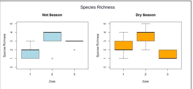
FIGURE 9 Species richness distribution among zones by season.. ... 51
FIGURE 10 Species diversity (Shannon Index) among zones and season ... 52
FIGURE 11Rodent density estimate (in animal/ha) per zone by season ... 53
FIGURE 12 & FIGURE 13 Correspondence Analysis Graphs for the rainy season (left) and the dry season (right). ... 55
FIGURE 14 Between-class correspondence analysis of species abundance among the three zones. ... 57
FIGURE 15 Between-class correspondence analysis of species abundance among zones during the dry season. ... 57
FIGURE 16 Habitat diversity measured with the Shannon Index, per species... 58
FIGURE 17 Phylogenetic analysis for the rrs gene of Leptospira sp. isolated from rodents based on BIONJ method. ... 62
9 LIST OF TABLES
TABLE 1 Advantages and disadvantages of common diagnostic tests for leptospirosis SOURCE : Musso, Scola, (2013) ... 29 TABLE 2 Species identity and total number of captured individuals by zone and by season. Species are ordered according to their total abundance.. ... 50 TABLE 3 Pathogenic Leptospira prevalence among rodent species and zones per season .... 59 TABLE 4 Leptospira prevalence (from the Leptospira species essay) among rodent species and zones per season. ... 60 TABLE 5 Pathogenic Leptospira species identified in the rodent species ... 61
10
PART 1 : RESUME EN FRANÇAIS
11 PART 1: RESUME EN FRANCAIS
1. Introduction
Le taux de déforestation au Cambodge se classe parmi les plus élevés du Sud Est de l’Asie et du Monde. La conversion des forêts en cultures agricoles entraîne des changements brusques et irréversibles de ces écosystèmes. De tels changements peuvent modifier la distribution des rongeurs et donc des pathogènes qu’ils hébergent. Entre autres, la leptospirose représente un problème de santé majeur au Cambodge où elle est endémique. L’hypothèse posée dans cette étude est que la déforestation modifie la population des rongeurs et, par conséquent, la transmission de leptospires.
La leptospirose est une zoonose mondialement répartie. La maladie est due à des espèces de leptospires pathogènes, bactéries appartenant à l’ordre des Spirochetacae et du genre
Leptospira. Ces bactéries peuvent infecter tous les mammifères dont l’Homme. Certains
serovars de leptospires démontrent une spécificité d’hôte. En particulier, les rongeurs sont considérés comme des réservoirs majeurs de leptospires pathogènes pour l’Homme. L’infection est maintenue chez les animaux infectés par une colonisation des tubules rénaux par les leptospires, qui sont ensuite relâchées dans l’environnement dans leurs urines. La plupart des espèces de leptospires survivent dans un environnement aqueux ou dans un sol humide. La présence de ces leptospires pathogènes dans l’environnement constitue ainsi le principal mode de contamination de l’Homme : par contact avec un environnement souillé d’urine d’animaux infectés. Dans les pays tropicaux où la leptospirose est endémique, la riziculture, l’élevage, les pluies abondantes ou encore la présence de rats dans les habitations ont été identifiés comme des facteurs de risque de la maladie. La circulation de leptospires pathogènes pour l’Homme a été mise en évidence chez les rongeurs en Asie du Sud-Est, en particulier chez des espèces synanthropiques mais également chez des espèces forestières peu étudiées.
Les taux élevés de déforestation en Asie du Sud-Est coïncident avec une augmentation d’émergence de maladies infectieuses. Un certain nombre d’études sur les maladies infectieuses émergentes s’accorde à établir qu’un des mécanismes d’émergence est la modification de l’habitat par l’homme. Les modifications de l’habitat par l’Homme peuvent impacter négativement l’intégrité de l’écosystème et la biodiversité. Ces changements dans la structure de l’écosystème peuvent également conduire à des modifications du système hôte-pathogène. Ainsi les modifications de l’habitat ont le potentiel de modifier la dynamique d’une maladie directement ou indirectement en modifiant l’abondance, la démographie, le comportement, la
12 réponse immune ou encore le contact entre espèces et la composition des espèces réservoirs (Gottdenker et al., 2014).
En effet, la modification des paysages par l’Homme se traduit par une perte de l’habitat des espèces forestières et sa fragmentation. Cela peut entraîner des conséquences potentielles sur la transmission des agents infectieux tels que l’augmentation de la densité d’une espèce réservoir de l’agent pathogène ; l’augmentation de contact interspécifique, c’est-à-dire entre des espèces différentes et en particulier en mettant en contact la faune sauvage avec l’homme directement ou indirectement avec les animaux domestiques ; une augmentation des contacts intra-spécifiques (au sein d’une même espèce), due à la fragmentation de l’habitat et au regroupement des ressources (Brearley et al., 2013). Ce sont des exemples de mécanismes hypothétiques par lesquels la modification de l’habitat peut aboutir à une modification de la prévalence des maladies. Dans le cas des agents infectieux transmis par les rongeurs, des études sur les hantavirus décrivent une telle modification (Suzán, Marcé, et al., 2008 ; Blasdell et al., 2016). Ces virus se transmettent par contact direct avec des fluides corporels infectés entre rongeurs et peuvent infecter l’Homme. Des épidémies à hantavirus étaient survenues dans un contexte de modification de l’habitat, où la biodiversité était réduite. L’explication proposée par ces articles était que la modification de l’habitat favorisait des espèces de rongeur opportunistes qui étaient réservoirs du virus et qui en l’absence de compétition, pouvaient atteindre des densités élevées conduisant à une augmentation de contacts intra-spécifique et consécutivement à une augmentation de la prévalence (Suzán, Marcé, et al., 2008). Il est donc important de déterminer quels sont les agents pathogènes infectieux transmis par les rongeurs qui pourraient suivre la même dynamique, suite à la perturbation de l’habitat.
Peu d’études se sont intéressées à l’impact de modification d’habitat sur la leptospirose, mais montrent une corrélation entre les espèces de leptospires et la topographie (Ivanova et al., 2012). En particulier, une étude suppose que l’infection des rongeurs par Leptospira spp. serait corrélée à la fragmentation de l’habitat forestier (Morand et al., 2015).
La conversion de forêt en zone agricole n’est pas seulement le passage d’un écosystème à un autre, c’est un processus progressif dans le temps. Pour comprendre les changements dans la transmission d’agents pathogènes zoonotiques dus à la déforestation, il est nécessaire de s’intéresser à l’enchaînement complexe des événements qui interviennent pendant cette période transitoire, ou chronotone. Le chronotone, terme proposé et défini par (Bradley, 2004), est « l’interface temporelle entre deux types de paysage », « c’est une période relativement rapide
13
de transformation qui sépare deux types d’utilisation prolongée des terres ». C’est cette période
transitoire qui représente un risque pour la santé publique et qui est étudiée dans cette thèse. 2. Méthode
L’étude se base sur un design de « space-for-time substitution» dans lequel le gradient géographique de déforestation dans un même site est appréhendé comme une substitution à la dynamique temporelle de la déforestation, le chronotone. Trois zones, correspondant à trois étapes de la déforestation, sont définies : « Forêt intacte » (Forêt indemne d’activité de déboisement ou d’activité faible) ; « Forêt perturbée » (Forêt où l’activité de déboisement est intense) et « Plantation récente » (Champs agricoles divers : rizière sèche, plantations de cassava ou de maïs, moins de un an après le début de la déforestation). Ce modèle de chronoséquence pose l’hypothèse que ces trois zones correspondent à une même zone à trois temps différents au cours du processus de déforestation. Pour respecter cette hypothèse, les trois zones sont choisies géographiquement proches les unes des autres au sein de chaque site et ayant moins d’un an depuis le début de la déforestation, afin de limiter les différences spatio-temporelles autres que celles dues au processus de déforestation. L’étude a été réalisée dans cinq sites au Cambodge dans les provinces du Mondulkiri et de Kampong Thom et fut répétée en saison des pluies et en saison sèche pour ces cinq mêmes sites. Les rongeurs ont été capturés simultanément dans les trois zones d’un même site pendant huit nuits consécutives. Les rongeurs capturés sont prélevés, marqués avec une boucle auriculaire unique et relâchés à leur lieu de capture. Les espèces de rongeurs ont été déterminées par marqueur moléculaire et séquençage (barcoding).
La première partie des résultats de cette thèse s’intéresse aux effets de la déforestation sur la communauté de rongeur. Les mesures de composition, richesse et diversité d’espèce, ont été comparés entre les trois zones. Le modèle de capture-marquage-recapture a permis d’estimer la densité de population de rongeur dans les trois habitats.
La deuxième partie des résultats porte sur les taux d’infections de la leptospirose chez les rongeurs capturés. L’infection par Leptospira spp a été testée par PCR en temps réel. Le gène
rrs, universellement présent chez les leptospires, a été amplifié afin de détecter les infections
par des espèces pathogénes et intermédiaires (PCR1). Le gène LipL32, présent uniquement chez les leptospires pathogènes, a permis de détecter les infections par des leptospires pathogènes uniquement (PCR2). Les leptospires pathogènes détectées ont été séquencées.
14 3. Résultats
A l’issu des deux saisons de captures, 522 rongeurs ont été capturés et pu être identifiés. Onze espèces ont pu être déterminées par marqueur moléculaire. Mus cervicolor Rattus sp R3 et
Maxomys surifer étant les espèces les plus fréquemment capturées et représentent 95% du total
d’animaux capturés.
3.1. Déforestation et rongeurs
Les indices de diversité et de richesse d’espèce ressortent significativement différents entre les trois zones de déforestation au sein de chaque site. La zone correspondant à la forêt perturbée contenait le plus grand nombre d’espèces de rongeurs et une plus grande diversité d’espèces que la forêt intacte et les champs récents. La richesse et diversité des espèces de rongeur augmentent donc de façon transitoire pendant la déforestation, puis diminuent après conversion agricole.
La densité de rongeur augmente également au cours de la déforestation, avec des valeurs significativement plus élevées en forêt perturbée qu’en forêt intacte. La densité de rongeurs dans les zones agricoles récentes apparaît significativement dépendante de la saison, atteignant des valeurs élevées (de 15 à 48 animaux par hectare) en saison des pluies mais des valeurs faibles en saison sèche.
Les analyses statistiques d’écologie (analyse de correspondance et matrice de dissimilarité) ont montré que la déforestation s’accompagnait d’un changement progressif de composition d’espèce de rongeur. Certaines espèces sont capturées exclusivement en forêt intacte (Rattus
andamanensis¸ Leopoldamys sabanus, Berylmys bowersi) supposant leur disparition avec la
déforestation, bien que le faible effectif de capture de ces espèces ne permette pas de conclure. En revanche, des espèces forestières telles que Maxomys surifer, Berylmys berdmorei et
Niviventer fulvescens sont également capturées dans les zones perturbées indiquant une
persistance de ces espèces malgré le déboisement. Aucune espèce forestière ne persiste à la fin de la déforestation et à sa conversion en champs agricoles (à l’exception de Rattus sp R3 qui fut capturé dans toutes les zones). La population de rongeurs dans les champs est alors caractérisée par très peu d’espèces (montré par des indices de richesse et diversité faibles) dominés par une espèce majoritaire Mus cervicolor et une densité élevée.
Des espèces agricoles, notamment Mus cervicolor, étaient également présentes dans les forêts perturbées indiquant une invasion précoce, exacerbée pendant la saison sèche. En effet, les dissimiliarités de composition entre la forêt perturbée et les champs agricoles récents sont
15 minimes pendant la saison sèche. Les conditions difficiles thermiques et l’absence de nourriture dans les champs en saison sèche peuvent expliquer une migration des espèces agricoles vers la forêt alentour. La forêt perturbée se compose donc à la fois d’espèces forestières et d’espèces agricoles qui n’étaient jusque-là pas en contact.
3.2. Déforestation et Leptospira spp.
L’infection par des espèces pathogènes et intermédiaires de Leptospira spp a été mise en évidence chez sept espèces de rongeurs avec des prévalences variant de 1 à 100%. Les valeurs extrêmes étant expliquées par des effectifs très faibles d’animaux pour certaines espèces. La prévalence totale chez les rongeurs était de 13.5% en saison de pluies et 7.3% en saison sèche mais la différence entre les deux saisons n’était pas significative à l’analyse multivariée. La prévalence n’était pas significativement différente entre les espèces ni entre les trois niveaux de déforestation. Les rongeurs mâles étaient significativement plus à risque d’être infectés par des espèces pathogènes et intermédiaires de Leptospira spp.
L’infection par des leptospires pathogènes uniquement n’a été trouvée que chez trois espèces :
Mus cervicolor capturés dans les champs en saison des pluies et Maxomys surifer en forêt
perturbée et Berylmys bowersi en forêt intacte en saison sèche. Les prévalences respectives chez ces espèces étaient de 4.5% (12/267), 3.8% (1/26) et 50% (1/2).
L’isolement et le séquençage des leptospires pathogènes chez ces animaux infectés a permis l’identification de trois espèces de leptospires pathogènes pour l’Homme : L. borgpetersenii, L.
weilii et L. interrogans.
4. Discussion
Les résultats de cette étude montrent une modification complète de la composition d’espèces de rongeur, engendrée par la déforestation. Les indices de richesse et de diversité d’espèces augmentent transitoirement pendant la déforestation pour atteindre des minimums dans les champs récemment convertis à l’agriculture. La déforestation et conversion semblent favoriser une espèce : Mus cervicolor, espèce majoritaire dans les champs et qui atteint des densités élevées. La présence de cette espèce dans les zones en cours de déforestation indique une invasion précoce de cette espèce (moins de un an après le début de la déforestation).
L’infection par L. borgpetersenii chez Mus musculus fait d’elle une espèce à risque. Cette leptospire nécessite une transmission directe, contrairement aux autres espèces de leptospires
16 qui ont la capacité de survivre dans un environnement humide et se transmettent principalement par contact indirect. La transmission de cette leptospire peut donc dépendre de la proportion d’animaux infectés dans la population mais également de la densité de sa population d’hôte. Les fluctuations de densité et de migration de cette espèce, induites par les saisons et exacerbées par les pratiques agricoles peuvent donc modifier la dynamique de la leptospirose.
L’infection par L. borgpetersenii d’un Maxomys surifer capturé en forêt perturbée pendant la saison sèche où se retrouve préférentiellement Mus cervicolor, pourrait indiquer un passage de leptospire d’une espèce agricole hôte à une espèce forestière. L’infection d’un animal seulement ne nous permet pas de conclure mais permet toutefois de soulever l’hypothèse que l’assemblage transitoire d’espèces en forêts perturbées, en augmentant la probabilité de contact entre espèces différentes pourrait entraîner un potentiel changement d’hôte des leptospires.
5. Conclusion.
La leptospirose est un problème de Santé Publique majeur au Cambodge. Bien qu’endémique dans ce pays et d’incidence supposée élevée, la leptospirose est négligée car sous-diagnostiquée et souvent confondue pour des cas de paludisme et de dengue, majoritairement présents dans le pays. Elle y est peu étudiée et les facteurs de risques de transmission à l’Homme ne sont pas identifiés au Cambodge.
Les résultats de cette thèse ont permis d’identifier quelles étaient les modifications écologiques qui survenaient au cours de la déforestation. Ainsi, la déforestation aboutit à une réduction de la diversité et richesse d’espèces de rongeurs, et favorise l’introduction et la persistance d’une espèce Mus cervicolor. Les résultats sur la leptospirose ne montraient pas de modification de la prévalence au cours de la déforestation, ni entre les espèces de rongeur. Toutefois les leptospires pathogènes séquencées correspondaient à trois espèces différentes dans les trois stades de déforestation.
La présence de leptospires zoonotiques dans les trois niveaux de déforestation indique un potentiel risque de leptospirose humaine associé à des activités forestières et agricoles ou encore la consommation de rongeurs sauvages. Il est donc nécessaire de déterminer par la suite le risque associé à chacune de ces activités.
17
PART 2: LITERATURE REVIEW
18 PART 2: LITERATURE REVIEW
1. DEFORESTATION IN CAMBODIA
Home to some of Southeast Asia’s oldest and most diverse forests, Cambodia is a recognized hotspot of biodiversity but is also a place of massive deforestation and of emerging infectious disease (Myers et al., 2000 ; Morand et al., 2014).
The rate of deforestation in Cambodia has accelerated in the last few years and has now reached about 208,000 hectares a year (Forest, 2015). The proportion of primary forest, characterized by naturally regenerated forest of native species without any ecological disturbance or any visible human activities, decreased to barely 3% of total forest cover in Cambodia in 2015. All presented data on deforestation in Cambodia are excerpted from a Forest Trend Report from 2015 (Forest, 2015). Cambodian forest is increasingly converted to other land use, with rubber plantations the most common plantation type, followed by sugar, pulp and paper plantations and then cassava and rice fields. These conversions are enabled by economic land concessions, though the legality of these land allocations is questionable. By 2015, 2.2 million hectares, twelve percent of the whole country had been allocated to economic land concessions (ELC) with no legal framework to justify these allocations. Tragically, they often overlap with protected areas and in 2013, 14 percent of protected forest land had been allocated resulting in even more loss of evergreen and primary forest (Forest, 2015). Aggravating the situation, the collection of timber on ELC, by becoming the main source of wood harvested in Cambodia, is believed to facilitate illegal logging in nearby areas, exacerbating the disputably legal logging from land concessions (Forest, 2015). There is a concerning lack of effective regulatory framework regarding land conversion and logging in Cambodia that will inevitably lead to tremendous loss of forest ecosystem and biodiversity (Wilcove et al., 2013). The link between forests and human health has been highlighted on several aspects, from the known role of forest in improving the human environment (for example by absorbing airborne pollution), its role as a source of bioactive medicinal compounds (Skirycz et al., 2016), to a recently explored causal link between forest change and emergence of infectious diseases which often originate in animals (Wilcox, Ellis, 2006). Understanding the relationship between deforestation and its potential impact on human health could help promote responsible forest management and control of forest-linked disease to lessen the impact.
19 Deforestation, by disrupting the natural environment of rodent species, will affect the species distribution and their densities and could thus have important consequences on the pathogens they carry. Rodents have been implicated in the emergence and spread of infectious diseases of importance to human health such as plague, murine typhus, scrub typhus, leptospirosis and hantavirus haemorrhagic fever, etc...(Herbreteau et al., 2012) Among them, leptospirosis, represents a major threat to public health in South-East Asia.
20 2. LEPTOSPIROSIS REVIEW
Leptospirosis is a worldwide zoonosis caused by pathogenic bacteria, the leptospires that are transmitted directly or indirectly from animals to humans.
2.1. Leptospira biology
Leptospires are bacteria that belong to the Spirochetacae order, Leptospiraceae family and
Leptospira genus. Leptospires are thin, highly motile, slow-growing spirochetes, about
0.1-0.3µm in diameter by 6–20µm in length (Levett, 2001). Too thin to be visible under the ordinary microscope, dark-field microscopy is most often used to observe leptospires after staining (figure 1).
FIGURE 1 Scanning electron micrograph of L. interrogans serovar icterohaemorrhagiae strain RGA SOURCE : Levett (2001)
They can be distinguished from other bacteria on the basis of their unique helical shape and the presence of periplasmic flagella. Leptospires have distinctive hooked ends. Two periplasmic flagella with polar insertions are located in the periplasmic space and are responsible for motility. Leptospires can exhibit two distinct forms of movement, translational and nontranslational that enable them to move in aqueous media (Goldstein, Charon, 1988).
21 Leptospires have a typical double membrane structure in common with other spirochetes, in which the cytoplasmic membrane and peptidoglycan cell wall are closely associated and are overlain by an outer membrane. They are obligate aerobes with an optimum growth temperature of 28–30°C. Leptospires are catalase and oxidase positive. Leptospiral lipopolysaccharide has a composition similar to that of other gram-negative bacteria, but has lower endotoxic activity (Levett, 2001).
2.2. Leptospira classification
Leptospira family contains both pathogenic leptospires, having the potential to cause disease in
animals and humans and saprophytic leptospires that are free living bacteria in wet environment and generally considered not to cause disease.
Initially, two serological species were recognized, namely pathogenic Leptospira interrogans and saprophytic Leptospira biflexa. Both complexes (L. interrogans and L. biflexa) have been divided into several serovars using the cross-agglutinin adsorption test and antigenically related serovars were grouped into serogroups. The Leptospira classification was historically based on serological characteristics and comprised over 260 pathogenic serovars (See Appendix 1). Recent genetic research has resulted in the reclassification of Leptospira spp., on the basis of DNA relatedness and has led to 12 pathogenic species and 5 saprophytic species (Bharti, 2003). However, there are still many new species that are believed to exist and yet to be discovered. The two classification systems based on the serovar and genetic concepts do not correspond as strains belonging to the same serovar may belong to different Leptospira species (Appendix 2). However, both the antigenic and genetic classification systems are in common use (Morey et al., 2006 ; Levett, 2001).
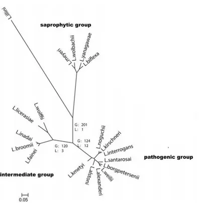
Currently, twenty Leptospira species have been identified on classical DNA-DNA hybridization studies and 16S ribosomal RNA gene phylogeny (Xu et al., 2016). They comprised of pathogenic, intermediate and saprophytic groups as detailed on the phylogenetic tree (Figure 2). The intermediate group consists of five species that occasionally cause disease in humans and animals. The six saprophytic species are not pathogenic and among them, L.
22 Recent genomic and phylogenetic studies of Leptospira spp. supported that the actual biodiversity of Leptospira spp inferred that host adaptation might be the driving force of
Leptospira diversification and evolution (Xue et al., 2008 ; Xu et al., 2016 ; Ko et al., 2009).
Complete genome sequencing of different Leptospira species showed a high diversity among species and found a range of genome size diversity higher than any other zoonotic pathogens (Xu et al., 2016). This high genomic variability was attributed to massive gene gain and loss events that allowed for adaptation to specific niche conditions and changing host environments (Xue et al., 2008). Saprophytic species are closer to the most recent common ancestor while intermediate and pathogenic species formed the two deepest branches, suggesting that virulent genes favoring host infection have been acquired during the evolution of the genus. Loss of genes involved in metabolic pathways and gains of virulent genes, for example these responsible for motility and chemotaxis required to colonize and invade a host, could thus explain the evolution from strains capable of surviving in complex ambient environments into those adapted for pathogenic life (Xu et al., 2016).
FIGURE 2 Phylogenetic tree of Leptospira species
Phylogenetic analysis based on the maximum likelihood of the concatenated core genes of the Leptospira genome with Leptonema illini as the outgroup. Scale bar indicated an evolutionary distance of 0.05 amino acid substitutions per position.
23 2.3. Host species
Leptospirosis affects both humans and animals and has been reported in most mammal groups worldwide (Levett, 2001).
Leptospira has been found in many mammal species worldwide: from common animals
(rodents, canidae, bovidae…) to more exotic discoveries (racoon, polar bear, whales)(Duncan, 2012 ; Grune Loffler et al., 2015 ; Richardson, 2003 ; Jabłoński, 2016 ; Junge et al., 2007). Some serovars are commonly associated with particular animal reservoirs. Certain host-serovar specificity exhibits relatively high fidelity, for example Rattus species and serovar Icterohaemorrhagiae, and mice with serogroup Ballum serovars (Bharti, 2003). More examples are given in Appendice 3.
Leptospires are usually adapted to their primary hosts and cause little illness in these. Clinical signs can be seen when a different serovar is introducer to a host species and the symptoms are variable, depending on the serovar or Leptospira species, the host and the animal immune system.
Leptospirosis in dogs can be asymptomatic or range from a transient fever to an acute, fulminant illness with fever, anorexia, vomiting, liver and renal failure. Four syndromes were described in canine leptospirosis: icteric, hemorrhagic, uremic and reproductive syndromes (Faine et al., 1999).
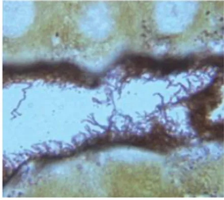
Cattle are the maintenance host for hardjo-bovis, infection with this serovar will often produce a carrier state in the kidneys associated with long-term urinary shedding. In addition, infections with hardjo-bovis can persist in the reproductive tract. Many leptospiral infections in cattle are subclinical, particularly in nonpregnant and nonlactating animals. In cattle and pig, clinical signs of leptospirosis are mostly reproductive symptoms such as abortion, mummified fetus, fertility loss, agalaxia... In horses, leptospirosis can result in chronic recurrent uveitis. During the healing phase, animals can be asymptomatic carrier of leptospires. Present in the renal tubules for short to long period of times, leptospires are excreted into the environment in the urine (see Figure 3).
24
FIGURE 3 Photomicrograph of a Warthin-Starry stained section of kidney tissue from sewer rat. Leptospires are seen as silver-impregnated filamentous structures within the proximal renal tubule lumen (400x
magnification) SOURCE : Ko et al., 2009.
Animal reservoirs that may pose a risk for human exposure include livestock, dogs (Mgode et al., 2015) but also wildlife: raccoons, prairie dogs (Olds et al., 2015 ; Richardson, 2003) and rodents.
Rodents are considered a major reservoir of human leptospirosis as they can be asymptomatic carriers of the bacteria and may continually excrete leptospires into the environment throughout their life (Faine et al., 1999). Leptospirosis is maintained by persistent Leptospira colonisation of the proximal renal tubules of infected animals (Figure 3) who can thus shed the bacteria in their urine and discharge them into the environment intermittently, regularly for months or years, or even for their lifetime in the case of rodents (Faine et al., 1999).
2.4. Environmental reservoir
Once excreted in the urine into the environment, leptospires survival depends on their biological properties and on the environmental conditions.
Saprophytic leptospires are naturally found in many types of wet or humid environment ranging from surface waters and moist soil to tap water. Some leptospires were even found in seawater. Both pathogenic and saprophytic species can be isolated from surface water and soils (Wynwood et al., 2014).
In warm and humid conditions, most pathogenic leptospires can survive several weeks to months in muddy soils or rivers, by mean of cellular aggregation (Trueba et al., 2004). Viable cells may persist up to 20 months after excretion and their virulence was fully preserved (Andre-Fontaine et al., 2015). Wet environments are thus an important source of leptospires, contributing to the transmission cycle of leptospirosis.
25 2.5. Transmission cycle
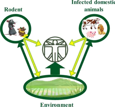
From this survival in the environment results the main transmission pathway of leptospirosis to humans: through contact with urine-contaminated environment or, less commonly, through direct contact with urine of infected animals (see Figure 4).
Leptospires may enter the body through cuts, abrasion on the skin or through the mucous membranes of the mouth, nose and eyes during swimming or (Wynwood et al., 2014). Exposures that pose a risk of transmission include splashes of infected material into the eyes, the ingestion of food or water contaminated with urine (Mwachui et al., 2015). Inhalation of water or aerosols also may result in infection via the mucous membrane of the respiratory tract. Infection may follow animal bites as it generates a skin lesion, enabling the leptospires from the urine to enter the organism. Leptospira may also be able to penetrate intact skin that has been in water for a long time (Levett, 2001).
There is some evidence that leptospires could be transmitted to infants through breastfeeding, causing infection. However, person-to-person transmission is rare (Koe et al., 2014 ; Bolin, Koellner, 1988).
FIGURE 4 Leptospirosis transmission cycle
Leptospires are excreted in the urine of infected animals (yellow arrows) and can be in direct contact with humans or disposed in the environment. Most of the leptospires can survive in humid environment and will be able to infect animals or humans in contact with the contaminated environment (green arrows). This is the main
26 2.6. Risk factors
Human infections may be acquired through occupational or recreational activities. The prevalence of different leptospiral serovars within a human population depend on the reservoir animals present and the serovars that they carry, as well as local environmental conditions, occupation, and agricultural practices (Bharti, 2003).
Leptospirosis is associated with activities such as livestock farming, butchering and veterinary medicine in which human are directly in contact with infected animals and their urine (Wasiński, Dutkiewicz, 2013 ; Kamath et al., 2014). For instance, assisting the delivery of a new-born from an infected animal or milking infected cows may be high risk of infection from
Leptospira interrogans serovar hardjo and pomona (White et al., 1981).
Leptospirosis is associated as well with mining, sewer maintenance where contact with urine-contaminated environment is important (Wasiński, Dutkiewicz, 2013 ; Kamath et al., 2014). In developed countries, many cases occur in association with recreational activities involving immersion in water kayak, swimming or adventure race (Stern et al., 2010 ; Mwachui et al., 2015).
In tropical countries where the temperature and moisture enables a longer survival of the leptospires, leptospirosis is endemic with an increase of the incidence during high seasonal rainfall and outbreaks following flooding (Dechet, 2012 ; Lau, 2010 ; Amilasan et al., 2012). In these countries where the incidence is already high, occupational exposure such as rice-farming, taro farming (Vinetz et al., 2005) and other agricultural activities increase the risk of leptospirosis (Mwachui et al., 2015).
2.7. Leptospirosis, the disease
The clinical symptoms of leptospirosis in humans varies greatly from benign forms with flu-like signs, fever, myalgia, headache and cough, to more severe forms. Weil’s syndrome is characterized by jaundice, renal failure, haemorrhage and myocarditis with arrhythmias (Haake, Levett, 2015).
Disease severity varies with the infecting serovar and the dissemination of leptospires to various organs such as kidney, lung, liver, brain. This dissemination to other organs and their damage can result in hepatic, pulmonary or renal forms of leptospirosis, characterised by haemorrhages and the organ failure.
27 These symptoms are not pathognomonic and in endemic countries, are easily attributed to other more common diseases with similar symptoms that are distributed in the same areas, such as malaria, dengue and enteric fever (Costa et al., 2015). Such misdiagnosis lead to an underestimation of leptospirosis cases and little is actually known about the true disease burden. Clinical diagnosis is difficult because of the varied and nonspecific presentation and undiagnosed leptospirosis can progress to more severe forms with poorer prognosis. Causes of death include renal failure, cardiopulmonary failure and widespread haemorrhage. The mortality rate varies from 5-10% for the symptomatic cases to 20-50% for the more severe forms with complications, especially in case of pulmonary haemorrhage (Haake, Levett, 2015 ; Segura, 2005).
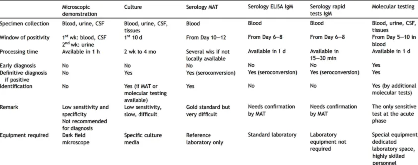
2.8. Diagnostic tests
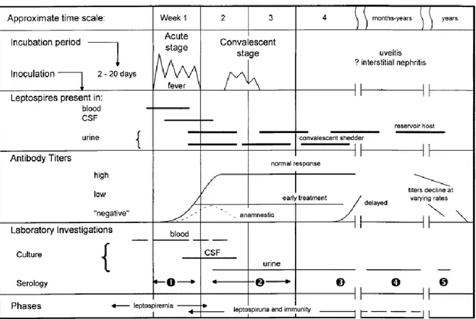
Numerous tests have been developed, but availability of appropriate laboratory support is still a problem. Table 1 presents a summary of the different tests and their advantages and disadvantages from Musso and Scola, (2013). Figure 5 from (Levett, 2001) is an illustration of the biphasic dynamic of leptospirosis and the relevant tests at the different stages of the disease.
FIGURE 5 Biphasic nature of leptospirosis and relevant investigations at different stages of disease SOURCE: LEVETT et al (2011).
28 Leptospires may be visualized in clinical material by dark-field microscopy or by immunofluorescence or light microscopy after appropriate staining. Dark-field microscopy to see organisms in blood or urine is fraught with false-positives and false-negatives, is unreliable and therefore, not recommended (Musso, Scola, 2013).
Culture and isolation of leptospires from clinical samples gives a definitive diagnosis. Blood should be cultured as soon as the patient’s presentation as leptospiremia occurs before the onset of symptoms and ends by the first week of the illness. Cerebrospinal fluid and dialysate fluid can also be cultured during the first week of illness and urine from the second week of symptomatic illness. The use of cultures to confirm diagnosis is rare as it is very tedious, expensive and requires prolonged incubation that can take up to months and does not contribute to early diagnosis (Musso, Scola, 2013).
Serology is the most frequently used diagnostic approach. The current gold standard is the microscopic agglutination test (MAT). Patient sera are reacted with live antigen suspensions of leptospiral serovars. After incubation, the serum-antigen mixtures are examined microscopically for agglutination and the titers are determined. This method, however, relies on the maintenance of panels of Leptospira serovars through culture. The MAT is complex to control and perform; it cannot be standardized because live leptospires are used as antigens (Chappel et al., 2004).
Enzyme-linked immunosorbent assay (ELISA) detects antibodies reacting with a broadly reactive genus-specific antigen and thus is not suitable for identification of the causative serovar or serogroup. Leptospiral DNA can be amplified from serum and urine. PCR detects DNA in blood in the first 5-10 days after the onset of the disease and up to the 15th day. Several primer pairs for PCR detection of leptospires have been described, some based on specific gene targets or repetitive elements. PCR is based on the detection of genes universally present in bacteria as
gyrB, rrs (16S rRNA gene), secY or genes restricted to pathogenic Leptospira spp. as lipL32, lfb1, ligA, ligB. Real-time quantitative PCR, combining amplification and detection of
amplified product in the same reaction vessel has an excellent sensitivity and specificity and low contamination risk. They can either be performed using SYBR Green or fluorescent TaqMan probes. Depending on the target gene, these PCR allow detection and differentiation of pathogenic and non-pathogenic leptospires from clinical and environmental samples. PCR can rapidly confirm the diagnosis in the early phase of the disease when bacteria are present and before antibody titres are at a detectable level. However, they require special equipment
29 and skilled technicians, lacking in some areas. Laboratory diagnosis tests are not always available, especially in developing countries.
2.9. Treatment
In Human, treatment of leptospirosis differs depending on the severity and duration of symptoms. Patients with mild, flu-like symptoms require only symptomatic treatment. Hospital admission is required for patients with icteric leptospirosis and dialysis for patients with acute renal failure. Severe cases of leptospirosis can be treated with high doses of intravenous penicillin. Doxycycline, ampicillin and amoxicillin are recommended in mild cases (Levett, 2001).
Vaccinations for dog and cattle are available to prevent illness but do not prevent the shedding of the bacteria and thus their transmission to human. Canine vaccines generally contain serovars canicola and icterohaemorrhagiae and can include serovars grippotyphosa and pomona (Klaasen et al., 2014). However immunized dogs may be infected with serovars other than those contained in commercial vaccines (Llewellyn et al., 2016).
Vaccines to prevent human leptospirosis are available in some countries but are not authorized in other countries. None of them protect against all circulating strains and focused only on important serovar. For instance in France, a monovalent vaccine against L. interrogans from Icterohaemorrhagiae serogroup is available only for sewers workers (Institut Pasteur, 2015).
TABLE 1 Advantages and disadvantages of common diagnostic tests for leptospirosis SOURCE : Musso, Scola, (2013)
30 Moreover, these killed bacteria vaccines are likely to provide only short-term and possibly incomplete protection (Haake, Levett, 2015).
31 3. LEPTOSPIROSIS EPIDEMIOLOGY
Leptospirosis is considered the most common and widespread bacterial zoonosis (Levett, 2001). The global burden of leptospirosis is estimated at around 1.03 million cases per year and 58 900 deaths per year (Costa et al., 2015). These estimates emphasize that leptospirosis is one of the greatest zoonotic causes of morbidity and mortality. Leptospirosis takes an even greater importance in tropical and subtropical regions where the disease is endemic. Over 73% of the total cases of leptospirosis worldwide are believed to occur in the tropics (Victoriano et al., 2009).
3.1. Leptospirosis epidemiology in humans, in SEA
Leptospirosis incidence in the Asia-Pacific is estimated between 10 and 100 cases per 100 000 habitants per year (Costa et al., 2015). Although transmission is endemic and large outbreaks have been reported, there is currently no routinely performed surveillance in South East Asian countries and therefore no official data on the incidence of the disease. In tropical regions, where humid and warm conditions enable a longer survival of Leptospira, leptospirosis is significantly associated with occupational exposure such as rice farming and other agricultural activities (Bharti, 2003 ; Mwachui et al., 2015) and with heavy rainfall or extreme weather events such as floods and cyclones (Dechet, 2012 ; Lau, 2010 ; Wasiński, Dutkiewicz, 2013). In Vietnam and Laos, leptospirosis was shown to occur in a seasonal pattern in these countries, with a peak incidence during the rainy season and outbreaks occurring after flooding (Lau, 2010).
In Cambodia, few studies give an insight into the leptospirosis situation in the country: in 2003, a survey in Takeo provincial hospital estimated the annual incidence to be 7.65 cases per 100 000 habitants, with Javania and Australis as the main serogroups (Seng et al., 2007). In 2012, fever surveillance in South-Central Cambodia found a seroprevalence of 20.8% IgM (Kasper et al., 2012). A more recent study by Institut Pasteur du Cambodge from 2007 to 2009, found a seroprevalence of 26.7% of febrile cases in patients younger than 20 years old, in Kampong Cham province. Of these, 15.8% had seroconversion illustrating the incident infection in this province. They also showed that probability of having a fever caused by leptospirosis was at 1% of all fevers per semester (Hem et al., 2016).
3.2. Leptospirosis epidemiology in rodents, in SEA
Little is known about human leptospirosis in Southeast Asia, but the lack of knowledge about leptospirosis circulation in rodents is even greater, though they are believed to be important reservoirs and could be a source of infection to humans. In a study conducted in the Mekong
32 Delta of Vietnam (Loan et al., 2015), 5.8% of captured rats were tested positive by RT-PCR and 18.3% by Microscopic Agglutination Test (MAT). They observed a higher prevalence of detection among older rats, suggesting a long-term carriage of leptospires and that Leptospira infection does not result in increased mortality in rats. In Thailand, leptospires were found in the following species: Rattus argentiventer, R. exulans R. losea, R. norvegicus, R. tanezumi but also in the less studied Bandicota indica and B. savilei (Herbreteau et al., 2012).
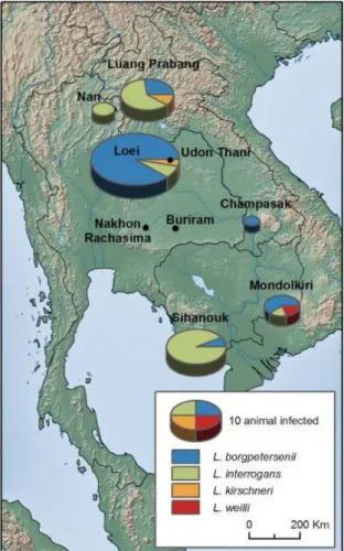
A recent study conducted in Thailand, Lao PDR and Cambodia assessed Leptospira prevalence in rodent species using RT-PCR, and found that detection varied from 0 to 18% across localities, and from 0 to 19% across species (figure 6, Cosson et al., 2014). In Cambodia, the prevalence was 4% in Mondolkiri province and 8.33% in Sihanoukville province and the leptospira species found, were Leptospira borgpetersenii, L. interrogans and L. weilli.
Leptospira borgpetersenii and L. interrogans were the most abundant species in Lao, Thailand
and Cambodia but were found in different habitats.
FIGURE 6 Geographic distribution of Leptospira infection in rodents from Thailand, Lao PDR and Cambodia. SOURCE : Cosson et al (2014)
33
L. borgpetersenii was more abundant in dry habitats than L. interrogans, suggesting a difference
in ecological niche for these Leptospira species.
However, the study found no difference of prevalence between floodable areas, forests and dry agricultural fields. Such a result illustrates the two transmission routes of leptospirosis and suggests that direct transmission could explain the circulation of leptospires in the dry habitats while the indirect transmission via wet environments occurs in floodable areas. Leptospira prevalence did not significantly vary across rodent species, though higher prevalence was observed in wild mice (Mus sp.) and in rarely investigated forest species (Berylmys sp.,
Maxomys sp.).
In Cambodia, the forest species Maxomys surifer and Niviventer fulvescens were also found to carry Leptospira and high prevalence was observed in Rattus argentiventer, a species found in rain-fed cultivated areas. Leptospira were also detected in Bandicota savilei and Berylmys
berdmorei which are both present in paddy fields; and B.berdmorei which is found in forest
areas near crops. High prevalence of Leptospira in rodents in cultivated areas and degraded forest suggests that these habitats may present a high risk of leptospirosis for humans (Svilena Ivanova, 2012).
Rodents may move among habitats, either as part of the natural dispersal process (from their birth place to their first breeding site or from one breeding site to another) or in response to the seasonal variation in habitat quality (i.e. amount of food, shelter availability, competition with other rodents, predation etc.). In Lao PDR, movements of rodents between field and village habitat were shown in response to the availability of food resources (Douangboupha et al., 2009). Because these movements may involve rodents infected with Leptospira, this process could have important consequences on Leptospira distribution within Southeast Asian landscapes. It seems therefore likely that deforestation, by disrupting rodent habitats, would have consequences on Leptospira distribution. But knowledge on the impact of deforestation on pathogen circulation is currently lacking. The following paragraphs summarize the current literature on how human land-use change impacts infectious disease transmission in general and rodent-borne pathogens.
34 4. IMPACT OF LAND-USE CHANGE ON PATHOGENS IN SEA
South East Asia is both a hotspot of land use change and zoonotic disease outbreaks. Indeed the current increase of infectious disease emergences coincides with accelerating rates of tropical deforestation (Wilcox, Ellis, 2006). An increasing number of studies on emerging infectious diseases points to changes in land cover and land use, including forest cover change such as deforestation and forest fragmentation, as major factors contributing to the surge in infectious diseases. Some examples of pathogens whose current emergence patterns show an association with forest degradation and clearing are Ebola virus, Nipah virus, malaria and Lyme disease (Wilcox, Ellis, 2006).
4.1. Land use changes and disease prevalence
Brearley et al. (2013) reviewed the influence of human-induced landscape change on wildlife disease prevalence. Half of the papers reviewed found an increase in disease prevalence due to human-induced landscape change, while 21% identified a decrease in disease prevalence and the remainder 26% indicated that disease prevalence varied (Brearley et al., 2013). Similarly, the Gottdenker et al. review (2014) about land use change impact on infectious disease showed that more than 56.9% of reviewed studies documented increased pathogen transmission in response to anthropogenic land use change, 10.4% found decreased pathogen transmission secondary to land use change, 30% observed variable pathogen response while 2.4% showed no changes at all (Gottdenker et al., 2014). These results (illustrated in Appendix 4) clearly indicate that the issue of wildlife disease in human-modified landscape is complex and highly variable and no general trend of disease prevalence response to land-use changes has been determined. Richness of infectious diseases was found positively correlated with the richness of mammals depicting biodiversity as a source of pathogens (Morand et al., 2014). The same study also found an association between an increase of zoonotic disease outbreaks and the loss of biodiversity as measured by the number of species at threat and proportion of forest cover. The current evidence suggests several hypothesized mechanisms that would lead to infectious disease changes after land use alteration. They include modification of habitat structure, microclimate and resource availability for both the host and its pathogens. Other proposed mechanisms were changes in the host community composition, and host jumping of the pathogen. Anthropogenically driven land use changes can produce ecological conditions that facilitate geographic expansion of pathogens via the modification of spatial distribution of the host or of their behaviour and movements (Gottdenker et al., 2014).
35 Deforestation and forest fragmentation can have two possible outcomes: on one hand the highly fragmented environments may reduce the disease prevalence due to lack of connectivity between the patches of forest, resulting in less contact rates between hosts and thus reducing the infection rates; conversely, such fragmentation may also increase contact rates and disease prevalence by clumping the resources and the hosts (Bradley, Altizer, 2007). Or, in case of indirectly transmitted diseases such as leptospirosis, fragmentation may result in overlapping distributions of species and might increase their probability of getting infected from each other, from the spread of germs along the tracks they now share. Furthermore, deforestation leads to ecological changes such as increased edge habitat and local extinction of predators that may favour some species that happen to be disease reservoir (Wilcox, Ellis, 2006).
4.2. Land use changes and rodent-borne diseases
The conversion and loss of forest in SEA is presumed to affect rodents in terms of their diversity and species composition. Several studies have established that rodent species may differ in their response (presence/absence or abundance) to habitat modification. The importance of habitat modification in shaping small mammal communities is now recognized and was assessed by world-wide studies (Bernard et al., 2009 ; Morand et al., 2015 ; Lynam, Billick, 1999 ; Umetsu, Pardini, 2007). Bernard et al. (2009) found that habitat types (forest versus plantation) were important determinant of small mammals’ species occurrences and assemblage composition in Borneo.
Morand et al. (2015) showed that habitat structure and fragmentation affected the spatial distribution of rodent species in SEA. In particular, alteration of the habitat (decreasing forest cover, increasing fragmentation and urbanization) was found to favour the presence of synanthropic rodent species such as Rattus tanezumi. Similarly, Suzán et al. (2008) highlighted that fragmentation of the habitats resulted in lower diversity of small mammals and higher densities of populations of rodents. All these studies focusing on forest fragmentation in tropical areas have emphasized that species richness or diversity are affected in disturbed ecosystems. Changes in land use in Southeast Asia are therefore expected to alter rodent species distribution and diversity (Morand et al., 2015). As rodents are important reservoirs of human diseases, it is likely that their incidence is linked to fluctuations in rodent population. Habitat fragmentation and decreasing forest cover seemed to favour the presence of synanthropic rodent species that are important host of rodent-borne diseases (Suzán, Marcé, et al., 2008). In particular,
36
Bartonella spp. and hantavirus were associated with disturbed landscapes with ongoing
fragmentation (Morand et al., 2015).
Outbreaks of hantavirus diseases, were found to occur in anthropologically disturbed habitats, where natural biodiversity had been reduced to low levels (Mills, 2006). This was explained by the fact that disturbed habitats favour opportunistic species that were reservoir of hantavirus and due to the lack of competitive pressure, these species could reach higher densities. It is therefore important to understand which rodent-borne pathogens would have a similar response to land use changes as those observed for hantavirus.
4.3. Land-use changes and leptospirosis
Studies conducted in SEA have found leptospires in all types of rodent habitats: from forest to floodable areas and dry agricultural fields (Cosson et al., 2014 ; Ivanova et al., 2012).
Ivanova et al. (2012) described a high prevalence in rodents trapped in newly cultivated areas and in degraded forests and that species from these two areas had similar level of infection. This study suggested that both rice fields and forests were also areas of potential risk for leptospirosis. Though it is important to note that Leptospira was detected in this study with the PCR protocol from Mérien et al (1992) that detects both pathogenic and non-pathogenic species.
Morand et al (2015) observed associations between habitat structure (forest, settlement, agricultural fields and fragmented habitats) and the prevalence of rodent-borne pathogens with
Leptospira prevalence linked to fragmented habitat (Figure 7). This discovery highlighted