COMPARATIVE STUDY: THE ANTIBACTERIAL
ACTIVITY OF MELISSA IN RELATIONS TO
OTHER PLANTS IN THE REGION OF SETIF,
ALGERIA
Adimi L. Z.
Guechi A.
Laidoudi O.
Chelil S.
Kicheh I
.Laboratory of applied Microbiology, Faculty of nature and life sciences University of Ferhat Abbas, Setif1, Algeria
Abstract
Antibacterial properties of essential oils, hydro ethanoicextracts, and aqueous bactericidal extracts of four medical plants, known for their therapeutic effects: Melissa officinalis , Origanum vulgare , Lavandula
angustifolia and Mentha piperita. The last three are largely used in
Algeria; however, Melissa appears to be less famous. The following work has been executed in a laboratory for the purpose of, yet again, revealing and confirming the benefits of Melissa, and comparing it to other plants. Two microorganisms have been used: Escherichia coli and Staphylococcus
aureus. Using the method of Aromatogram with essential oils, Melissa was
active the most with zones of inhibition of 8 mm for Escherichia coli and 7 mm for Staphylococcus aureus. Applying the same method for hydro ethanoicextracts, Melissa was effective the most with a 50 mm zone of inhibition for Escherichia coli, followed by mint (40 mm zone of inhibition). As for Staphylococcus aureus, Melissa presented a 14 mm zone of inhibition, while mint displayed a zone of inhibition of 13 mm. Using the bioassay method with essential oils, Melissa has the best effect with a 40 mm zone of inhibition for Escherichia coli and 35mm for Staphylococcus aureus. Always with the same technique but with hydro ethanoicextracts, both
Melissa and Mint presented the best inhibition value of 50 mm for Escherichia coli. As for Staphylococcus aureus, Melissa presented a 26 mm
zone of inhibition, followed by mint with 24 mm. For the spectrophotometry technique, almost all four plants, witht heir aqueous extracts, presented a antibacterial activity for both bacteria. These results confirm the strong
anti-bacterial and bactericidal activity of the four plants, especially Melissa, and their diverse traditional use.
Keywords: Melissa officinalis, Origanum vulgare, Lavandula angustifolia,
Mentha piperita, antibacterial activity,essential oils
Introduction
Today, according to the World Health Organization (WHO), nearly 80% of the population depend on traditional medicine for primary health care (Muthu and al., 2006).
In recent decades, there has been a growing interest in the study of medicinal plants and their traditional use in different parts of the world, because the important problems have accompanied the treatment of disease by conventional drugs, for examples: their inaccessibility especially because of their high costs, progressive resistance of the pathogen vis-à-vis the active substances and the manifestation of severe side effects or even toxic in some cases. (Bah and a1., 2006). Hence the need for a valuation of traditional medicine.
Among the medicinal plants found balm that is consumed by humans since ancient times for its sedative and relaxing virtues antioxidants ... etc. But Melissa is a bit neglected by our community.
The aim of our work is to demonstrate and approve the antibacterial effects of balm and compared with three other plants in the Setif region, and are Oregano, Lavender and Mint (Table 1)
It is essential to test the oil and aqueous extracts of these ethanoic four plants in vitro on the growth of certain microorganisms and to deduce the best plant with its powerful bacterial effect.
Table 1: Studied medicinal plants
Botanical name used Organs Some uses
Melissa officinalis-
Aerial parts (leaves) Nervousness and sleep disturbances - Gastrointestinal disorders
(Babulka.,2005)
- Anti-herpes activity (Schnitzler and al., 2004)
Origanum vulgare Aerial parts (leaves + flowers)
- Toned stomach - Expectorant and sedative - Antiseptic (Daniel., 1997) Lavandula
angustifolia
Aerial parts (leaves + Flowering tops)
- analgesic - anti-inflammatory
- antibacterial (Umezu and a1., 2006) Mentha piperita Aerial parts (leaves) antispasmodic
Materials and methods Plant Material
Plants {Table 1} were collected from the surroundings of County of Setif (village of Beni Aziz)
The botanical identification was carried out in the Botanical Laboratory of pharmacy department at the University of Ferhat Abbas 1.
Microorganisms
Employed Germs are responsible for several diseases for which our plants are frequently used. They were obtained from the laboratory of microbiology, pharmacy department at the University of Ferhat Abbas 1. Bacteria were maintained by subculture on agar media.
Obtaining essential oils
Essential oils of each plant were obtained by hydro distillation technique but with different amounts for each:
- Melissa --- 36 g in 300 ml of distilled water - The Oregano --- 48 g in 300 ml of distilled water - The lavender --- 15 g in 300 ml of distilled water - The --- 48g mint in 300 ml of distilled water
Obtaining extracts of maceration
Plant extracts were obtained by maceration (48 hours) 100 g of 800 plants sprayed in ethanol-water mixture (V/V). The filtrate obtained on filter paper are then evaporated using a rotary evaporator. The resulting solutions were dried in an oven (50°).
The obtained residues were stored at 4° C with their use.
Plant juices preparation for spectrophotometry
Different plants, balm, mint, lavender and oregano, were ground and sterilized.
A teaspoon of each ground plant was added to 100m1 of sterile distilled water. We allow to react for half an hour and then we filter. We thus obtained four vials. A fifth vial is prepared with only the sterilized nutrient stock (100ml) and which serve to compare and another as witness comprising only sterile distilled water.
Methods
Technique of aromatogramme (Cohen, M., 2013)
The technique consists of deposit on the surface of an agar medium previously inoculated filter paper disks impregnated with essential oils and Maceras to be tested for each plant.
Modus operandi
From the suspension of the germ to be tested for 24 hours, we realize a 1/10 dilution in sterile Physiological water with which we seed five agar boxes.
Muller-Hinton in which we apply soaked disks each 1 ml of essential oil to be tested. The fifth box will serve as a witness.
These boxes are left for 30 minutes at laboratory temperature, and then incubated in an oven at 37° C for 24 hours. Same work was performed for the plant extracts.
Bioassay technique: bioassay (method of wells) (In Adimi., 1990)
The technique consists to dig wells to the surface of an agar medium previously inoculated. These wells will receive quantities of essential oils and maceras test.
Modus operandi
From the suspension germ to be tested for 24 hours, we realize a 1/10 dilution in sterile Physiological water to which is seeded five Muller-Hinton agar plates boxes, four will be drilled with the drill for wells in level of each well 1 ml of the essential oil to be tested is placed. The fifth box is not pierced and shall be a witness. All boxes shall be incubated at 37° C for 24. Same work was performed for the plant extracts.
Technical spectrophotometry
Spectrophotometry is a quantitative analytical method of measuring the absorbance or optical density of a given substance, typically in solution. Over the sample is concentrated, the more it absorbs light in the proportionality limits set by the Beer-Lambert law.
From the suspension of germ to be tested for 24 hours, 101 dilution is performed in sterile Physiological water with 1 ml of which different vials already prepared were separately seeded.
The vials are incubated at 37° C for 24 hours during which period the optical density measurements were made.
Results and discussion
Technique of aromatogramme
Table 2: Action of various essential oils (E.O) on growth of E. coli and S. aureus (inhibition zones in mm).
Bacterium E. coli S. aureus
E.O Hydrolat E.O Hydrolat
Balm 8 mm 7 mm 7 mm 5 mm
Oregano 6 mm 4 mm 6 mm 5 mm
Lavender 7 mm 4 mm 4 mm 4 mm
Mint 6 mm 0 mm 6 mm 0 mm
Witness 0 mm 0 mm 0 mm 0 mm
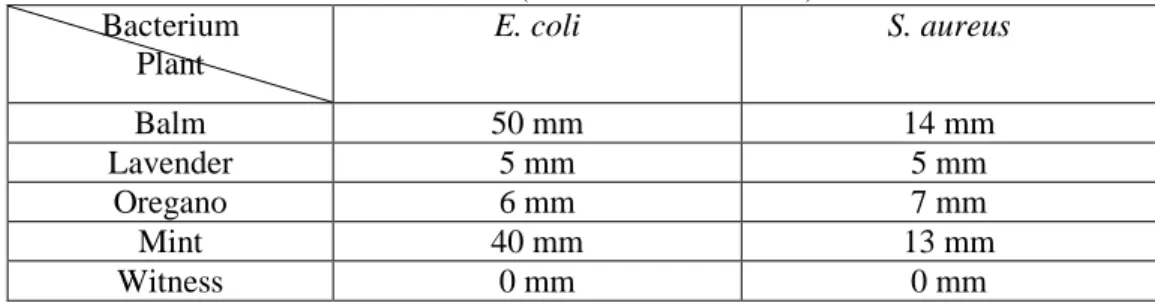
Table 3: Effect of different solutions of the alcoholic maceration on the growth of E. coli and S. aureus (inhibition zones in mm)
Bacterium Plant E. coli S. aureus Balm 50 mm 14 mm Lavender 5 mm 5 mm Oregano 6 mm 7 mm Mint 40 mm 13 mm Witness 0 mm 0 mm
It is apparent that the four plants have antibacterial activity against two bacteria. With essential oils diameters of inhibition zones
Escherichia coli vary from 6 mm to 8 mm and for Staphylococcus aureus, the diameters of the inhibition zones vary from 4 mm to 7 mm. With
maceration extracts, the diameters of zones of inhibition for Escherichia coli vary from 5 mm to 50 mm, and Staphylococcus aureus, the diameters of zones of inhibition vary from 5 mm to 14 mm.
Even with hydrolats inhibition effects totaled present which explains that the active ingredient is also present in these solutions.
Balm is as the best plant antibacterial activity which has already been confirmed by the work (Neda., M, D., and al 2004 and Anicic .,and al 2005)
Bioassay technique: bioassay (wells method)
It appears as though the four plants have antibacterial activity against two bacteria.
With Essential oils, the diameters of the inhibition zones for
Escherichia coli vary from 14 mm to 24 mm, for Staphylococcus aureus the
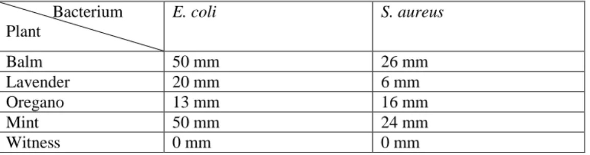
diameters of zones of inhibition vary from 12 mm to 18 mm(Tab 4). With maceration extracts, the diameters of inhibition zones for Escherichia coli vary from 13 mm to 50 mm, and for Staphylococcus aureus, the diameters of the zones of inhibition vary from 16 mm to 30 mm(Tab 5).
The extracts of balm macerating have the best antibacterial activity which has already been confirmed (Teresa M and a1., 2007)
By comparing the two techniques, bioassay provides greatest inhibition diameters.
Table 4: Effect of various essential oils on growth of E. coli and S. aureus (inhibition zones in mm) Bacterium Plant E. coli S. aureus Balm 24 mm 18 mm Lavender 18 mm 12 mm Mint 14 mm 14 mm Oregano 16 mm 15 mm Witness 0 mm 0 mm
Table 5: Action of different solutions of alcoholic maceration on the growth of E. coli and
S. aureus (inhibition zones in mm)
Bacterium Plant E. coli S. aureus Balm 50 mm 26 mm Lavender 20 mm 6 mm Oregano 13 mm 16 mm Mint 50 mm 24 mm Witness 0 mm 0 mm Technical of spectrophotometry We noticed
Concerning E coli Oregano,Balm,Lavender and Mint have a bactericide activity with the optical density’s values which are :0,014,0,017,0,08 and 0,190 after 1 hour 30 minutes of incubation(Tab 6)
Table 6 Different values of the evaluation of the optical density for E. coli
Time (hours) Do of Plant T=13 T= 1430 T=16 T=24 Balm 0.900 0.017 0.016 0 Lavender 0.280 0.080 0.055 0.012 Oregano 0.026 0.014 0.005 0 Mint 0.250 0.190 0.056 0 Nutritive stock 0.060 0.390 0.550 0.922 Distilled water 0.120 0.090 0 0 Concerning S aureus
A bactericide activity was noticed with the optical density’s values for Balm ,Oregano, Lavender and Mint which are 0,015,0,02,0,026 and 0,056 after 1 hour 30 minutes of incubation
The Lavender’s bactericide activity occurs only after three hours of incubation(Tab 7)
Table 7 : Different values of the evaluation of the optical density of S. aureus
Time (hours) Do of Plant T=13 T= 1430 T=16 T=24 Balm 0.016 0.015 0.005 0 Lavender 0.175 0.026 0 0 Oregano 0.064 0.02 0.015 0 Mint 0.067 0.056 0.042 0 Nutritive stock 0.002 0.005 0.039 0.819 Distilled water 0.01 0.009 0 0
It appears that the four plants exhibit bactericide activity after 24 hours incubation is for Escherichia coli and Staphylococcus aureus.(Tab 6 and Tab 7)
Conclusion
This preliminary work has helped to highlight the antibacterial properties of essential oils, extracts of maceration and bactericides of aqueous extracts of four medicinal plants.
The results reveal the presence of antibacterial and bactericides active ingredients in each plant, which explains their traditional use in several infections compared, balm has the best antibacterial effect in the first two techniques.
References:
ADIMI L Z, (1990): Activité antibactérienne de certaines plantes médicinales vis-à-vis d’Escherichia coli .Thèse d’ingéniorat contrôle de qualité et analyse, Université Ferhat Abbas de Sétif (p72).
ANICIC NV, DIMITRIJEVIC S, RISTIC MS, PETROVIC S S ,PETROVIC SD,(2005) Antimicrobialactivity of essentiel oil of Melissa officinalis
L.,Lamiaceae. Hemijskaindustija, (vol 59) (N9/10) 243-247.
BABULKA P (2005): La mélisse (Melissa officinalis L.), Phytothérapie, 3 - p. 114-117.
BAH S, DIALLO D., DEMBELE S, PAULSEN BS, (2006) Ethnopharmacological survey of plants used for the treatment of schistosomiasis in Niono District, Mali. Journal of Ethnopharmacology, 105, 387–399.
COHEN M, (2013) : Laboratoire d’analyse médicale, Paris.
DANIEL JOURDAIN, (1997) : Dictionnaire des plantes médicinales, Québec, Quebecor, P 120-122-136.
MUTHU C, AYYANAR M, RAJA N, IGNACIMUTHU S,(2006) : Medicinal plants used by traditional healers in Kancheepuram District of Tamil Nadu, India. Journal of Ethnobiology and Ethnomedicine, 2:43, 10.1186/1746-4269-2-43.,2006.
NEDA M D, BILJANA B, MARINA S, SATANA S,(2004) : Antimicrobial and antioxidant activities of Melissa officinalis L (Lamiaceae) Essential oil. J.Agric.FoodChim ., 52, 2485-2489.
SCHNITZLER P,SCHUHMACHER A,ASTANI A,REICHLING J,(2004) Melissa officinalis oil affects infectivity of enveloped herpesviruses. Phytomedicine,(2004 May Volume 15, Issue 9- 734-740 de Mimica-Dukic N, Bozin B, Sokovic M, Simin N. Antimicrobial and antioxidant activities of Melissa officinalis L. (Lamiaceae) essential oil. J Agric Food Chem. 5; 52(9):2485-9
TERESA M, PATRIZIA P, CARLA S, RITA A,(2007)
Triterpen,antioxidant and antimicrobial compouds from Melissa officinalis. J.nat .Prod., 70, 1889-1894.
UMEZU T, NAGANO K, ITO H, KOSAKAI K, SAKANIWA M, MORITA M.(2006 Dec)
Anticonflit effects of lavender oil and identification of its active constituents. Pharmacol Biochem Bhav ;85(4):713-21