Reporting nuclear cardiology: a joint position
paper by the European Association of Nuclear
Medicine (EANM) and the European Association of
Cardiovascular Imaging (EACVI)
Elin Tra¨ga˚rdh
1*
†, Birger Hesse
2†, Juhani Knuuti
3, Albert Flotats
4, Philipp A. Kaufmann
5,
Anastasia Kitsiou
6, Marcus Hacker
7, Hein J. Verberne
8, and Lars Edenbrandt
1Document Reviewers: Victoria Delgado, Erwan Donal, Thor Edvardsen, Maurizio Galderisi, Gilbert Habib,
Patrizio Lancellotti, Koen Nieman, Raphael Rosenhek (for EACVI) and Denis Agostini, Alessia Gimelli, Oliver Lindner,
Riemert Slart, and Christopher U¨ bleis (for EANM)
1
Department of Clinical Sciences, Clinical Physiology and Nuclear Medicine Unit, Lund University, Ska˚ne University Hospital, Inga Marie Nilssons gata 49, 205 02 Malmo¨, Sweden;
2
Department of Clinical Physiology, Nuclear Medicine & PET, Rigshospitalet, Copenhagen, Denmark;3Turku PET Centre, University of Turku and Turku University Hospital, Turku, Finland;4
Nuclear Medicine Department, Hospital de la Santa Creu i Sant Pau, Universitat Auto`noma de Barcelona, Barcelona, Spain;5
Cardiac Imaging, University Hospital Zurich, Zurich, Switzerland;6Cardiology Department, Sismanoglio Hospital, Athens, Greece;7Division of Nuclear Medicine, Medical University of Vienna, Vienna, Austria; and8Department of Nuclear Medicine, Academic Medical Center, University of Amsterdam, Amsterdam, The Netherlands
Received 20 November 2014; accepted after revision 24 November 2014; online publish-ahead-of-print 24 January 2015
The report of an imaging procedure is a critical component of an examination, being the final and often the only communication from the interpreting physician to the referring or treating physician. Very limited evidence and few recommendations or guidelines on reporting imaging studies are available; therefore, an European position statement on how to report nuclear cardiology might be useful. The current paper combines the limited existing evidence with expert consensus, previously published recommendations as well as current clinical practices. For all the applications discussed in this paper (myocardial perfusion, viability, innervation, and function as acquired by single photon emission computed tomography and positron emission tomography or hybrid imaging), headings cover laboratory and patient demographics, clinical indication, tracer administration and image acquisition, findings, and conclusion of the report. The statement also discusses recommended terminology in nuclear cardiology, image display, and preliminary reports. It is hoped that this statement may lead to more attention to create well-written and standardized nuclear cardiology reports and eventually lead to improved clinical outcome.
-Keywords Cardiac imaging † Nuclear cardiology † Nuclear medicine reports † Practice guidelines
Preamble
This position paper on reporting nuclear cardiology examinations has been developed under the auspices of the Cardiovascular Commit-tee of the European Association of Nuclear Medicine (EANM) and the Section on Nuclear Cardiology and Cardiac Computed Tomog-raphy of the European Association of Cardiovascular Imaging (EACVI) of the European Society of Cardiology (ESC), highlighting the importance for close collaboration and bridging between the two specialties.
In the daily routine, this collaboration is particularly obvious in two areas: referral for the nuclear cardiology examination and the com-munication of the outcome of the examination. The former has more recently received a lot of attention with discussions of appro-priate use criteria, classes of indications of the examinations, and varying reimbursement in some European countries, etc. In contrast, the communication of the results has received much less attention, though of equal significance. It is therefore important that, by reading the report, the results of the examination are understood as closely and accurately as possible reflecting the interpretation of
*Corresponding author. Tel:+4640338701; Fax: +4640336620, Email: elin.tragardh@med.lu.se
†E.T. and B.H. shared first authorship.
Published on behalf of the European Society of Cardiology. All rights reserved.&The Author 2015. For permissions please email: journals.permissions@oup.com.
by guest on April 24, 2015
the nuclear medicine physician. Ideally, the information presented should be uniform and independent of individual physician’s prefer-ences or patient-specific parameters.
The information presented below is specifically adapted to Euro-pean practice. A significant limitation of our recommendations is the lack of evidence from original scientific studies on the influence of the report on the use of the results of the examination. Another important limitation for the present, English written, paper is related to the great variations within Europe both regarding national traditions and regulations, and the differences in the languages. With those limita-tions, the authors wish to give some recommendations regarding structure and standards for the nuclear cardiology report: the goal of the report must be to transfer from the interpreting physician to the referring physician a message that in a coherent clinically relevant
and predictable format1and in an easily readable way that concisely
reflects the nuclear medicine interpretation of the examination.
Introduction
‘The report of an imaging procedure is often the only communication from the interpreting physician to the caregiver, and is the final
and perhaps the most critical component of an imaging procedure’.1
It may occasionally also may become legal evidence.2In a way, nuclear
cardiology studies undergo two interpretations: the first one being performed by the physicians who make a report based on the ana-lyses and interpretation of the images, stress data, etc. The second is the interpretation made by the physician who reads the report and from this reading draws his or her conclusions for further clinical action. Although sometimes the referring and image report making person are the same, the information in the report should be uniform and as accurately possible reflecting the interpretation. Guidelines on reporting imaging procedures in nuclear cardiology, to optimize the communication of the information from reporter to reader, are essential.
Ideally, guidelines should be based on evidence from clinical
studies,3but in practice mostly on expert opinions. Owing to a lack
of published evidence, all available recommendations on reporting nuclear cardiology are largely or totally based on expert opinions.
Fortunately, there is wide consensus on most of the issues,4–6
includ-ing the critical need of structured reportinclud-ing, as opposed to free text descriptions, so that key report components and data elements are not omitted. Increased standardization would facilitate the reading
of reports.1Terminology must be accurate, but always expressed
in a reader friendly style.
The present paper combines existing evidence with expert opinions
and previously published guidelines and recommendations1,4–11with
current clinical practices. This joint expert statement focuses on: (i) Myocardial perfusion imaging (MPI) with single photon emission
computed tomography (SPECT) or positron emission tomog-raphy (PET).
(ii) Equilibrium radionuclide ventriculography (ERNV).
(iii) Viability imaging evaluated by18F-fluoro-deoxyglucose (FDG)
PET/MPI.
(iv) Hybrid imaging with coronary artery calcium score (CACS) and/ or coronary CT angiography (CCTA) and MPI.
(v) 123I-metaiodobenzylguanidine (MIBG) imaging.
The paper also includes discussion of terminology, use of preliminary reports, and the selection of images accompanying the report. In
con-cordance with the previously published guidelines,10three levels of
importance are used, including ‘must’ (information required in the report), ‘should’ (information highly recommended), and ‘may’ (optional information).
The minimum information recommended to be included in the report appears as ‘must’ and encompasses unequivocal identifica-tion of patient, study, date, and signature (often digital), as well as a description of findings, whether normal, abnormal or inconclusive, and finally a conclusion presenting the clinical interpretation of the findings. The remaining data that can be added in the report depend on a number of factors, including national and local traditions and ‘culture’, national legislation, and relation between the referring and reporting physicians and/or institutions. Examples of reports are presented that include both ‘must’ and ‘should’ information to present the level recommended by the authors, i.e. in-between the minimum (‘must’) and, ‘may’ be included information. Reports must include sufficient data of relevant detailed elements to describe the findings, but too lengthy reports should be avoided. Compared
with US guidelines,1,10 those presented in this statement allow
for more degrees of freedom, which is related to European vs. US traditions, the national variations within European countries, and possibly also related to the more common use of medical legal litigat-ing in the USA and thereby the related over-completeness/defensive medicine.
It is hoped this paper may lead to improvement in the clinical value of nuclear cardiology for patients and physicians as well as to facilitate
and improve research in nuclear cardiology.12However,
recommen-dations presented are neither infallible nor substitute for good clinical judgment.
Terminology in the report
It is crucial that the referring or treating physician understands the report as intended by the interpreter of the images. This implies careful attention to the terminology used in the report.
Regarding general language, it is strongly recommended that: (i) The report is written in a simple way, if possible, without the use
of technical terms.
(ii) The use of abbreviations and technical information not import-ant for the referring physician should be avoided or extremely limited.
(iii) Qualitative descriptions (e.g. small, medium-sized, large or slightly, moderately, severely reduced) should be replaced, if possible, by quantified data since qualitative words are used
and understood differently.13
(iv) Protective expressions (e.g. is likely, cannot be excluded) are used as little as possible. However, relevant doubt about the clinical implication of the interpretation must be communicated.
The report must cover clinically relevant information, but not technically irrelevant details. Terms should be used that are widely recognized and approved both in nuclear medicine and in cardiology. The section on findings must give a precise description of the images. Some expressions may appear equally good: a perfusion defect can be
by guest on April 24, 2015
reversible (stress-induced) or irreversible (non-reversible, fixed, and permanent). Depending on the context, one expression may appear more correct than another one. In the description of SPECT findings, an ischaemic perfusion defect is less accurate than a reversible or stress-induced perfusion defect, but is more relevant in the clinical conclusion of the study. Likewise, in the conclusion, an expression like a fixed or permanent perfusion defect, relevant in the section on find-ings, should be translated to infarction or scar tissue, provided that viable tissue is unlikely. The more standardized format of accurate and relevant information is provided, the better the reader’s inter-pretation will be minimizing misunderstandings of the report leading to subsequent better clinical decisions.
The preliminary report
In communications other than the final report, the preliminary report is the most important type of message given about an imaging study. Preliminary reports, typically given in order to direct immediate patient management, may be written, transmitted electronically, or given verbally. It is not expected to include all information of the
final report.14The person responsible for the preliminary
communi-cation must assure the receipt of it. The preliminary report should be reproduced into a permanent format and archived as a preliminary communication, since clinical decisions may very well be based on a preliminary report. Subsequently, it must be documented in the final report. The documentation is important, as recently shown
for pulmonary scintigraphy.15If it has been given as a
person-to-person communication, it must specifically name the person-to-person to whom the communication was delivered. If the message of the final report deviates from that of the preliminary report, this discrepancy should be clearly stated in the final report. Immediate transfer of a
preliminary report has been shown in radiology to result in a small, but important number of adverse outcomes. However, if edited in the final report, the benefits of rapid information transmission may
outweigh the additional risks.16
Oral communications
Sometimes other forms of communication may occur, e.g. during a clinical conference or by a verbal comment to an outside study. Occa-sionally, such an interpretation does not result in a ‘formal’ report. That type of communication carries an inherent risk by missing compari-son with previous studies, adequate patient history, etc., and is therefore not recommended. Ideally, discussion in multidisciplinary meetings (i.e. more than one medical specialty present) and the subsequent clinical decisions should be reported in a separate report.
The structured nuclear cardiology
report
A structured report, in contrast to free text, with adequate headings should be used, since a well-structured report is more easily
access-ible for the referring or treating physician.1,4In the present paper, a
number of headings have been used to describe the different aspects of the report. They were chosen since they are widely used in clinical imaging practice and recommended for nuclear cardiology
by others as well.7,9Headings may differ between different
institu-tions and different countries due to local tradition and legislation. The headings used here include: demographics; clinical indication; tracer administration and image acquisition; findings; conclusion; as
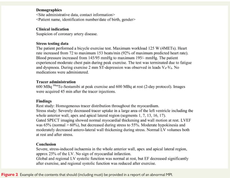
well as date and signature. Figures 1 and 2 show examples of
Figure 1 Example of the contents that should (including must) be provided in a report of a normal MPI.
by guest on April 24, 2015
reports from a normal and from an abnormal MPI, with information that ‘should’ (including ‘must’) be included in the report.
Demographics
Site administrative data (physical address), contact information, as well as name and affiliation of the referring physician must be pro-vided, as well as patient name, unique identification number (date of birth, etc.), and gender.
Clinical indication(s)
The clinical indication for the study should be reported, both to show the appropriateness of the study and to focus the examination and the report. Cardiac history and symptoms and prior cardiac investigations may be summarized, active medications may be noted, and pre-test probability of coronary artery disease may be calculated.
Tracer administration
The tracer administered and amount of radioactivity should be reported. According to legislation in some countries, the amount
of radioactivity must be reported. For 99mTc-labelled tracers, it
should be noted if a 1-day or a 2-day protocol is followed. The
timing in relation to termination of a stress test procedure and the time interval between tracer injection and image acquisition should be noted.
For an FDG study, blood glucose at the time of FDG injection must be reported in diabetic patients; in other patients, it may be noted. Oral glucose load, insulin – glucose clamp, acipimox administration etc., must also be reported.
Image acquisition
If the routine procedure is followed, it should not be described in the report. If the default procedure has not been chosen, the reason must be presented, e.g. changes from the usual protocol regarding rota-tion, position of patient, gating, or attenuation/scatter correction problems, etc.
Findings
Myocardial perfusion imaging
The stress testing procedure and findings must be briefly described,
also if the stress test is normal (Table1). The perfusion distribution is
the key information: Does the activity distribution in the myocardium appear normal, abnormal only at stress or also at rest, or is the study
non-diagnostic? The findings should be described as shown in Table2.
Figure 2 Example of the contents that should (including must) be provided in a report of an abnormal MPI.
by guest on April 24, 2015
Left ventricular function
If gated studies have been acquired (rest and/or stress), left
ventricu-lar (LV) function data should be reported, as also shown in Table3.
Reference values of LV ejection fraction (EF) should accompany the report, either as values from the department (preferable) or as values referred from the literature obtained with similar technique and software tools. Possible discrepancies between regional perfu-sion and regional myocardial functional data must be discussed. Equilibrium radionuclide ventriculography
LVEF must be presented (Table4), and reference values of LVEF
should accompany the report, either as values from the department (preferable) or as values referred from the literature obtained with
similar technique and software tools. It should be noted (cf. section
on reference values in ref. [8]) that LVEF values differ between men
and women and between gated MPI and ERNV.
Viability with FDG in combination with MPI (by PET or SPECT)
Regional FDG uptake must be described in relation to reduced re-gional perfusion (SPECT or PET): is FDG uptake reduced (match between reduced metabolism and reduced perfusion), or is it normal or enhanced (mismatch in relation to reduced regional perfu-sion)? The evaluation compares the uptake in the hypoperfused myo-cardial region with that in the remote myocardium. Quantification of mismatch is recommended.
. . . . Table 2 Findings of tracer distribution in the report of a gated myocardial perfusion SPECT study
Tracer distribution Must be included Should be included May be included
Normal Brief description
Abnormal Presence of defect(s) Other comments to perfusion distribution abnormalities Location of defect(s) Relation to LV segments, relation to the
patient’s coronary artery distribution if known
Preferably using the 17-segment model.9
Suggestion of single- or MV disease
Relation to standard coronary anatomy with reservations regarding anatomy variations Extent of defect(s) Description of defect size(s). ‘Large’,
‘small’, etc. is a minimum
Quantification as percentage or a percentage interval of the LVa; alternatively in summed
scores Severity of defect(s) Description of defect severity. ‘Mild’,
‘severe’, etc. is a minimum
Quantified in summed stress/rest/difference scoresb
Reversibility of defect(s)
Reversible (stress-induced), fixed (permanent and irreversible), or mixed (partially reversible) defect(s)
Quantified in summed difference scoresb
Quantification of regional perfusion in PET
Absolute values in ml/min/g tissue at rest/ during hyperaemia, including reference values. Coronary flow reserve in units Other abnormalities Incidental extracardiac findings Deviations in tracer distribution (locally
increased/decreased uptake, LV cavity dimensions)
Non-diagnostic study Describe the reason
LV, left ventricular; MV, multivessel.
a
A reversible defect .10% of the LV has prognostic information.17,18
b
Scores vary with software systems used.19
. . . . Table 1 Findings related to the stress test in the report of an myocardial perfusion SPECT study
Stress test type Must be included Should be included May be included
Symptom limited exercise test
Reason for termination of test Exercise capacity (MET), peak HR, and BP, changes vs. rest.
Stress-induced symptoms and abnormal ECG findings (rest and stress-induced)
Type of protocol: Bruce, modified Bruce, etc.
Pharmacological stress + exercise
Vasodilators or dobutamine (+atropine). Reason for premature termination. Other drugs including doses administered
during the test (anti-anginal, etc.)
Dose of stress agent and timing of administration Symptoms and ECG changes.
HR and BP baseline/peak
BP, blood pressure; HR, heart rate; METs, metabolic equivalents.
by guest on April 24, 2015
Hybrid imaging with CACS and/or CCTA
The description of MPI is similar to the stand-alone study. The additional information obtained from CACS or CCTA must be reported and integrated with the MPI results. With CCTA, report-ing should follow the recommendations of stand-alone CCTA, as described in detail in part B of the guidelines of the Society of
Car-diovascular Computed Tomography.20The severity of a stenosis
can be assessed in qualitative terms (minimal, mild, moderate, severe, and occluded), or the length and luminal reduction may be quantified. The location and severity of detected lesions must be set in relation to the regional MPI findings, and a conclusion drawn regarding agreement or disagreement between findings by
the two modalities. The clinical interpretation must be discussed in case of a possible disagreement, e.g. is a stenosis detected by CCTA hardly of haemodynamic significance since stress perfusion is normal in that region; or could the perfusion findings be falsely normal, maybe due to balanced ischaemia in multivessel coronary disease? Further diagnostic examinations or invasive angiography may be recommended.
123
I-metaiodobenzylguanidine
Normal or reduced123I-MIBG cardiac uptake must be described, and
comment on the clinical significance should be included (Table5). If
available, possible perfusion/innervation mismatch should be discussed. . . . . Table 3 Findings of LV function in the report of a gated myocardial perfusion SPECT study
LV function Must be included Should be included
LVEF Numerical values Reference values
LV volumes Numerical values (with reference values)
Presence of TID (visual evaluation and/or quantified)
WM Visual evaluation: normal, hypokinesia (mild, moderate, and severe), akinesia, or dyskinesia
WT Visual evaluation: normal, decreased (mild, moderate, and severe), or absent
Phase analysis Dyssynchrony
Differences between stress and rest global and regional LV function
Stress-induced LV dilatation (TID) Comment on differences
Findings that may reduce the accuracy of the assessment of LV function
Other comments (i.e. cardiac arrhythmias)
Local perfusion/WM or WT relationship A comment Non-diagnostic study Describe the reason
LV, left ventricular; EF, ejection fraction; TID, transient ischaemic dilatation; WM, wall motion; WT, wall thickening.
. . . . Table 4 Findings in the report of an ERNV
Must be included Should be included May be included
LV functional evaluation
LVEF value Reference values, either as values from the department (preferable) or as values referred from the literature obtained with similar technique and software toolsa
Regional LV function and volumes, if relevant (e.g. suspicion of ischaemic aetiology of a cardiomyopathy). Description of a very dilated or a very small LV cavity in
qualitative terms.
An artificially high LVEF value (.70%) may be related to a small LV
LVEF monitoring
Add comment on a significant change for LVEF determination (if applicable)
RV functionb
Presence of a large RV or tricuspid regurgitation into the splanchnic area
ERNV, equilibrium radionuclide ventriculography; LVEF, left ventricular ejection fraction; RVEF, right ventricular ejection fraction.
a
LVEF values differ between men and women and between gated myocardial perfusion SPECT and ERNV.
b
RVEF can be determined accurately only from a tomographic ERNV or by a first-pass technique (not discussed in this paper).
by guest on April 24, 2015
Conclusion of the report
The conclusion must address and as clearly as possible answer the clin-ical question from the indication. A statement must be given whether the study is normal, abnormal, or inconclusive. Results from the present study should be compared with previous studies if available. In-formation about technical errors, sub-optimal quality, or abnormal extracardiac tracer uptake should be mentioned. Further diagnostic in-vestigation may be suggested, dependent on the relationship between the referring and interpreting physician and based on the extent and severity of present perfusion and functional abnormalities.
For the different study types, specific points are presented in
Table6.
Images in the report
Images accompanying the report must illustrate and support the con-clusion. Care should be taken not to present images that may cast doubt on the interpretation of the study (e.g. images with artefacts, reported as normal). If not, they can be confusing or even lead to mis-interpretation for the clinical reader of the report. Technical images (a raw image from a screen capture of a cine loop, etc.) and text (matrix, filter information, etc.) are superfluous and should not be included. Several colour scales are available in current reporting environments. It is important to use the same, standardized scale
for each type of study9and to present a limited number of images
since the referring physician rarely wants to look at too many images. . . . . Table 5 Findings in the report of a cardiac123I-MIBG study
Cardiac images Must be included Should be included May be included
Planar Visual description (normal, abnormal, or non-diagnostic study)
Quantified in early and late H/M ratios and washout rate, with reference to normal values
Prognostic information
SPECT Description of regional defects regarding location, extent, and severity
Relation to perfusion when MPI is availablea
H/M, heart-to-mediastinum ratio.
a
Cf. Table2for the findings to include in the report regarding the gated MPI part of the study.123
I-MIBG uptake should follow the same nomenclature.
. . . . Table 6 Conclusions in the report of nuclear cardiology study types
Must be included Should be included May be included
Myocardial perfusion SPECT
Defect suggesting stress-induced ischaemia or scar tissue. Location and extension/severity
Defect: Extent and severity quantified. Relation of defect to coronary anatomy
and/or stenosis if reported/available Functional data from gated
myocardial perfusion SPECT
Stress and rest (if available) LVEF and change from rest to stress. Reference values for LVEF. LV dilatation, TID.
Concordances and discrepancies between perfusion and wall motion, if observed
LV volumes and regional function. Synchrony
Other quantitative values
ERNV LVEF value with reference values. Significant change from a previous
EF value
LV volumes Regional LV abnormalities
Viability imaging Viable or non-viable tissue. Summary of the location and extent
of viable tissue (% of LV)
Extracardiac FDG accumulations LV function
Hybrid imaging Integration of both imaging modalities. Otherwise similar to stand-alone
studies
Comparison between quantified stenosis and quantified stress-induced perfusion defect. Integrated risk stratification
123I-MIBG Normal or reduced123I-MIBG uptake.
Significantly abnormal H/M ratios and/or washout rate. Possible perfusion/innervation
mismatch
Prognostic information (if relevant)
EF, ejection fraction; ERNV, equilibrium radionuclide ventriculography; FDG,18
F-fluoro-deoxyglucose; LV, left ventricular; TID, transient ischaemic dilatation.
by guest on April 24, 2015
Gated MPI
Images showing both tomograms (stress and rest slices correctly aligned) and polar plots are recommended. An image display has been discussed in further detail in the European procedure guidelines
on myocardial perfusion.9
Equilibrium radionuclide
ventriculography
A printer/reader friendly screen capture can be used showing ‘best septal’ separation of the LV in end-diastole and end-systole with regions of interest superimposed (including background) with an LV time/activity curve. Parametric amplitude and phase images (the
latter with its histogram) may be included.8
Viability imaging
Relevant slices or polar plot images (cf. above under MPI images) showing perfusion and FDG images should be shown side by side, correctly aligned.
Hybrid imaging
Perfusion images are displayed as discussed above. The CCTA images should be analysed and displayed according to the standard method-ology for CCTA. Specific software tools for hybrid displays are cur-rently not yet standardized. The general aim, however, is that the perfusion distribution is overlaid with individual coronary vasculature to allow precise localization of perfusion abnormalities with coron-ary anatomy.
123
I-MIBG cardiac imaging
Anterior, planar, early, and late images should be presented.21In case
SPECT or PET images are presented, the display should follow the same rules for slice presentation and polar plots as described for
MPI.9Images showing ROIs may be added to show the quality of
quantified data.
Conclusion
Over the years, a lot has been done to achieve optimal data and images by the best protocols, tracers, and cameras, and to improve their interpretation by training and the use of sophisticated hardware and software tools. However, little attention has been paid to the transmission of the image information from the report-ing physician to the referrreport-ing physician: the creation of the good report. Efforts must be made to improve the report by increased standardization and by an appropriate written communication, using simple, clinically relevant, and accurate terminology. In general, the reports should be brief. Information that is of little value for the referring physician should be omitted and the use of protective expressions limited to the doubt in interpretation that sometimes must be communicated.
The present joint paper may hopefully lead institutions and teachers of nuclear cardiology to better recognize, underwrite, and instruct the importance of a good report. In addition, this joint expert statement may trigger studies on the effect of different report-ing manners and systems on clinical decision-makreport-ing, thereby
generating scientific evidence on this final, important component of nuclear cardiology examinations.
Conflict of interest: none declared.
Funding
The study was financed by Lund University Medical Faculty (ALF grant).
References
1. Douglas PS, Hendel RC, Cummings JE, Dent JM, Hodgson JM, Hoffmann U et al. ACCF/ACR/AHA/ASE/ASNC/HRS/NASCI/RSNA/SAIP/SCAI/SCCT/SCMR 2008 Health Policy Statement on Structured Reporting in Cardiovascular Imaging. J Am Coll Cardiol 2009;53:76 – 90.
2. Schwartz PJ, Breithardt G, Howard AJ, Julian DG, Rehnqvist Ahlberg N. Task Force Report: the legal implications of medical guidelines—a Task Force of the European Society of Cardiology. Eur Heart J 1999;20:1152 – 7.
3. Steinbrook R. Guidance for guidelines. N Engl J Med 2007;356:331 – 3.
4. Hendel RC, Wackers FJ, Berman DS, Ficaro E, DePuey EG, Klein L et al. American Society of Nuclear Cardiology consensus statement: reporting of radionuclide myo-cardial perfusion imaging studies. J Nucl Cardiol 2006;13:e152 – 6.
5. Cerqueira MD. The user-friendly nuclear cardiology report: what needs to be con-sidered and what is included. J Nucl Cardiol 1996;3:350 – 5.
6. Wackers FJ. The art of communicating: the Nuclear Cardiology Report. J Nucl Cardiol 2011;18:833 – 5.
7. Anagnostopoulos C, Harbinson M, Kelion A, Kundley K, Loong CY, Notghi A et al. Procedure guidelines for radionuclide myocardial perfusion imaging. Heart 2004; 90(Suppl 1):i1 – 10.
8. Hesse B, Lindhardt TB, Acampa W, Anagnostopoulos C, Ballinger J, Bax JJ et al. EANM/ESC guidelines for radionuclide imaging of cardiac function. Eur J Nucl Med Mol Imaging 2008;35:851 – 85.
9. Hesse B, Tagil K, Cuocolo A, Anagnostopoulos C, Bardies M, Bax J et al. EANM/ESC procedural guidelines for myocardial perfusion imaging in nuclear cardiology. Eur J Nucl Med Mol Imaging 2005;32:855 – 97.
10. Tilkemeier PL, Cooke CD, Ficaro EP, Glover DK, Hansen CL, McCallister BD Jr. American Society of Nuclear Cardiology information statement: standardized reporting matrix for radionuclide myocardial perfusion imaging. J Nucl Cardiol 2006;13:e157 – 71.
11. Tilkemeier PL, Serber ER, Farrell MB. The nuclear cardiology report: problems, pre-dictors, and improvement. A report from the ICANL database. J Nucl Cardiol 2011; 18:858 – 68.
12. Khorasani R, Bates DW, Teeger S, Rothschild JM, Adams DF, Seltzer SE. Is ter-minology used effectively to convey diagnostic certainty in radiology reports? Acad Radiol 2003;10:685 – 8.
13. Tragardh E, Hoglund P, Ohlsson M, Wieloch M, Edenbrandt L. Referring physicians underestimate the extent of abnormalities in final reports from myocardial perfusion imaging. EJNMMI Res 2012;2:27.
14. American College of Radiology (ACR). ACR Practice Guideline for Communication of Diagnostic Imaging Findings. Reston (VA): American College of Radiology, 2010, 6 p. 15. Toney LK, Lewis DH, Richardson ML. Ventilation/perfusion scanning for acute pul-monary embolism: effect of direct communication on patient treatment outcomes. Clin Nucl Med 2013;38:183 – 7.
16. Holman BL, Aliabadi P, Silverman SG, Weissman BN, Rudolph LE, Fener EF. Medical impact of unedited preliminary radiology reports. Radiology 1994;191: 519 – 21.
17. Hachamovitch R, Rozanski A, Shaw LJ, Stone GW, Thomson LE, Friedman JD et al. Impact of ischaemia and scar on the therapeutic benefit derived from myocardial revascularization vs. medical therapy among patients undergoing stress-rest myo-cardial perfusion scintigraphy. Eur Heart J 2011;32:1012 – 24.
18. Wijns W, Kolh P, Danchin N, Di Mario C, Falk V, Folliguet T et al. Guidelines on myo-cardial revascularization. Eur Heart J 2010;31:2501 – 55.
19. Ather S, Iqbal F, Gulotta J, Aljaroudi W, Heo J, Iskandrian AE et al. Comparison of three commercially available softwares for measuring left ventricular perfusion and function by gated SPECT myocardial perfusion imaging. J Nucl Cardiol 2014;21: 673 – 81.
20. Raff GL, Abidov A, Achenbach S, Berman DS, Boxt LM, Budoff MJ et al. SCCT guide-lines for the interpretation and reporting of coronary computed tomographic angi-ography. J Cardiovasc Comput Tomogr 2009;3:122 – 36.
21. Flotats A, Carrio I, Agostini D, Le Guludec D, Marcassa C, Schafers M et al. Proposal for standardization of123
I-metaiodobenzylguanidine (MIBG) cardiac sympathetic imaging by the EANM Cardiovascular Committee and the European Council of Nuclear Cardiology. Eur J Nucl Med Mol Imaging 2010;37: 1802 – 12.
by guest on April 24, 2015