University of Montreal
The role of Microsomal prostaglandin synthase-1 (mPGES-1) and Ephrin B2 in Scleroderma
By
Parisa Ghassemi-Kakroodi
Department of Biomedical Sciences Faculty of Medicine
Thesis presented to the Faculty of Medicine To obtain the distinction of Master’s of Science (M.Sc.)
In Biomedical Sciences Musculoskeletal option
September 2012
University of Montreal
Faculty of Graduate and Postdoctoral Studies
This Thesis entitled:
The role of Microsomal prostaglandin synthase-1 (mPGES-1) and Ephrin B2 in Scleroderma
Presented by: Parisa Ghassemi-Kakroodi
Evaluated by a jury composed of the following people:
Dr. Marc Servant President-Reporter Dr. Mohit Kapoor Research Director Dr. Johanne Martel-Pelletier Research Co-Director Dr. Muhammad Zafarullah Member of Jury
RÉSUMÉ
La sclérodermie (sclérose systémique, ScS) est une maladie auto-immune du tissu conjonctif caractérisée par l’épaississement de la peau, l’apparition spontanée de lésions cicatricielles, des maladies des vaisseaux sanguins, divers degrés d’inflammation, en association avec un système immunitaire hyperactif. La pathogénèse exacte de cette maladie est inconnue et aucun traitement approprié n’est disponible. La fibrose est un élément distinctif de la maladie de ScS et est considérée résulter d’une incapacité à mettre fin de façon appropriée à la réponse normale de réparation des plaies. L’analyse histologique du stade initial de la ScS révèle une infiltration périvasculaire de cellules mononucléaires dans le derme, associée à une synthèse accrue de collagène dans les fibroblastes environnants. Ainsi, la compréhension des moyens de contrôler le stade inflammatoire de la ScS pourrait être bénéfique pour contrôler la progression de la maladie peu après son apparition. La mPGES-1 est une enzyme inductible qui agit en aval de la cyclo-oxygénase (COX) pour catalyser spécifiquement la conversion de la prostaglandine (PG) H2 en PGE2. La mPGES-1 joue un rôle clé dans l’inflammation, la douleur et l’arthrite;; toutefois, le rôle de la mPGES-1 dans les mécanismes de fibrose, spécifiquement en rapport avec la ScS humaine, est inconnu. Mon laboratoire a précédemment montré que les souris à mPGES-1 nulle sont résistantes à la fibrose cutanée induite par la bléomycine, à l’inflammation, à l’épaississement cutané, à la production de collagène et à la formation de myofibroblastes. Sur la base de ces résultats, j’ai formulé l’hypothèse que l’inhibition pharmacologique de la mPGES-1 régulera à la baisse la production de médiateurs pro-inflammatoires et pro-fibreux au cours de la maladie de ScS. Afin d’explorer le rôle de la mPGES-1 dans l’inflammation et la fibrose associées à la maladie de ScS, j’ai d’abord examiné l’expression de la mPGES-1 dans la peau normale comparativement à des biopsies de peau extraites de patients atteints de ScS. Mes résultats ont montré que la mPGES-1 est nettement élevée dans la peau de patients atteints de ScS en comparaison avec la peau humaine normale. De plus, les niveaux de PGE2 dérivés de la mPGES-1 étaient également significativement plus élevés dans les fibroblastes cutanés isolés de patients atteints de ScS comparativement aux fibroblastes isolés de témoins sains. J’ai également étudié l’effet de l’inhibition pharmacologique de la mPGES-1 sur l’expression de marqueurs pro-fibreux. Mes études ont montré que l’expression de médiateurs pro-fibreux clés (α-SMA, endothéline-1, collagène de type 1 et facteur de croissance du tissu conjonctif (FCTC)) est élevée dans les fibroblastes cutanés ScS en comparaison avec les fibroblastes cutanés normaux. Un traitement avec un inhibiteur de la mPGES-1 a eu pour effet de réduire significativement l’expression de l’α-SMA, de l’endothéline-1, du collagène de type 1 mais pas du FCTC dans les fibroblastes ScS, sans effet significatif sur les fibroblastes normaux. J’ai en outre examiné l’effet de l’inhibition de la mPGES-1 sur des cytokines pro-inflammatoires clés impliquées dans la pathologie de la ScS, incluant IL-6, IL-8 et MCP-1. L’inhibition pharmacologique de la mPGES-1 a eu pour effet de réduire significativement les niveaux de production de cytokines pro-inflammatoires IL6, IL8 et MCP-1 dans les fibroblastes avec lésion ScS comparativement à des fibroblastes non traités. De plus, les patients atteints de ScS ont présenté des niveaux plus élevés de p-AKT, de p-FAK et de p-SMAD3 en comparaison avec les fibroblastes cutanés normaux. L’inhibiteur de la mPGES-1 a pu réguler à la baisse cette expression accrue de AKT et de p-FAK, mais pas de p-SMAD3, dans les fibroblastes ScS. Ces résultats ont suggéré que l’inhibition de la mPGES-1 pourrait être une méthode viable pour réduire le développement de sclérose cutanée et constituent une cible thérapeutique potentielle pour contrôler les mécanismes fibreux et inflammatoires associés à la pathophysiologie de la maladie de ScS.
L’un des autres processus critiques reliés à l’évolution de la réponse fibreuse associée à la maladie de ScS est la différenciation des fibroblastes en des cellules activées spécialisées
appelées myofibroblastes, responsables de déclencher une signalisation adhésive excessive et le dépôt excessif de matrice extracellulaire, conduisant à la destruction de l’architecture de l’organe. Ainsi, l’identification des facteurs endogènes qui initient/ favorisent la différenciation fibroblaste-myofibroblaste peut mener à des stratégies thérapeutiques prometteuses pour contrôler l’excès de signalisation adhésive et de fibrose associé à la maladie de ScS. Des études antérieures dans le domaine de la biologie du cancer ont suggéré que l’éphrine B2, une protéine transmembranaire appartenant à la famille des éphrines, est impliquée dans la signalisation adhésive et le remodelage extracellulaire. Cependant, son rôle dans la fibrose n’a jamais été exploré. Dans la deuxième partie de mon étude, j’ai donc étudié le rôle de l’éphrine B2 dans la fibrose. Mes études montrent que l’expression de l’éphrine B2 est significativement augmentée dans la peau humaine ScS comparativement à la peau normale. Plus important encore, le traitement in vitro de fibroblastes de la peau humaine normale avec de l’éphrine B2 recombinante est capable de transformer des fibroblastes en cellules myofibroblastiques manifestant toutes les caractéristiques myofibroblastiques typiques, incluant la formation accrue de fibres de tension, des adhérences focales, l’activation accrue de la FAK, un accroissement de l’expression et de la migration de fibroblastes et de leur adhérence à la fibronectine à la fois chez les fibroblastes cutanés normaux et ScS. En outre, j’ai traité des souris avec de l’éphrine B2 recombinante et montré que ces souris ont développé une fibrose cutanée significative associée à une épaisseur dermique et à une synthèse de collagène augmentées, une teneur en hydroxyproline (teneur en collagène) accrue et un nombre accru de myofibroblastes exprimant de l’α-SMA, une activation augmentée de la FAK et de marqueurs pro-fibreux incluant le collagène de type 1 et le FCTC.
Dans l’ensemble, mes études ont identifié deux médiateurs endogènes cruciaux impliqués dans la propagation de l’inflammation et de la fibrose associées à la maladie de ScS. L’inhibition de la mPGES-1 pourrait représenter une bonne stratégie alternative pour contrer l’inflammation et la fibrose au moins durant les stades précoces de la maladie de ScS. De plus, une signalisation excessive de l’éphrine B2 favorise la signalisation adhésive et fibreuse en déclenchant la différenciation de fibroblastes en myofibroblastes par l’activation de la voie de signalisation de la FAK. Ainsi, l’inhibition d’éphrine B2 bloquera la formation de fibroblastes-myofibroblastes et régulera à la baisse la fibrose associée à la maladie de ScS. En somme, la mPGES-1 et l’éphrine B2 semblent toutes deux des cibles attrayantes pour le traitement de la ScS et des troubles fibreux qui y sont reliés.
Mots-clés. Sclérose systémique, Microsomal prostaglandin synthase-1 (mPGES-1), Fibroblaste, Myofibroblaste, Éphrine B2, Éphrine B4.
SUMMARY
Scleroderma (Systemic sclerosis, SSc) is an autoimmune disease of the connective tissue featuring skin thickening, spontaneous scarring, and blood vessel disease, varying degrees of inflammation, associated with an overactive immune system. The exact pathogenesis of this disease is unknown and there is no appropriate treatment available. Fibrosis is a hallmark of SSc disease and is considered to arise due to an inability to appropriately terminate the normal wound repair response. Histological analysis of the initial stage of SSc reveals perivascular infiltrates of mononuclear cells in the dermis, which is associated with increased collagen synthesis in the surrounding fibroblasts. Thus understanding how to control the inflammatory stage of SSc may be of benefit in controlling the progression of early onset disease. mPGES-1 is an inducible enzyme that acts downstream of cyclooxygenase (COX) to specifically catalyze the conversion of prostaglandin (PG) H2 to PGE2. mPGES-1 plays a key role in inflammation, pain and arthritis; however, the role of mPGES-1 in fibrotic mechanisms especially with respect to human SSc is unknown. My laboratory has previously shown that mPGES-1-null mice are resistant to bleomycin-induced skin fibrosis, inflammation, cutaneous thickening, collagen production and myofibroblast formation. Based on these results I hypothesized that pharmacological inhibition of mPGES-1 will downregulate the production of pro-inflammatory and pro-fibrotic mediators during SSc disease. To explore the role of mPGES-1 in inflammation and fibrosis associated with SSc disease, I first investigated the expression of mPGES-1 in normal skin compared to skin biopsies extracted from SSc patients. My results showed that mPGES-1 is markedly elevated in SSc skin compared to normal human skin. In addition, the levels of mPGES-1-derived PGE2 were also significantly higher in skin fibroblasts isolated from SSc patients compared to fibroblasts isolated from healthy controls. I further investigated the effect of pharmacological inhibition of mPGES-1 on the expression of pro-fibrotic markers. My studies showed the expression of key pro-fibrotic mediators (α-SMA, endothelin-1, collagen type 1 and connective tissue growth factor) are elevated in SSc skin fibroblasts compared to normal skin fibroblasts. Treatment with mPGES-1 inhibitor resulted in significant reduction in the expression of α-SMA, endothelin-1, collagen type 1 but not CTGF in SSc and normal fibroblasts. Further, I investigated the effect of mPGES-1 inhibition on key pro-inflammatory cytokines implicated in SSc pathology including IL-6, IL-8 and MCP-1. Pharmacological inhibition of mPGES-1 resulted in significant reduction in the production levels of pro-inflammatory cytokines, IL6, IL8 and MCP-1 in SSc-lesioned fibroblasts compared to untreated fibroblasts. In addition, SSc patients exhibited higher levels of p-AKT, p-FAK and p-SMAD3 compared to normal skin fibroblasts. mPGES-1 inhibitor was able to down regulate this increased expression of p-AKT, p-FAK but not p-SMAD3 in SSc fibroblasts. These results suggested that inhibition of mPGES-1 may be a viable method to alleviate the development of cutaneous sclerosis and is a potential therapeutic target to control fibrotic and inflammatory mechanisms associated with the pathophysiology of SSc disease.
One of the other critical processes associated with the evolution of fibrotic response associated with SSc disease is the differentiation of fibroblasts into specialized activated cells called myofibroblasts responsible for triggering excessive adhesive signaling and deposition of excessive extracellular matrix (ECM) leading to the destruction of organ architecture. Thus identifying endogenous factors which initiate/promote fibroblast-myofibroblast differentiation can lead to promising therapeutic strategies to control excessive adhesive signaling and fibrosis associated with SSc disease. Previous studies in cancer biology have suggested that ephrin B2, a transmembrane protein belonging to the family of ephrins, is involved in adhesive signaling and extracellular remodeling. However its role in fibrosis has never been explored. Therefore, in second part of my study, I investigated the role of ephrin B2 in fibrosis. My studies show ephrin
B2 expression is significantly enhanced in human SSc skin versus normal skin. Most importantly, in vitro treatment of normal human skin fibroblasts with recombinant ephrin B2 is able to transform fibroblasts into myofibroblastic cells exhibiting all typical myofibroblastic-characteristics including increased stress fibre formation, focal adhesions, increased activation of FAK, increased expression of and enhanced fibroblast migration and adhesion to fibronectin in both normal and SSc skin fibroblasts. Further, I treated mice with recombinant ephrin B2 and showed that these mice developed significant skin fibrosis associated with enhanced dermal thickness and collagen synthesis, increased hydroxyproline content (collagen content) and increased number of α-SMA-expressing myofibroblasts, enhanced activation of FAK and pro-fibrotic markers including type-I collagen and CTGF.
Overall, my studies have identified two crucial endogenous mediators involved in propagating inflammation and fibrosis associated with SSc disease. mPGES-1 inhibition may present a good alternative strategy to counteract inflammation and fibrosis at least during early stages of SSc disease. Further, excessive ephrin B2 signaling promotes adhesive and fibrotic signaling by triggering fibroblast to myofibroblast differentiation via activation of the FAK signaling pathway. Thus, inhibition of ephrin B2 will block fibroblast-myofibroblast formation and downregulate fibrosis associated with SSc disease. Overall, both mPGES-1 and ephrin B2 seems to be attractive targets for treatment of SSc and related fibrotic disorders.
Keywords. Systemic sclerosis, Microsomal prostaglandin synthase-1 (mPGES-1), Fibroblast, Myofibroblast, Ephrin B2, Ephrin B4.
TABLE OF CONTENTS
RÉSUMÉ...iii SUMMARY...v LIST OF FIGURES ...x LIST OF TABLES...xi LIST OF ABBREVIATIONS...xii DEDICATION...xiii ACKNOWLEDGEMENTS...xiii INTRODUCTION...1 Systemic sclerosis ...1 Subtypes...3 Symptoms...6 Etiology...7Possible Genetic Association...7
Prognosis...8
Pathogenesis of systemic sclerosis...9
Microvascular changes...9
Inflammation and Immune cell activation...10
Pro-inflammatory cytokine...12
Fibrosis...14
Fibroblasts and Myofibroblasts...15
TGFβ activation and Signaling…...16
SMAD pathway...17
The Non-SMAD pathways...17
Focal adhesion kinase (FAK)……….……….. 18
Endothelin-1……….………...…...18
Connective Tissue Growth Factor………..………..…....19
Current treatment, drugs in market and drugs in clinical trials………….………..….…..20
Arachidonic acid Pathway………21
Microsomal Prostaglandin E Synthase-1 (mPGES-1) and PGE2………..…..24
Ephrins………..………...27
Purpose of the study………...………29
Aims and Hypothesis……….30
Microsomal Prostaglandin synthase-1 and Scleroderma………..………...……..…32
Author contributions………...………...32
Abstract………..………...33
Introduction...34
Materials and methods………....……….……….…...36
Human fibroblasts culture………..……….…….……36
Western blotting……….………..……….……...37
Histological and Immunohistochemistry studies………...………..…….…37
RNA isolation and real time PCR………...…….………..38
ELISA………..……..…39
Statistical analysis………...……….………..40
Results………..……….……..40
Ephrin B2 and Scleroderma……….44
Authors Contributions………..……….………44
Abstract……….………..……44
Introduction………46
Materials and Methods……….…………...47
Normal human and SSc skin biopsies……….………....47
Immunohistochemistry………..……….48
Western blotting……….……….48
RNA isolation and real time PCR……….……….………..49
Adhesion, migration and cell spreading assay……….…….……50
Subcutaneous treatment of mouse with Ephrin B2……….………….….51
Histological assessment of Collagen content……….………….…..52
Assessment of Inflammation……….……….…52
Hydroxyproline assay……….……….…..52 α-SMA Immunohistochemistry……….………....52 Statistical analysis……….……….53 Results……….53 Discussion………...……….………58 Acknowledgment………..……….………...60 Figure Legends……….………. 61 General Discussion………....…….65 Conclusion………..68 REFERENCES………...69
LIST OF FIGURES
Manuscript #1 attached: Pharmacological Inhibition of mPGES-1 downregulates the expression of pro-fibrotic markers and the production of pro-inflammatory cytokines.
Figure 1: Upregulation of mPGES-1 and PGE2 in SSc patients.
Figure 2: Downregulation of Pro-fibrotic markers in the presence of mPGES-1 inhibitor in SSc skin fibroblasts.
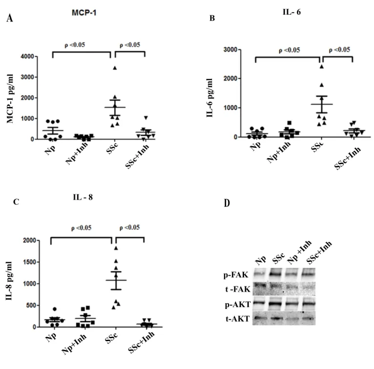
Figure 3: Downregulation of Pro-inflammatory cytokines in the presence of mPGES-1 inhibitor in SSc skin fibroblasts/ Downregulation of p-FAK and p-AKT in the presence of mPGES-1 inhibitor in SSc skin fibroblasts.
Ephrin B2 and Scleroderma
Figure 1: Ephrin B2 is overexpressed in human SSc skin
Figure 2: Ephrin B2 treated fibroblasts exhibit myofibroblast phenotype with enhanced actin stress fibre formation, cell spreading, and increased phosphorylation of FAK and α-SMA expression
Figure 3: Ephrin B2 treated fibroblasts exhibit enhanced migration and adhesion to Fibronectin
Figure 4: Mice treated with mouse recombinant Ephrin B2 Fc exhibit dermal fibrosis Figure 5: Mice treated with mouse recombinant Ephrin B2/Fc exhibit enhanced collagen
content and α-SMA-expressing myofibroblasts
Figure 6: Mice treated with mouse recombinant Ephrin B2/Fc exhibit enhanced expression of p-FAK, type-I collagen and CTGF
LIST OF TABLES
Table 1: Differences between Limited cutaneous sclerosis (lcSSc) and diffuse cutaneous sclerosis (dcSSc)
LIST OF ABBREVIATIONS
bFGF: Basic fibroblast growth factor BSA: Bovine serum albumin
CR- CHUM: University of Montreal Hospital Research Center
CTGF: Connective tissue growth factor COX: Cyclooxygenase
DAB:Diaminobenzidine
dcSSc: Diffused cutaneous systemic sclerosis
DMEM: Dulbecco’s Modified Eagle Medium
ECM: Extracellular Matrix ET-1: Endothelin -1 Eph B2: Ephrin B2 FBS: Fetal Bovine Serum FGF: Fibroblast Growth Factor IHC: Immunohistochemistry
IL: InterleukinlcSSc: Limited cutaneous systemic sclerosis
MCP: Monocyte chemotactic protein-1 MMP: Matrix Metalloproteinase
mPGES : Microsomal Prostaglandin synthase
PCR: Polymerase Chain Reaction PGE2: Prostaglandin E2
PGH2: Prostaglandin H2
PDGF: Platelet-derived growth factor SMA: Smooth muscle actin
SSc: Systemic sclerosis TBS:Tris Buffered Saline
TGF: Transforming growth factor TNF: Tumor Necrosis Factor SDS: Sodium dodecyl sulfate
VEGF: Vascular Endothelial Growth Factor WT: Wild Type
DEDICATION
This thesis is dedicated to my parents who have supported me all the way since the beginning of my studies.
ACKNOWLEDGEMENTS
I would like to express my deepest appreciation to my thesis supervisor, Dr. Mohit Kapoor, for offering his research expertise, astute insights and constructive assistance to me throughout my graduate studies at university of Montreal. His consistent support, guidance and understanding have been valuable in the completion of this work. He patiently helped me navigate my way through my daily experiments and gave much appreciated moral support and environment.
Also, I would also like to thank my co-director Dr. Johanne Martel Pelletier who was always willing to help me and give me her best suggestions during the past two years.
Last, but by no means least, I would like to thank all of the directors, members and friends in Osteoarthritis research unit in Notre-Dame hospital for their support and encouragement.
INTRODUCTION
SCLERODERMA (SYSTEMIC SCLEROSIS)
Scleroderma (Systemic sclerosis, SSc) is a fibroproliferative disorder associated with the production and accumulation of excessive fibrous connective tissue [1]. Primary features of SSc disease include autoimmunity, inflammation, obliterative vasculopathy and fibrosis. SSc does not just affect single organ and it spreads in large area of the skin and one or more internal organs such as kidneys, esophagus, heart, and lungs[2].
SSc has a global distribution. More women suffer from the disease than men[3] . The factors behind this gender disposition have not yet been elucidated and the overall incidence rate of SSc among adults in America is in the region of 20 for every 1 million persons annually. Statistics indicate that there was an increase in this rate between 1943 and 1973 but since then the rate has remained more or less constant. It is also reported that the prevalence rate of SSc among adults in America has remained more or less constant at 240 per million[4] . The frequency of occurrence of SSc in America is higher than that seen in UK, Asia, and continental Europe.There are between 75,000 and 100,000 people suffering from SSc in U.S. There is also a racial factor in the incidence and prevalence of SSc. The incidence of SSc is much higher in black women than in white women [4]
SSc is a leading cause of morbidity and case-specific mortality amongst all autoimmune rheumatic illnesses. Majority of the morbidity and mortality associated with SSc disease arises due to the development of complications that include gastrointestinal, renal, or cardiopulmonary diseases. Common organ involvement and manifestations in SSc disease include skin lesions,
gastrointestinal manifestations, cardiac involvement, pulmonary arterial hypertension (PAH), interstitial pulmonary fibrosis, and renal disease. Skin tightening and subcutaneous thickness may be the initial complaint that causes patients to seek for help.Generalized pruritus and cutaneous vasculitis are two more common cutaneous presentations where an underlying systemic disease may be present and will influence management[5]. Although skin manifestations are one of the most important components of clinical diagnosis and classification, studies have shown that life-threatening complications are independent from skin fibrosis [6]. Gastrointestinal manifestations represent the most common organ complications in patients suffering from SSc [7]. It is estimated that close to 90% of all patients with SSc manifest some form of gastrointestinal involvement with the most common of these being gastro-esophageal manifestations [8]. However, most of the gastrointestinal manifestations in patients with SSc disease are non-life threatening. Patients who have established SSc usually present with serious small intestine involvement causing dilation of small bowel loops leading to frequent bouts of intestinal pseudo-obstruction. Overgrowth of bacteria in the small intestine can then lead to reduced motility causing bloating, diarrhea, weight loss, cachexia, and malnutrition[7].
Involvement of the cardiac system is also one of the key determinants of mortality in patients with SSc which is largely seen in patients with the diffused cutaneous SSc form of systemic sclerosis[1] . It is however difficult to establish the precise percentage of cardiac involvement in patients with SSc due to diagnostic limitations[9]. Estimates suggest that the percentage of patients with SSc who have pericardial effusions is 35%. Involvement of the myocardium in patients with SSc has been attributed to fibrosis, ischemia, and myocarditis[1]. PAH is yet another organ complication that may occur in patients with SSc. PAH refers to elevated mean pulmonary artery pressure exceeding 25 mmHg when a person is resting, the right
complication occurs in both the limited systemic sclerosis (lcSSc) and the diffuse cutaneous sclerosis (dcSSc) forms of SSc and is a major cause of death in patients with SSc [10].
Interstitial pulmonary fibrosis has also been reported in patients with systemic fibrosis. The main types of interstitial lung disease observed in patients with SSc are the non-specific interstitial pneumonia (NSIP) and the usual interstitial pneumonia (UIP). Development of interstitial lung disease in people with SSc occurs insidiously and usually culminates in irreversible fibrosis of the lung. Reduced lung function is witnessed in only 15% of patients with SSc and this reduction usually occurs during the initial 4 years of disease [10] . The main types of renal manifestations that afflict patients with SSc are inflammatory renal pathology, chronic kidney disease, and scleroderma renal crisis (SRC). The most significant renal complication in SSc is SRC and it is seen in 10-15% of patients who have dcSSc. It is however very uncommon in patients with lcSSc as only 1-2% get to have SRC [11]. There is a high mortality of patients with SSc due to SRC [12]. Some evidence suggests that SRC may be triggered by intake of corticosteroids [13].
Subtypes of SSc disease
Systemic sclerosis is divided into specific mutually exclusive subsets because of their variable prognostic and diagnostic characteristics. The 2 main subsets of SSc are the limited systemic sclerosis (lcSSc) and the diffuse cutaneous sclerosis (dcSSc). The 2 subtypes are distinguished by the degree and scope of skin thickening and susceptibility to visceral involvement[14]. The autoantibody profile is also used to distinguish between the limited and diffuse disease forms[15] .
Limited Systemic Sclerosis (LcSSc)
In the lcSSc form of SSc disease, fibrosis is largely limited to the distal portions of the elbows or knees[16] . Skin involvement may also be witnessed in the face. Progress of fibrosis is usually
slow. Patients with this form of disease have a somewhat smaller risk of developing serious involvement of the interstitial lung [17].
Table 1: Differences betweenLimited cutaneous sclerosis (lcSSc) and Diffuse cutaneous sclerosis (dcSSc) [9]
Limited cutaneous sclerosis (lcSSc) Diffuse cutaneous sclerosis (dcSSc) Fibrosis limited to the distal portions of the
elbows or knees
Fibrosis limited to the proximal portions of the trunk and extremities
Heart involvement is minimal Heart involvement is severe in 10% of patients Interstitial lung disease is severe in 15% of
patients
Interstitial lung disease is severe in 15% of patients
PAH is seen in 10-15% of patients PAH is seen in 5-10% of patients
Minimal kidney involvement Kidney involvement is severe in 10-15% of patients
Concurrent primary biliary cirrhosis in 6-8% of patients
Large joint contractures
The overall survival rate is better than that of dcSSc
Diffuse cutaneous sclerosis (dcSSc)
In the dcSSc form of SSc, fibrosis is largely limited to the proximal portions of the trunk and extremities [16]. Patients with dcSSc stand a higher risk of getting serious heart and kidney involvement than patients with the lcSSc form of disease [18] . Table 1 above depicts the primary distinguishing features between the 2 forms of SSc.
The main disadvantage of this subset classification method is that patients who have the early disease but show no visceral involvement and having or lacking minimal skin thickening fit nowhere in this scheme. To address this shortcoming, a new scheme for classifying subsets of SSc was proposed. In this scheme, it is possible to classify patients with early disease based on particular autoantibodies, changes in the nail fold capillary, and Reynaud’s phenomenon. (An exaggeration of vasomotor responses to cold or emotional stress causes skin discoloration).
They can be grouped in the limited form of systemic sclerosis also termed as pre-scleroderma [16] . However, validation of the proposed scheme is yet to be done [15] .
Barnett et al [19] and Ferri et al [20] describe yet another scheme that can be used to subtype systemic sclerosis. In this scheme, 3 subtypes of systemic sclerosis have been described and these include limited cutaneous sclerosis, intermediate cutaneous sclerosis, and diffuse cutaneous sclerosis. In this scheme, the main feature of the limited cutaneous sclerosis subset is that the thickening of the skin is limited to digits and facial involvement may be present or absent. In intermediate cutaneous sclerosis, there is skin involvement in the limbs while in the diffuse cutaneous sclerosis; there is skin involvement in the trunk. This method of classifying subsets of SSc is better because its discrimination power is higher than that based on only 2 subsets [21]. Methods that only encompass skin changes and fail to factor in autoantibodies and imaging characteristics are inadequate and can hardly be used for the optimal classification of subsets of SSc. Classification of SSc can also be done based on the Preliminary Criteria for the Classification of SSc which was formulated by The American College of Rheumatology (ACR). This is a diagnostic criteria which has however been shown to be unsuitable for the diagnosis of SSc [10].
Symptoms
Reynaud’s phenomenon is one of the earliest symptoms observed in SSc. Reynaud’s phenomenon is a condition in which the toes and fingers undergo vasospasm due to cold. It occurs periodically, is reversible and manifests long before other signs of disease are observed [17]. In Reynaud’s phenomenon, the toes, fingers and other extremities become discolored and may cause acral ulcer upon persistence [22] . Other common symptoms of SSc are telangiectases or obviously dilated blood vessels, sclerodactyly, vasculitis, and calcinosis in the hands, fingers and bony regions. Calcinosis refers to deposits of calcium in these areas[23].
Other symptoms include ulceration which may result in dry gangrene and fingertip loss, thickening of the skin, changes in the nail fold capillaries, SRC, malignant hypertension, PAH, and gastric antral vascular ectasia [3] . Symptoms are also dependent on the type and extent of organ involvement. Patients with renal involvement and specifically SRC may show non-specific symptoms such as fever, headache, dyspnea, and malaise[3]. Acute renal failure, pulmonary edema, hypertensive retinopathy and encephalopathy may be symptomatic in patients with end-organ damage. Coagulopathy is an uncommon symptom but thrombocytopenia and microangiopathic hemolytic anemia are common, occurring in 50% and 60% of patients respectively[23] .
Patients with SRC and SSc also manifest reduced significant systemic hypertension and reduced renal function [11] . Dry cough, dyspnea, and rare hemoptysis and chest pain are the main symptoms in patients with SSc who have interstitial pulmonary fibrosis. Pulmonary arterial hypertension (PAH) can be asymptomatic until it becomes very advanced. A common symptom is dyspnea while less common symptoms are syncope and chest pain. Symptoms of patients with
and malnutrition in severe cases. Classic symptoms are bowel pattern changes accompanied with abdominal distension and regular loose, floating and foul-smelling stools [7].
Etiology
SSc disease is highly heterogeneous [17] and the exact etiology of this debilitating disease is largely unknown [15] . Possible causative agents of SSc include viruses such as cytomegalovirus (CMV), exposure to silica, vinyl chloride, and organic solvents, and drugs [24] . In particular, Namboodiri et al [25] and Lunardi et al [26] have detected antibodies against CMV in patients with SSc. These antibodies do not only enhance endothelial cell apoptosis but also activate fibroblasts in cell culture assays, implying that they play a direct role in the damage of tissues in SSc. In addition, human CMV infection also causes an increase in the production of the connective tissue growth factor (CTGF) which is associated with the activation of fibroblasts and has been shown to play a role in pathological fibrosis[27].
Possible Genetic Association
There is a genetic association with SSc. SSc is inherited but not in a Mendelian fashion. There is a low disease concordance rate that does not exceed 5% among both dizygous and monozygous twins. The disease is seen more in families and less in the general population. Among families, the rate of the disease is 1.6% and this far exceeds the rate among the general population which is just 0.026% [17] . A positive family history of SSc is the biggest risk factor for the disease [28]. It has been observed that there are geographic clusters of the disease. Such clusters of SSc include the Chocktaw Native American Cluster and Italy and London clusters. The former cluster suggests that the disease is caused by yet to be identified genetic factors. Familial clustering has also been reported in Australia and the United States[4] .
Gender and the major histo-compatibilty complex (MHC) are also important genetic factors associated with SSc. The female to male ratio of SSc is 3:6.1 while people with systemic
sclerosis have a higher frequency of class I and class II MHC alleles. It has been shown that SSc is primarily associated with the linkage of DRw52 with DR5 and DR3 and that lung fibrosis is largely associated with B8-DR3-DRw52-DQB2. Presence of Antitopoisomerase (ATA) and DR52a can be used to predict pulmonary disease. Black women are more prone to the disease than white women [1].
Prognosis
The prognosis of SSc is variable and this is largely due to the variability of the disease spectrum [29]. Prognosis is improved by optimal and early treatment. There is no effective cure for SSc and people with dcSSc have a higher risk of mortality. The survival rate of patients with this form of disease is 55% at 10 years [30]. Varga et al [2] assert that better management of systemic sclerosis has led to an improvement in clinical outcomes. However, a cure for the disease has not yet been found and dcSSc has a higher fatality risk than the lcSSc form of disease [23].
The leading cause of death in SSc is pulmonary disease. Renal and cardiac diseases are also associated with poor prognoses. Whereas morbidity due to gastrointestinal disease has been noted, it is not easy to quantify the degree to which this occurs. There has been an improvement in the overall survival of people with SSc over the past few decades. The mean survival from diagnosis is estimated at 12 years. The prognosis also varies depending on the type and extent of organ involvement [30]. Patients with SSc and PAH have a median survival of between 1 and 3 years [31]. The mean survival for patients with SSc and severe pulmonary fibrosis is less than 3 years [31].
Pathogenesis
Even though the pathogenesis of SSc is complex and heterogeneous, the large number of studies carried out in the recent past has helped to shed more light on the pathophysiological events associated with this condition. It is characterized by sequence of events that are common among all the subsets of the disease. During pathogenesis of SSc, microvascular change is followed by inflammation and immune activation which eventually leads to fibrosis[32] .
Vascular injury caused by physical trauma, autoantibodies, viruses, and oxidative stress leads to activation of endothelial cells, leukocyte adhesion, vascular obliteration, and tissue hypoxia. These events trigger inflammation and autoimmunity resulting in production of growth factors and cytokines which cause activation of fibroblasts causing fibrosis [23] .
Microvascular Changes
The first events to occur during pathogenesis of SSc involve vascular injury. Vascular injury can be caused by autoantibodies that are cell-specific, physical trauma, granzymes, reactive oxygen species (ROS) that are generated as a result of ischemia and reperfusion, inflammatory cytokines, and vasculotropic viruses. Vascular injury is manifested by changes in the nail fold capillaries, cutaneous telangiectasia, malignant hypertension, PAH, and gastric antral vascular ectasia [3] . Vascular injury leads to the activation of endothelial cells and renders them dysfunctional as well [33] . It also results in changes in the permeability of capillaries, modified secretion of vasoactive mediators, and enhanced expression of the endothelial leukocyte adhesion molecule1 and VCAM-1[34] . Vascular injury also causes fibrinolytic and platelet pathways to become activated [35].
Once activated, the endothelial cells release endothelin-1 (ET-1). Endothelin-1 (ET-1) is a powerful vasoconstrictor, which also activates fibroblasts, enhances the proliferation of smooth
muscle cells, and induces the adhesion of leukocytes to the endothelium. Due to the vascular injury, vascular remodeling occurs and hypertrophy of the medial and intimal layers is seen together with adventitial fibrosis to result in the gradual narrowing of the lumen and its eventual elimination[36] . Apoptosis of the endothelial cells combines with the preceding processes to lead to cause the gradual loss of blood vessels and disruption of angiogenesis. Impaired formation of blood vessels has been attributed to the impaired differentiation and reduction of the CD34+ cells originating from the bone marrow [37, 38]. Consequently, hypoxia occurs and causes a significant increase in the expression of the vascular endothelial growth factor (VEGF) and its receptors [39], [34] .
Inflammation and Immune cell activation
The onset of inflammation in dcSSc is longer than that of lcSSc. In addition, there is an extensive spread of inflammation in the musculoskeletal and skin in the dcSSc form of SSc. This inflammation is accompanied by edema, which is indicative of changes in the permeability of the endothelium. As the disease progresses, widespread fibrotic changes occur and Reynaud’s symptoms can manifest simultaneously with skin changes or can occur after the changes[4] .In contrast, the onset of lcSSc is much slower and there may be a pre-existence of Reynaud’s phenomenon for a couple of years. In addition, the prominence of the vascular component is a characteristic feature of lcSSc and accounts for most of the manifestations of the disease including renal involvement, digital ulceration, and PAH. There is nevertheless a small amount of fibrosis and this is usually observed in the face, skin, gastrointestinal tract, and extremities[4].
Vascular injury leads to inflammation and autoimmunity. Following vascular injury, leukocytes are recruited. Chemokines such as monocyte chemoattractant protein 3 (MCP-3) and monocyte chemoattractant protein 1 (MCP-1) lead to the accumulation of mononuclear cells
(IL-1) and TGFβ, which activate fibroblasts thereby initiating fibrogenesis. These cytokines also activate resident fibroblasts produced by pericytes and mesenchymal progenitor cells thereby further enhancing fibrinogenesis. Production of resident fibroblasts by pericytes is induced by the activity of the platelet-derived growth factor (PDGF), the basic fibroblast growth factor (bFGF), and the endothelin-1 (ET-1) on the pericytes. According to Varga et al [2] PDGF is a powerful mitogen and chemoattractant for fibroblasts and can stimulate them to produce TGFβ, IL-6, and MCP-1 and to generate collagen, proteoglycans, and fibronectin. Activated fibroblasts are also acted upon by TGFβ and connective tissue growth factor (CTGF) produced by the T helper cell 2 (TH2) leading to the formation of myofibroblasts which cause permanent scarring through tissue remodeling and fibrosis[4] .
There is a delicate balance between TH2 and TH1 cells and alteration of this balance is associated with increased fibrinogenesis. There is a shift towards TH2 predominance in SSc. Whereas TH1 cells predominantly secrete interferon gamma (IFNγ) and interleukin 2 (IL2), TH2 cells largely produce IL4, IL13, and IL5 [40]. The IFNγ inhibits the expression of collagen-encoding genes and abolishes the stimulatory actions of TGFβ. As such, IFNγ is a powerful inhibitor of the contraction of fibrogenesis since it inhibits the trans-differentiation of fibroblasts into myofibroblasts, the contraction of the extracellular matrix, and the proliferation of fibroblasts. The regulatory T cell also activates Myofibroblasts.
MAJOR PRO-INFLAMMATORY CYTOKINES IMPLICATED IN SSc
DISEASE
Interleukin- 6 (IL- 6)
IL- 6 is a cytokine, which is produced by local tissues and later released in the circulation system. It is a polypeptide that comprises of 212 amino acids and is produced by various cells such as the T- cells and the monocytes. The molecular weight of IL- 6 ranges from 21- 29 kDa due to variable and extensive glycosylation and phosphorylation [41]. IL- 6 is vital in almost homeostatic perturbation situations such as trauma and acute infections [42]. It is also a multifunctional cytokine with a vital role in host defense due to its various ways of immune and hematopoietic activities [43]. IL-6 modulates various functions in the body such as apoptosis, cell differentiation and proliferation, and inflammation. Apart from its main function, IL- 6 also influences various body systems such as neural and endocrine systems, skeletal muscles and bone metabolism [44].
Studies by Yu et al showed that IL-6 contributes to the initiation and extension of the inflammatory process. During inflammation process, IL- 6 activates B and T lymphocytes and also stimulates hepatocytes to produce acute phase proteins [45]. Studies demonstrated that IL- 6 has anti-inflammatory and protective properties too. These properties include the ability to inhibit production of tumor necrosis factor (TNF), IL- 1 and macrophage inflammatory proteins [45]. IL- 6 is vital in activating fibroblasts to produce extracellular matrix whose excessive accumulation leads to SSc. Some Studies demonstrated that IL- 6 is highly expressed in patients with SSc especially during the early stages of the disease in inflammatory phase [46]. High IL- 6 expression is associated with more severe skin involvement at early stages. Fibroblasts isolated
and cultured from SSc lesional skin involvement produced higher level of IL- 6 compared with non lesional SSc samples [47].
Interleukin- 8 (IL- 8) and SSc
Interleukin- 8 (IL- 8) is a member of a family of structurally related proinflammatory factors that have a low molecular weight and are referred as the chemokine [48]. IL- 8 has a low molecular weight of approximately 8kDa. It is a non-glycosylated protein comprising of 72 amino acids. A number of studies have been carried out in the past to determine the possible changes in the serum levels of IL-8 in patients with SSc disease. According to a study by Reitamo and others [49], the levels of IL-8 and autoantibodies to IL-8 were significantly higher in patients with SSc disease [49]. In fact, the levels were undetectable in normal serum, but highly detectable in more than 12.5% of the patients. A study by Guang-bin Cui et al have also shown similar results [50] where the levels of the IL-8 were determined in mice induced with persistent inflammatory pain, such as the one experienced in SSc. In this case, it was found that in all the mice samples used, the level of serum IL-8 had raised significantly. The study indicates that the up-regulation of the IL-8 in mice is related to the activation of fibroblasts or mononuclear phagocytes and other immune cells [50]. In addition, such activation may be related to the production of the autoanitibodies targeting IL-8 molecules that are now detectable in the serum.
Monocyte chemo attractant protein- 1(MCP- 1) and SSc
MCP- 1 is an inflammatory chemokine that is produced predominantly by macrophages and endothelial cells. The expression of this chemokine increases in patients who have atherosclerotic lesions, thus, MCP-1 plays a vital role in artherogenesis [51]. MCP- 1 secretion is induced by the cytokine activation and also interaction of activated platelets with monocytes or endothelial cells. Research shows that MCP- 1 is a chemokine that links monocyte activation to vascular inflammation of patients with SSc [52] . Studies revealed that MCP- 1 has both proinflammatory
role and pro fibrotic role is SSc patients. In the early stages of SSc, MCP- 1 is released. It then attracts the T cells and mononuclear cells to the affected area. This leads to the production of pro- fibrotic cytokines such as IL- 4, which then activate the synthesis of ECM in dermal fibroblasts, and causes fibrosis in later stages of SSc [52]. Studies have shown that the inhibition of MCP-1 reduces the extent of SSc as well as atheroma in mice induced with hypercholesteroma [53]. Many other studies on targeting MCP-1 as a possible treatment for SSc patients revealed promising result in animal models [52]. Ongoing clinical trials are testing the MCP-1 antagonists on various disease as well as SSc patients.
Fibrosis
Fibrosis is the definitive feature of SSc [54]. It involves the gradual replacement of tissue architecture by the extracellular matrix (ECM), which is rich in collagen and other fibrotic components. Excessive ECM deposition in fibrotic organs leads to organ dysfunction [55]. Fibrosis commonly occurs in the lungs, skin, heart, gastrointestinal tract, endocrine glands, ligaments, and tendons, and it constitutes a large part of the mortality and morbidity that are associated with SSc[56].
Typically, the extracellular matrix is made up of 2 compartments, the cellular and connective tissue compartments [4]. According to Namboodiri et al. [25], the former compartment consists of inhabitant and infiltrating cells while the latter compartment consists of adhesion molecules, collagens, fibrillins, and proteoglycans. The extracellular matrix is also a reservoir for matricellular proteins as well as growth factors such as CTGF and TGFβ. These proteins regulate the differentiation, function continued existence of mesenchymal cells in concert with the connective tissue compartment.
Fibroblasts and Myofibroblasts
Fibroblasts are cells found in the connective tissue throughout the body which produce collagen and other proteins found in the extracellular spaces of cell [57]. They have a vital role in matrix deposition, matrix degradation and also in growth factor secretion as well as inflammatory response and control [58]. Fibroblasts migrate within tissues through a process known as cell migration; Cell migration is a cellular process that has a role in disease and health such as wound healing, immune response and tissue development.[59] Fibroblasts take part on wound healing since they have the ability to move to the site of the wound to repair damaged tissue and eventually heal the wound [60]. Tissue injury and microenvironmental changes are important stimuli for phenotype transition of fibroblasts to myofibroblasts. In response to tissue injury and as a change in normal intracellular environment, fibroblasts acquire actin fibers, the stress fibers which are the hallmark of stable protomyofibroblasts. The final step of fibroblast differentiation to myofibroblasts is the expression of α-SMA in protomyofibroblasts. The generation of α-SMA needs TGF-β 1and β2, ED-A fibronectin (an isoform de novo expressed during wound healing and fibrotic changes) and the high extracellular matrix stress.
Fibrosis is largely executed by the differentiation of fibroblasts to myofibroblasts [61]. Myofibroblasts express the stress fibers that lead to ECM contraction. Myofibroblasts also produce α-SMA (α-smooth muscle actin), which is an important contractile factor [62]. Moreover, fibroblasts produce collagen in response to stimuli from inflammatory cells, platelets, and epithelial and endothelial cells. Stimuli also cause fibroblasts to secrete other molecules of the ECM that attach, contract, organize, and remodel connective tissue. In fibrotic disorders, myofibroblasts exhibit defective apoptosis process resulting in the maintenance of the fibrosis [63]. Cytokines and growth factors are also produced by fibroblasts, which can also undergo trans-differentiation to form contractile myofibroblasts [64]. Several growth factors and signaling
pathways have been shown to be implicated in the pathogenesis of fibrosis. Pro-fibrotic proteins, including TGF-β, Endothelin-1, and connective tissue growth factor, are believed to play an important role in the pathogenesis of fibrosis [65],[33].
Transforming growth factor-β (TGF-β)
TGF-β is an important regulatory cytokine that has diverse effects on cell differentiation, proliferation, survival, and remodeling [66, 67]. At least three isoforms of TGF-β have been identified in mammals, but only TGF-β1 has been shown to play a pivotal role in wound healing and fibrosis. TGF-β is stored in a latent form in ECM, and it binds to the latent TGF-β binding factor (LTF). When the proteolysis of the carboxyterminal in LTF occurs, TGF-β is converted to its active form and signaling starts through the TGF-β specific receptors. TGF-β has 2 types of receptors with several subtypes. There are five Activin receptor–like kinases (ALKs) for type II. There are seven type I receptors [68]. When TGF-β binds to the receptors, the aggregation of both receptors occurs consequently, and TβRII (TGF-β Receptor type II) activates TβRI (TGF-β Receptor type I). The signaling cascade occurs through the phosphorylation of the SMAD proteins. Moreover, TGF-β also acts through other signaling pathways. The mitogen-activated protein kinase (MAPK), P38, and Jun-kinase (JNK) cascade are other pathways [69], [70]. Upon the activation of TGF-β, the expression of collagens and fibronectin increases, which causes the matrix deposition. Furthermore, TGFβ inhibits the activation of matrix metalloproteinases (MMPS) that degrade the ECM [71], [72]. Studies by Desmouliere et al show that TGF plays an important role in the differentiation of myofibroblasts through α-smooth muscle actin (α-SMA) activation [73]. In their study, the administration of TGF-β in rats induced the formation of granulation tissue with high expressions of α-SMA in myofibroblasts, which is specific for TGF-β. Choi et al demonstrated that knocking down TGF-β expression using anti-sense RNA
decreases the fibrotic tissue after injury [74]. Studies by Bonniaud et al show that an overexpression of TGF-β in lungs leads to lung fibrosis in mice [75].
Signaling Pathways
SMAD signaling pathwayThe SMAD pathway is the major pathway involved in transmitting signals from the TGFβ receptors. As already indicated before, the extracellular matrix is a reservoir of inactive or latent TGFβ. The dormancy of the latent TGFβ is maintained by the latent TGFβ binding proteins (LTBPs). The TGFβ is activated by plasmin, integrins, THY-1, and thrombospondins and attaches to the cell surface receptors namely TGFβRII and TGFβRI. An intracellular signal transduction cascade is triggered due to the binding of the TGFβ to the receptors and this leads to activation of the target genes. The receptors are serine-threonine kinases and they cause phosphorylation of SMAD proteins [76].
When SMAD2 and SMAD3 are phosphorylated, they create hetero-complexes with SMAD4 and move into the nucleus from the cytoplasm. In the nucleus, the hetero-complexes attach to the cis-acting DNA sequence (CAGAC), which characterizes the consensus SMAD-binding element (SBE). The SBE is present in the promoters of a large number of genes that can be induced by TGFβ. After attachment to the SBE, recruitment of transcriptional factors to the DNA by the activated SMAD proteins occurs and this induces the transcription of the collagen-encoding genes. Inhibition of SMAD-dependent signal transduction is mediated by SMAD7. Studies have shown that SSc is associated with changes in the activation and inhibition of specific cofactors and proteins involved in the SMAD signaling pathway [2], [23] ,[77].
The non-SMAD pathways also play a critical role in the pathogenesis of SSc. The non-SMAD pathway involves the activation of the focal adhesion kinase (FAK), MAPK, Jun kinase (JNK), calcineurin, TGFβ activated kinase 1 and lipid kinases such as AKT and PI3K. . Bujor et al studied the role of AKT in deposition of collagen by normal dermal fibroblasts. They discovered that the basal production of collagen was hindered when AKT was inhibited. Inhibition of AKT also led to elevated production of matrix metalloproteinase 1 (MMP1) and elimination of the inhibitory effect of TGFβ on MMP1. The findings demonstrate that AKT is profibrotic as it increases the synthesis and reduces the degradation of collagen. It was thus concluded that AKT plays a role in fibrosis in SSc [78].
Focal adhesion kinase (FAK)
Focal Adhesion kinase (FAK) is a 125 kD protein which plays an important role in the focal adhesion dynamics between cells, as well as in motility and cell survival. FAK is phosphorylated in response to growth factors, integrin, and other stimulation [79]. Studies demonstrate that the phosphorylation of the FAK (p-FAK) is involved in myofibroblast differentiation and plays a role in the pathogenesis of SSc[80]. In SSc patients, myofibroblasts have the ability to produce α-SMA with the stimulation of TGF-β. For this induction, TGF-β needs focal adhesion kinase phosphorylation on the Tyr-397 site [81]. Moreover, studies by Mimura et al [80] demonstrate that P-FAK on the Tyr-397 site is also higher in the myofibroblasts of SSc patients as compared to that in normal fibroblasts. These results also confirm the possible role of p-FAK in the pathogenesis of SSc by TGF-β signaling and α-SMA expression in myofibroblasts.
Endothelin-1
Endothelin-1 (ET-1), which is a potent vasoconstrictor secreted from endothelial cells, plays a critical role in the pathogenesis of SSc[82]. Studies by Abraham el al [83] show that ET-1 is
fibrotic disorders. Studies by Mutsaers et al [84] demonstrate that ET-1 levels are elevated in animal models of lung fibrosis. ET-1 binds to ETA and ETB receptors on fibroblasts directly to induce the differentiation of myofibroblasts [85]. Moreover, studies of lung fibrosis also show that ET-1 induces elevated levels of α-SMA through Akt and the ras/MEK/ERK signaling pathway [86, 87].
Connective Tissue Growth Factor
Connective tissue growth factor (CTGF) is a cysteine-rich protein and a member of the CCN superfamily, plays a direct role in fibrosis as well as an indirect role through the creation of a favorable environment [88] for other factors that induce the fibrosis in fibrotic disorders. CTGF is a promoting factor for the adhesion of fibroblasts to fibronectin [84, 89]; it also helps TGF-β to induce cell adhesion to fibronectin and other ECM components [90]. Moreover, CTGF increases the effect of the ET-1 and TGF-β signaling pathway and indirectly increases the fibrosis effect [91]. Studies by Sato et al [92] show that serum CTGF is higher in SSc patients as compared to control samples. Furthermore, CTGF has a positive correlation with skin fibrosis and pulmonary fibrosis in SSc patients. CTGF seems to be involved in maintaining the fibrotic phase of SSc [92].
Table 2: Pathways and signaling molecules dysregulated in SSc [15]
Molecule Changes in SSc fibroblasts
Cofactors and transcription factors
SMAD2/3 Accumulates in the nucleus and becomes phosphorylated
constitutively
PPARγ Expression is reduced
SP1 Becomes phosphorylated constitutively
P300/CBP Expression is increased, binds constitutively to SMAD2/3
FLI-1 Expression is reduced
Kinases
FAK1 Activated in a constitutive manner
PKC-δ Expression is increased
ERK Activated in a constitutive manner
Surface receptors
TGFβ receptors Expression of TGFβR1 and TGFβR11 is elevated
Integrin αγβ3 Expression is increased
Integrin αγβ5 Expression is increased
PDGFRα Expression is increased
PDGFRβ Expression is increased
Current treatment, drugs in market and drugs in clinical trials for SSc disease
Treatment options for SSc remain a challenge because of the unclear pathogenesis of this autoimmune disease. However, those immune-modulators which target blood vessels and aid in recognition, management of end-organ damage, adjunctive therapies like light, physical and psychotherapy are considered most effective to treat this multi-factorial disease. The search for new drugs that work as anti-fibrotic agents is probably one of the most active areas of research in this field. Open label studies with Revimmune drug therapy have shown a desirable effect on the immune system of patients suffering from SSc but the trials are ongoing for more data to confirm efficacy of the drug[93]. Controlled clinical trials with Imatinib mesylate (Gleevec) have been carried out to determine the safety and tolerability in patients. Proven effectiveness (anti-fibrotic effect) and low incidence of side effects was observed as a result [94]. A platelet gel for treating digital ulcers is currently in clinical trials along with others (anti-fibrotic agents like interferon gamma, D-penicillamine, kolchichicine, calcitriol) which reduce excessive production of collagens and other connective tissue proteins to prevent and control symptoms like skin fibrosis[95]. Clinical trials are ongoing with drugs like Orencia, and MQX-503 whereas efficacy
even though these studies could not differentiate more efficacious form of drug in terms of dose[96]. Drugs like methotrexate, cyclosporine, nifedine, iloprost have all been studied in controlled trials with variable outcomes and a considerable number of trials have proved nifedipine, a calcium channel blocker as a gold standard. Randomized trials on the drug - cyclophosphamide confirm the moderate clinical benefits seen in patients with early, symptomatic disease[97]. Many studies are ongoing on finding an appropriate treatment for SSc; however there is not any approved treatment, which can stop this disorder completely. I anticipate that targeting inflammation during early phases of SSc disease could be a better therapeutic option. Therefore it’s essential to identify mediators, which are responsible for initiating inflammatory response during early phases of SSc disease. Another option is to identify endogenous mediators, which initiate the differentiation of fibroblasts to myofibroblasts and promote adhesive and fibrotic signaling. For instance, a variety of in vitro and in vivo studies using murine models of fibrotic diseases suggest that FAK inhibitors exhibit potent antifibrotic effects, thus making them attractive drugs for fibrotic disorders seen in the clinic. In recent years, several orally bioavailable ATP-competitive FAK inhibitors have been developed by pharmaceutical companies and have entered early into human clinical trials [98]. One of the first clinically available specific FAK inhibitors was PF-562, 271, which inhibited FAK phosphorylation in vivo in a dose-dependent fashion in several human s.c. xenograft models [99]. Recently the present authors showed that PF-562, 271 also prevented bleomycin-induced lung fibrosis in a mouse model. The Phase I study using PF-562, 271 was performed in patients with head and neck, prostatic and pancreatic cancer (clinical trial #NCT00666926, http://clinicaltrials.gov/). Clinically, PF-562, 271 prolonged disease stabilization in a subgroup of patients. Due to the low toxicity of this drug, combination therapies with blocking antibodies or antagonists/inhibitors of profibrotic factor receptors seem possible. However, to our knowledge, there are no clinical studies that have reported the effects of FAK inhibitors in any fibrotic diseases.
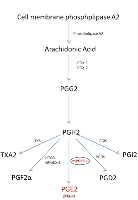
Arachidonic Acid Pathway
Arachidonic acid plays an important role in many physiological processes. The pathway has an important role on generation of pain and inflammation as well as for maintenance of homeostasis. Arachidonic acid is formed by the activity of phospholipase A2 on cell membrane phospholipids.
There are 2 main pathways for the metabolism of arachidonic acid and these are the 5-lipoxygenase (5-LO) and the cyclooxygenase (COX) pathways. In the 5-LO pathway, 5-HPETE is formed by the activity of 5-lipooxygenase enzymes on arachidonic acid and this is the precursor for several leukotrienes such as LTB4, LTC4, LTD4, and LTE4. In the COX pathway, the cyclo-endoperoxide PGG2 is formed in reactions catalyzed by the cyclooxygenase enzymes [100].
There are many different types of cyclooxygenases including COX-1, COX-2, and COX-3. The PGG2 is then catalyzed to PGH2, which is then converted into prostanoids such as thrombooxanes (TXA2), prostaglandins such as PGD2, PGE2 and PGF2α, and prostacyclins such as PGI2. Prostaglandins catalyze the modulation of immune function; leukotrienes add molecular oxygen to particular double bonds in polyunsaturated fatty acids and thrombooxanes are potent vasoconstrictors and enhance the aggregation of platelets[100] . PGE2 is the commonest prostanoid as it is produced by many cell and tissue types and its spectrum of activity is wide. It acts on the G-coupled EP1, EP2, EP3, and EP4 receptors and together with PGI2, it is the main prostanoid involved in inflammation and pain [101], [102] . Formation of PGE2 from PGH2 is catalyzed by the microsomal prostaglandin E synthase-1 (mPGES-1) and this is depicted in the diagram below [103].
Diagram 1: Illustration of the pathway involved in biosynthesis of prostaglandin E2 (PGE2).
In the diagram above, mPGES=microsomal prostaglandin E synthase, PG=prostaglandin, TPX=thromooxane A2 receptor, TXA2=thrombooxane A2. As shown, conversion of arachidonic acid to PGH2 is mediated by the cycloxgenases COX-1 and COX-2. TXA2, PGE2, PGD2, and PGI2 are synthesized in reactions mediated by TXA2 synthases and PGI2 synthases respectively[103]
(Major
Microsomal Prostaglandin E Synthase-1 (mPGES-1)
Microsomal prostaglandin E2 synthases (mPGES) are enzymes that catalyze the conversion of
PGH2 to PGE2 [104]. Thus far, three PGE synthases, namely cytosolic PGE synthase (cPGES),
mPGES-1 and mPGES-2, have been characterized [104-106]. cPGES is localized in the cytosolic region of cells and tissues under basal conditions and is most likely to be involved in the homeostatic production of PGE2 [106]. mPGES-2 is also constitutively expressed in wide variety
of tissues and cell types and is synthesized as a Golgi membrane associated protein [107]. In contrast, mPGES-1 is induced in response to inflammation, and acts downstream of cyclooxygenases (COX) [108, 109]. PGE2, the final metabolite of cyclooxygenase pathway, has a variety of endogenous functional effects [110]. Besides its role in the initiation and perpetuation of inflammatory processes, PGE-2 helps the blood clot formation, protect the gastrointestinal tract by increasing the mucus formation and also takes part in labor by constricting the uterine[111].
mPGES-1 and its derived PGE2 in inflammation and fibrosis
mPGES-1 has been shown to be a critical mediator of inflammation, pain, angiogenesis, fever, bone metabolism and tumorgenesis [102, 112-114]. Previous studies have shown that mPGES-1 expression is elevated in tissues and cells of various inflammatory diseases including rheumatoid arthritis (RA) and osteoarthritis (OA) [108, 109, 115, 116]. mPGES-1 null mice are resistant to chronic inflammation of joints in the models of collagen induced arthritis (CIA) and collagen antibody induced arthritis (CAIA) [102, 112]. We have also shown that mPGES-1 is induced during skin wound healing process in mice [117].
My laboratory is the first to investigate the role of mPGES-1 in fibrosis using animal models. In our recent study, [118] we investigated the effect of mPGES-1 genetic deletion in mice model of
![Table 2: Pathways and signaling molecules dysregulated in SSc [15]](https://thumb-eu.123doks.com/thumbv2/123doknet/2073206.6699/33.918.106.814.861.1046/table-pathways-signaling-molecules-dysregulated-ssc.webp)