Recognition of the Latency-Associated Immediate Early
Protein IE63 of Varicella-Zoster Virus
by
Human Memory
T Lymphocytes’
Catherine Sadzot-Delvaux,2* Paul R. Kinchington,+ Serge Debrus,* Bernard Rentier,* and
Ann
M. Arvin*
Varicella-zoster virus (VZV) is a human alphaherpesvirus that establishes latency in sensory ganglia. latency is characterized by the abundant expression of the immediate early protein 63 (IE63), whereas other viral proteins have not yet been detected during the quiescent phase of VZV infection. The IE63 protein is a component of the virion and is expressed very early in the infectious cycle. The IE63 protein is also expressed in skin during episodes of varicella and herpes zoster. We have evaluated the cell-mediated immune response against IE63 in naturally immune adults with a history of chickenpox, by T lymphoprolif- eration and cytotoxicity assays. Among donors who had
T
cell proliferation to unfractionated VZV Ags from infected cell extract, 59% had T cell recognition of purified IE63. The C T l response to IE63 was equivalent to CTL recognition of IE62, the major tegument component of VZV whose immunogenicity has been previously described. IgC Abs against IE63 were detected in serum from healthy immune adults. These results indicate that IE63 is an important target of immunity to VZV. T cell recognition of 1E63 is likely to be involved in controlling VZV reactivation from latency. The Journal of Immunology, 1997, 159: 2802- 2806.V
aricella-zoster virus (VZV)’ is a human herpesvirus that causes varicella (chickenpox) and herpes zoster (shin- gles) separated by an extended period of latency (1). After the primary infection, VZV reaches the sensory ganglia and persists for the life of the host. Although several regions of the viral genome are transcribed during latency (2, 3), the product of open reading frame 63 (ORF63) of VZV is the only viral protein that has been detected in latently infected ganglia in a rat model (4), and more recently in human tissues (5). The IE63 protein is associated with purified viral particles (6), and as a true immediateearly protein is expressed very early in the infectious cycle, ap- pearing first i n the nucleus and then in the cytoplasm (4). IE63 is
also abundant in the skin lesions of patients with acute varicella or herpes zoster (4, 7). The regulatory functions of this IE protein are being characterized in vitro, but its role in the establishment or maintenance of VZV latency is still unknown.
*Division of Infectious Diseases, Department of Pediatrics, Stanford University School of Medicine, Stanford, CA 94305; +Departments of Ophthalmology, Mo- lecular Genetics and Blochemistry, Unwersity of Pittsburgh, Pittsburgh, PA
of Liege, Liege, Belgium
1521 3; and *Laboratoire de Virologie fondamentale, Pathologie B23, Universite Recelved for publication April 14, 1997. Accepted for publication June 19, 1997.
The costs of publication of this article were defrayed in part by the payment of page charges. This article must therefore be hereby marked advertisement in accordance with 18 U.S.C. Section 1734 solely to Indicate this fact.
A136884, POLCA49605
’
(to A.M.A.), and ROl-EY 09397 (to P.R.K.). While per- This work was supported by National Institutes of Health Grants A120459, formlng thts work, C.S.-D. was a VZV Research Foundation and Belgian Amer- ican Educational Foundatlon fellow, and was supported by a N.A.T.O. and Francqui fellowship.’
Address correspondence and reprlnt requests to Dr. Catherine Sadrot-Delvaux, Laboratoire de Virologie fondamentale, Pathologie 823, Universite of Liege,4000 Litge, Belglum.
S-transferase; IE, immediate early; LCL, lymphoblastoid cell line; ORF, open
’
Abbreviatlons used i n this paper: VZV, varicella-zoster virus; CST, glutathione- reading frame; RCF, responder cell frequency; SI, stimulation index.Copyright 0 1997 by The American Association of Immunologists
Cell-mediated immunity is particularly important in preserving VZV latency. Clinical observations show that the frequency of viral reactivation increases significantly in patients, with waning of cellular immunity related to age, immunosuppressive treatments for cancer, transplantation, and HIV infection. Severe and pro- longed cellular immunodeficiencies are associated with severe lo- cal skin disease and visceral dissemination of the reactivated virus (8-1 1). Previous studies have shown that viral constitutive pro- teins, including IE62, the major tegument protein as well as the major glycoproteins, such as gE and gH, are important targets of the cellular immune response to VZV (12). T lymphocytes from most VZV naturally immune donors proliferate in vitro after stim- ulation with these proteins, and can lyse autologous target cells that express IE62, gC, gE, gG, and gI (for review, see Ref. 13). The immunogenicity of the IE63 protein is of particular interest be- cause it is expressed during latency as well as during acute infec- tion and could be an important target for host control of latent virus. Our experiments show that this immediate early protein elic- its Ab production and it is recognized by memory T cells with helper and cytotoxic functions that persist for decades after pri- mary VZV infection.
Materials and Methods
Study populationsB l o o d was obtained from 17 healthy adults with a h i s t o r y o f varicella, as confirmed by detection o f IgG A b s t o VZV b y E L I S A . A m o n g these do- nors, one has had repeated episodes of herpes zoster and one had acute herpes zoster at the time of evaluation. Two healthy adults who had no history of varicella and no measurable VZV IgG Abs were used as controls. Informed consent was obtained from all subjects according to the U.S. Department of Health and Human Services and Stanford University guide- lines for research involving human subjects.
Vaccinia virus recombinants
Vaccinia virus recombinants that express VZV IE62 or IE63 were con- structed as previously described (6, 14). The control vaccinia virus has a 0-galactosidase gene, but c o n t a i n s n o f o r e i g n v i r a l D N A .
The Journal of Immunology 2803 GST and GST-(€63 production and purification
The GST and GST-IE63 proteins were produced from the plasmids, pGex-3X and pGex-3X/ORF63, previously described (4). Briefly, bacteria transformed with the plasmid were induced by 1 mM of IPTG (isopropyl- P-D-thiogalactopyranoside) for 3 h at 3 7 T , pelleted, and sonicated in PBS Triton X-100 ( I % w/v). The GST or GST-IE63 proteins released into the supernatant were purified by affinity chromatography on glutathione- Sepharose-4B (Pharmacia-LKB, Uppsala. Sweden), followed by elution with reduced glutathione ( I O mM). The proteins were quantified by a Brad- ford assay (Bio-Rad, Hercules. CA) and loaded onto SDS-10% polyacryl- amide gel to verify their purity.
Whole VZV Ag preparation
Melanoma cells cultured in DMEM supplemented with 10% FCS were infected with a laboratory VZV strain Chase, dispersed with glass beads when 90% cytopathic effect was observed, washed with PBS, and sonicated for 60 s. After a centrifugation at 4°C for 10 min, supernatants were frozen and thawed three times and aliquots were stored at -70°C. An uninfected cell control was prepared in parallel.
Assays for cellular immunity to VZV
PBMC, separated from 50 ml of heparinized blood by Ficoll-Hypaque gradient, were used for T lymphocyte proliferation, cytokine, or cytotox- icity assays.
T lymphocyte proliferation assay
PBMC from each donor were cultured in 96-well plates in RPMI 1640 supplemented with 10% human serum (Sigma Chemical Co., St. Louis, MO). at a concentration of 3 X 10' cells/well. Cells were stimulated with VZV Ag or control, or with GST or GST-IE63 fusion proteins at concen- trations o f 5 , 2.5, I .25, and 0.6 pg/well. PHA (100 pgiml) and RPMI 1640 were used as positive and negative controls. After S days, T cell prolifer- ation was detected by [3H]thymidine uptake, as previously described (12). T cell proliferation was expressed as the stimulation index (SI), calculated as the ratio of mean cpm in VZV Ag-stimulated wells to control wells, and in GST-63-stimulated wells to cpm in GST-stimulated wells. A positive response was defined as an SI 2 2 .
Assays for cytokine production
Cytokine production was evaluated by stimulating 3 X IO' PBMC with GST-IE63 Ag or GST ( I .25 ,ug/well) in 96-well plates. Supernatants were harvested from replicate wells on days 2, 4, and 6, and tested for IL-2, y-IFN, and IL-4. Cytokine release was quantitated using commercial ELISA procedures, with sensitivities of detection defined by reference standards in each assay. The cytokine response was calculated as the amount of cytokine produced with GST-IE63 minus the amount elicited by GST. IL-2 and y I F N were measured using commercial assays from Gen- zyme Corp. (Cambridge, MA) and Endogen (Cambridge, MA), respec- tively. Presence of IL-4 was assessed using the Cytoscreen ultrasensitive assay from Biosource (Camarillo. CA).
Cytotoxicity assays
Cytotoxicity assays were performed using autologous EBV-transformed B cells that were infected with vaccinia recombinants as targets. Unfraction- ated T cells or purified CD4' and CD8+ T cells were used as effector cells. Preparation of lymphobkzstoid cell lines (LCL). For each donor, EBV- transformed LCL were prepared as described previously (IS). After 2 wk, cells were harvested and infected with vaccinia-IE63, vaccinia-IE62, or vaccinia-control (5 PFU/cell); uninfected targets were also used in each assay. After 14-h incubation at 3 7 T , targets were centrifuged, resuspended in RPMI/2% FCS, and incubated with 300 pCi of 5'Cr (Amersham, Buck- inghamshire, U.K.) for 2 h. Labeled cells were washed three times with RPMVIOB FCS resuspended in the same medium at a concentration of 3 X IO" cells/ml, and were added to 96-well V-bottom plates at 3 X 10' cells/well in 0.1 ml. The expression of IE62 and IE63 proteins by LCL was verified by Western blot analysis (data not shown).
Preparation of VZV-specific CTL. Effector T cells were recovered from PBMC by separation with Lymphokwik-T (One Lamba, Los Angela, CA), or CD4+ and CD8+ populations were prepared by magnetic cell sorting (MACS, Miltenyi Biotec, Sunnyvale, CA). PBMC (4 X IO7 cells) were resuspended in 320 pI of RPMI/4% FCS and incubated at 4°C for 15 min with 80 p1 of MACS anti-CD8 microbeads. The cells were washed, resus- pended in 500 pl of media, and loaded onto an RS+ column (MACS, Miltenyi Biotec) for positive selection. CD8- cells were recovered by 4X 500-1*.1 washes performed on magnet. CD8+ cells were recovered off mag-
net by injection of 1 ml of the same media. The CD8- cells recovered from the column were centrifuged and used for CD4+ selection, following the same protocol. FACS analysis was performed to determine the percentage of CD4+, CD8+, and CD16+ cells in each sample. Positive selection yielded 290% CD4+ or CD8+ cells. the contaminating population was
5 S % , and CD16+ cells constituted 510% of the effector cells in all experiments.
VZV-specific effector T cells were generated by incubation of 2 X 10' cells/well with whole VZV Ag, in 24-well plates, in the presence of 10' autologous irradiated PBMC (2700 rad) as feeders. The presence of IE63 and IE62 in the whole VZV Ag preparation was verified by Western blot analysis (data not shown). Cells were maintained for 2 wk and refed every 3 days with 10% FCS, VZV Ags, and I O U/ml of human rIL-2 (R&D Systems. Minneapolis, MN).
To measure cytotoxicity, effector cells were added to LCL targets at E:T ratios of 30:1, I5:l. 3:1, 1:1, and 1:3 in replicate of eight wells. The plates were centrifuged at 1000 rpm for 2 min, incubated at 37°C for 4 h, and centrifuged again; 100 pl of supernatant was removed from each well and samples were counted in a gamma counter. The spontaneous release of "Cr and the maximum release after lysis with 1% Nonidet P-40 detergent were determined for each target cell preparation. The percentage of lysis was calculated for each target as [(mean experimental release) - (mean spontaneous release)]/[(mean maximal release) - (mean spontaneous re- lease)] X 100.
The responder cell frequency (RCF) for each assay was determined by applying the maximum likelihood method to standard limiting dilution plots using a computerized analysis (16): the replicates for each effector cell concentration were scored as positive if cpm was higher than the mean cpm for the corresponding spontaneous release
+
3 SD.Results
IgG Abs to /E63 protein
The presence of IE63-specific Abs in sera was assayed by Western blot analysis using purified GST and GST-IE63 as Ags. To dif- ferentiate IgG binding to IE63 from the presence of anti-GST Abs, the fusion protein GST-IE63 was cleaved with factor Xa before loading onto the acrylamide gel. Serum from all immune donors analyzed by Western blot bound to a 45-kDa band, corresponding
to IE63, while no binding to IE63 was detected with sera that were negative by ELISA for IgG Abs to VZV Ags (data not shown). However, reactivity with the IE63 was limited, even when a large amount of protein was loaded, indicating that concentrations of IE63-specific Abs are low in naturally immune donors.
T cell proliferation assays
Preliminary experiments were performed using a broad range (3-
100 pg/ml) of purified GST-IE63 protein to determine the most effective concentration for detecting T cell stimulation, relative to GST-protein control. Optimal results were obtained with concen- trations of 3 p g to 25 pg/ml; higher concentrations were associ- ated with increased responses to GST (data not shown).
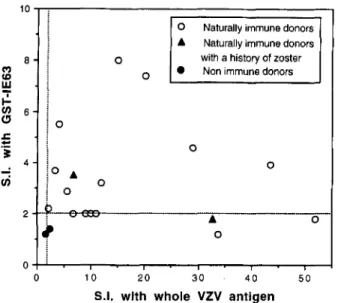
PBMC from I9 healthy adults were assayed for T cell prolifer- ation in response to stimulation with GST-IE63-purified protein and whole VZV Ag. The mean SI with whole VZV Ag was 15.5 2
3.3 SE, with values between 2.1 and 52. The mean SI after stim-
ulation by GST-IE63 was 3.4 2 0.48 SE (range 1.8-8). These responses were significantly higher than those observed in two nonimmune subjects who had an SI of 1.2 and 1.4 to IE63, and 1.6 and 2.3 to whole VZV Ag. T cell proliferation to GST-IE63 was compared with responses to VZV Ag. All 17 immune donors had an SI 2 2 to VZV Ag (Fig. 1). Among immune individuals, 10 of 17 (59%) had T cell proliferation to GST-IE63, with SI of 2.3 to 8 (Fig. 1). No correlation was found between the magnitude of the SI elicited by GST-IE63 and VZV Ag. The other 7 immune donors
had an SI 5 2 to GST-IE63, and an SI to VZV Ag ranging from 5.4 to 11. One of them had a history of multiple herpes zoster erup- tions. However, all of these donors with a low SI to GST-IE63 also had a significant response to GST alone (data not shown).
2804 CELLULAR IMMUNITY T O VZV IE63
Naturally immune donors Naturally immune donors Non immune donors 0 with a history of zoster
0
0
0
" I - I . I - I - , . I
0 10 20 30 40 5 0
S.I. with whole VZV antigen
FIGURE 1. Comparison of proliferation induced by stimulation of T cells after stimulation with IE63 protein and whole VZV Ag. SI was calculated for each Ag concentration as the ratio between the mean cpm for CST-IE63-purified protein or VZV Ag and CST-purified protein or control Ag, respectively. The highest SI obtained for each donor has been plotted in this graph.
Table I. y-IFN and IL-4 production b y PBMC stimulated with /E63
T Cell Proliferation in Response to IFN-y CST-IE63 iS.1.) Donor IL-4 ipdml) P F h I 1 2.9 750 0 2 2.2 1400 0 3 7.4 500 0 4 8.0 625 0 5 2.0 175 0 6 2 .0 50 0 Cytokine assays
Cytokine release by PBMC stimulated with IE63 was evaluated in seven VZV immune subjects. PBMC were stimulated with GST- IE63 or GST, and supernatants were tested for IL-2, IL-4, and y-IFN on days 2, 4, and 6. The results were expressed as the difference between the concentration of the cytokine detected after stimula- tion by GST-IE63 or GST alone. IFN-y was produced in signifi- cant amounts (175-1400 pg/ml) by T cells stimulated with IE63, with maximum concentrations on day 4, while IL-2 and IL-4 were not detected. IFN-y concentrations were lowest in two immune donors who had low T cell proliferation to GST-IE63 (Table I).
Cytotoxicity assays
IE63-specific CTL responses were detected in limiting dilution assays of T cells obtained from 10 VZV-immune donors and com- pared with CTL recognition of IE62 protein. Cytotoxicity assays were performed using autologous targets infected with vaccinia- IE63, vaccinia-IE62, or vaccinia-control; uninfected LCL were also included as control targets. Lysis of virus-specific targets was considered specific only when it was at least 10% higher than lysis of control targets (uninfected or infected with vaccinia-control).
When unseparated T cell populations from four donors were tested, the lysis of targets expressing IE63 was significantly higher than lysis of uninfected or control targets, and specific lysis of IE62 targets was also detectable, as illustrated in a representative
40 30 20 10 0 1 : 3 1:l 3: 1 15:l 30:l Effector:Target ratio
FIGURE 2. Cytotoxic T recognition oflE63 protein. T cells from an immune donor were stimulated for 2 wk with VZV Ag and used as effector cells in cytotoxicity assays against autologous target cells. Ef- fector and target cells were incubated at different ratios for 4 h. The percentage of specific lysis was calculated as I(mean experimental release) - (mean spontaneous release)l/[(mean maximal release) - (mean spontaneous release)] X 100.
assay (Fig. 2). The specific lysis of targets expressing IE63 ranged from 27 to 43% and was equivalent to the range of 25 to 48% lysis of targets expressing IE62. The mean effector frequency for T cells that recognized IE63 was 1 :3 1,000 ? 16,000 SE and is compared with 1:44,500 ? 22,500 SE for IE62; these values were not sig- nificantly different ( p = 0.8).
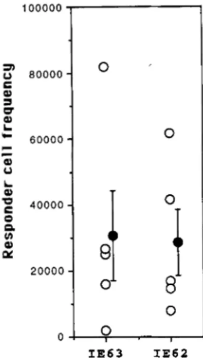
Limiting dilution cultures were also prepared using purified populations of CD4+ and CD8+ T lymphocytes as effectors. Both populations lysed IE63 and IE62 targets. The RCF of CD8+ cells recognizing IE63 was 1:30,500 ? 13,700 SE compared with 1 :
28,500 i- 10,100 SE with IE62 (Fig. 3). These frequencies were not statistically different ( p = 0.9), indicating that both viral pro- teins were recognized with the same efficiency by CD8+ cells. The frequency of T cells lysing IE63 targets was equivalent in CD4+ and CD8+ populations of effector cells: the mean CD4+ etfector cells were 1 :31,450 2 7,500 SE, which is the same as the fre- quency of CD8+ cells specific for IE63 ( p = 0.97).
Discussion
The essential role of VZV immunity in preserving viral latency is indicated by the low frequency of viral reactivation among the normal adult population compared with the increased frequency and the severity of the reactivated disease in immunocompromised patients (8-1 I ) . In healthy individuals, virus-specific T lympho- cytes persist for many years after primary infection, as detected in vitro by T lymphoproliferation assays (for review, see Ref. 17): all
subjects with evidence of VZV infection have T lymphocyte rec- ognition of IE62, the major virion tegument protein, and of the viral envelope glycoproteins, including gE (gpI), the most abun- dant glycoprotein (1 2). Long-term immunity to VZV is also char- acterized by the presence of CD4+ and CD8+ lymphocytes that have cytotoxic function with specificity for IE62 as well as the glycoproteins (1 8, 19).
The product of ORF63 is expressed very early in the productive infectious cycle in VZV-infected cells (4) and is also a component of purified virions (6). The IE63 protein is the only viral protein
The Journal of Immunology 2805 100000 a C al 3 w a L u 80000 I c 60000 Q u L a 40000 0 0. v)
u
a 20000 0 0 0 00
I IE63 IE62FIGURE 3. Precursor frequencies of CD8' T cells. Recognition of IE63 by CTL from nine donors was compared with lysis of targets ex- pressing IE62 under limiting dilution conditions. Responder cell fre- quencies (reported on y-axis as a reciprocal value) were estimated by computer analysis of limiting dilution data. Each open circle represents the data for one donor; the mean RCF 2 SE is indicated by the black circle with error bars.
that has been detected to date in latently infected ganglia, as was demonstrated for the first time in a rat model of VZV infection (4). In this model, inoculation of VZV-infected cells into the footpad of adult rats leads to the establishment of a latent infection in the lumbar ganglia, corresponding to the nerve pathways of the limb. During this quiescent phase, some other immediate early and early genes are transcribed, but no gene products are detected except the ORF63 protein, which is present in 50 to 80% of neurons. Late genes encoding glycoproteins, such as gE, are not transcribed or translated. Expression of the immediate early protein IE63 during latency has been confirmed in human ganglia, although fewer neu- rons are positive than in the rat ( 5 ) . The IE63 protein is also ex- pressed very early in the cutaneous phase of VZV reactivation (7). It is detected in keratinocytes in early, nonvesicular VZV lesions, in which gE is not detectable (4). While the contribution of IE63 to the pathogenesis of VZV latency and reactivation is not defined, its production during latency and early cutaneous infection indi- cates that it may be an important target for immune surveillance. Although it has been shown that IE62 and the glycoproteins (12, 18, 19) can be processed and presented to elicit humoral and cel- lular immunity, the immunogenicity of the IE63 protein has not been evaluated.
Our experiments demonstrate that most donors who have mem-
ory T cells specific for unfractionated VZV Ags also have memory immunity against IE63, as detected by proliferation of T cells stim- ulated with a GST-IE63 fusion protein. The analysis of cytokine production after in vitro stimulation of T cells with IE63 protein showed production of y-IFN, but not of IL-4, which is similar to the pattern observed with T cells stimulatkd in standard PBMC culture with unfractionated VZV Ags. Since supernatants were harvested at days 2, 4, and 6 in our experiments, it is likely that IL-2 was produced earlier or directly taken up by the TCR (19). It is also possible that a direct use of IL-4 as a growth factor makes
it hardly detectable. Moreover, Zhang et al. (20) found that under limiting dilution cultures, 85% of T cells that proliferated in re- sponse to VZV produced y-IFN, in contrast to IL-4, which was produced only by 10% of T cells. These results suggest that the number of cells producing IL-4 is much lower than the cells pro- ducing IFN, and that this difference can make difficult the detection of IL-4 i n bulk cultures. Although our observations have to be confirmed by a search for other cytokines, they indicate that the T cell stimulation by IE63 induces primarily a T h l response, as shown by the production of y-IFN.
The processing of IE63 by APC also results in a cytotoxic re- sponse that is mediated by CD4+ and CD8' T cells. As we have observed with IE62 and VZV glycoproteins, IE63 protein has amino acid sequence that can be presented by the MHC class I as well as class IT pathway (21). The lysis of autologous targets ex- pressing IE63 was qualitatively comparable with results obtained with cells expressing IE62, the highly immunogenic major tegu- ment protein of VZV. The frequencies of memory cytotoxic T cell recognizing IE62 and IE63 were 1:44,500 2 1:22,500 SE and 1 :3 1,000 -C 1 : 16,000 SE, respectively, which is not significantly different.
We conclude that IE63, the only VZV protein detected to date in latently infected cells, is highly immunogenic and elicits a long- term immune response after natural infection, which is comparable with immune response to other major VZV immunogenic proteins such as IE62 and gE (12). It is likely that its continued expression during the quiescent stage of VZV infection maintains humoral and cellular immunity to IE63 protein. It will be of interest to characterize the immune response to IE63 in the elderly and im- munocompromised patients who have a high incidence of herpes zoster to examine possible correlations between the risk of viral reactivation and diminished responses to IE63. Enhancing the im- mune response to IE63 protein may be an important strategy to prevent VZV reactivation from latency. If so, IE63 protein could be a suitable candidate for incorporation into a therapeutic VZV vaccine for administration to population at high risk for herpes zoster.
References
1. Weller. T. H., and H. M. Witton. 1958. The etlologic agents of varicella and herpes zoster: isolation, propagation and culture character~stics in vitro. J. Exp.
Med. 10X:X43.
2. Croen, K. D., I. M. Ostrove. L. J. Dragovic, and S. E. Straus. 1988. Patterns of gene expression and sites of latency in human nerve ganglia are different for varicella-zoster and herpes simplex viruses. P m c . Nut/. Acud. Sci. USA X5:977.?.
3. Cohrs, R. J . . M. Barbour, and D. H. Gilden. 1996. Varicella-zoster virus (VZV) genes 21, 29. 62, and 63 in a cDNA library enriched for VZV RNA. J. Virol. transcription during latency in human ganglia: detection of transcripts mapping to
70:2789.
4. Debrus. S., C. Sadzot-Delvaux, A. J. Nikkels. J. Piette. and 6. Rentier. 1995.
Varicella-zoster virus gene 63 encodes an immediate early protein abundantly expressed during latency. J. Virol. 69:3240.
5 . Mahalingam. R.. M. Wellish. R. Cohrs, S. Dehrus. J Piette, B. Rentier. and D. H. Gllden. 1996. Expression of protein encoded by varicella-zoster virus open reading frame 63 in latently infected human ganglionic neurons. Proc. Nuti.
Acud. Sei. USA 93:2122.
6. Kinchington, P. R., D. Bookey, and S . E. Turse. 1995. The transcrlptional reg- ulatory proteins encoded by varicella-zoster virus open reading frames (ORFs) 4
and 63, but not ORF61, are associated with purified virus particles. J . Vim[. 69:4274.
7. Nikkels. A. F., S. Debrus, C. Sadzot-Delvaux, J. Piette, B. Rentier, and G. E. Pikrard. 1995. Immunochemical identification of varicella-zoster virus gene 63- encoded protein (IE63) and late (gE) protein on smears and cutaneous biopsies: impl~cat~ons for diagnostic use. J. Med. V i m l . 47.342.
8. Hayward, A. R,, and M. Herderger. 1987. Lymphocyte responses 10 varicella-
zoster virus In the elderly. J. Ciin. Itnrnunoi. 7t174.
9. Berger. R., G. Florent, and M. Just. 1981. Decrease of the lymphoproliferative response to varicella-zoster virus antigen in the aged. Infect. Irnmun. 32:24. 10. Arvin, A . M., R. 6. Pollard, L. E. Ramussen, and T. C Merigan. 1980. Cellular
2806 CELLULAR IMMUNITY TO VZV IE63 I I . Sorensen, 0. S., S. Haahrs, A. Moller-Larson, and K. Wildenhoff. 1980. Cell
mediated and humoral immunity to herpes viruses during and after herpes zoster
12. Arvin, A. M., E. Kinney-Thomas, K. Shriver, C. Grose, C. M. Koropchak, E.
infections. Infecr. Immun. 29:369.
Scranton, A. E. Wittek, and P. S. Diaz. 1986. Immunity to varicella-zoster viral glycoproteins, gpl (90/58) and gpIIl (gpl l8), and to a non glycosylated protein,
13. Arvin, A. M. 1994. The T-lymphocyte response to varicella-zoster virus and it$
p170. J. Immunol. 137:1346.
14. Kinchington, P. R., I. K. Houghland, A. M. Arvin, W. T. Ruyechan, and J. Hay. relevance to vaccine development. Rev. Med. Virol. 4:161.
66359.
1992. Varicella-zoster virus IE62 protein is a major virion component. J. Virol. 15. Diaz, P. S., S. Smith, E. Hunter, and A . M. Arvin. 1989. T lymphocyte cytotox-
icity with natural varicella-zoster virus infection and after immunization with live attenuated varicella vaccine. J. Immunol. 142:636.
16. Fazekas de St. Groth, S. 1982. The evaluation of limiting dilution assays. J. Im-
rnrmol. Methods 49:1211.
17. Arvin, A. M., J . F. Moffat, and R. Redman. 1996. Varicella-zoster virus: aspects of pathogenesis and host response to natural Infection and varicella vaccine. Adv.
18. Giller, R. H., S. Winistorfer, and C. Grose. 1989. Cellular and humoral immunity Virus Res. 46.263.
to varicella-zoster virus glycoproteins in immune and susceptible human subjects. J. Infect. Dis. 160:919.
19. Sharp, M.. K. Terada, A. Wilson, S. Nader, P. E. Kinchington. W. T. Ruyechan, J. Hay, and A . M . Arvin. 1992. Kinetics and viral protein specificity of the cytotoxic T lymphocyte response in healthy adults immunized with live attenu- ated varicella vaccine. J. Infect. Dis. 165:852.
20. Zhang, Y., M. Cosyns, M. I. Levin, and A. R. Hayward. 1994. Cytokine pro-
duction in varicella zoster vlrus-stimulated limiting dilution lymphocyte cultures. 21. Arvin, A. M., M. Sharp, S. Smlth. C. M. Koropchak, P. S. Diaz, P. Kinchington.
Clin. E,rp. Immunol. YX:12X.
W. Ruyechan, and J . Hay. 1991. Equivalent recognition of a varicella-zoster virus either CD4+ or CD8+ phenotype. J. Immunol. 146257.