Der p 1 is the primary activator of Der p 3, Der p 6 and Der p 9 the
proteolytic allergens produced by the house dust mite
Dermatophagoides pteronyssinus
Julie Herman
a, Nicolas Thelen
b, Nicolas Smargiasso
c, Anne-Catherine Mailleux
d, André Luxen
e, Marie Cloes
b,
Edwin De Pauw
c, Andy Chevigné
f, Moreno Galleni
a,⁎
, Marie-Eve Dumez
a,fa
Macromolécules Biologiques, Centre for Protein Engineering, University of Liège, 4000 Liège, Belgium
b
Unit of Cell and Tissue Biology, GIGA-Neurosciences, University of Liège, 4000 Liège, Belgium
c
Mass Spectrometry Laboratory, GIGA-R, Department of Chemistry, University of Liège, 4000 Liège, Belgium
dEarth and Life Institute, Université catholique de Louvain, 1348 Louvain-la-Neuve, Belgium eCentre de Recherche du Cyclotron, University of Liège, 4000 Liège, Belgium
f
Laboratory of Retrovirology, CRP-Santé, 1526 Luxembourg, Luxembourg
a b s t r a c t
a r t i c l e i n f o
Article history: Received 17 June 2013
Received in revised form 30 October 2013 Accepted 18 November 2013
Available online 27 November 2013
Keywords: Mite allergen Protease Digestion
Dermatophagoides pteronyssinus
Background: The enzymatic activity of the four proteases found in the house dust mite Dermatophagoides pteronyssinus is involved in the pathogenesis of allergy. Our aim was to elucidate the activation cascade of their corresponding precursor forms and particularly to highlight the interconnection between proteases during this cascade.
Methods: The cleavage of the four peptides corresponding to the mite zymogen activation sites was studied on the basis of the Förster Resonance Energy Transfer method. The proDer p 6 zymogen was then produced in Pichia pastoris to elucidate its activation mechanism by mite proteases, especially Der p 1. The role of the propeptide in the inhibition of the enzymatic activity of Der p 6 was also examined. Finally, the Der p 1 and Der p 6 proteases were localised via immunolocalisation in D. pteronyssinus.
Results: All peptides were specifically cleaved by Der p 1, such as proDer p 6. The propeptide of proDer p 6 inhibited the proteolytic activity of Der p 6, but once cleaved, it was degraded by the protease. The Der p 1 and Der p 6 proteases were both localised to the midgut of the mite.
Conclusions: Der p 1 in either its recombinant form or in the natural context of house dust mite extracts specifically cleaves all zymogens, thus establishing its role as a major activator of both mite cysteine and serine proteases. General significance: This finding suggests that Der p 1 may be valuable target against mites.
© 2013 Elsevier B.V. All rights reserved.
1. Introduction
The house dust mite Dermatophagoides pteronyssinus is the primary source of allergens that are involved in the development of allergy leading to asthma, rhinitis or atopic dermatitis. Today, more than 20 groups of house dust mite (HDM) allergens have been identified, among which groups 1, 3, 6 and 9 correspond to secreted proteases[1,2]. The proteolytic
activity of these allergens has been shown to enhance the development and the chronicity of allergy either by cleaving receptors, leading to the activation of innate or adaptive immunity, or by increasing the perme-ability of the airway epithelium, promoting allergen penetration[3–6]. These proteases are expected to be involved in biological processes in the mite. Indeed, Der p 1[7,8]and Der f 3, the homolog of Der p 3 in Dermatophagoides farinae[9], are localised to the gut of mites and to HDM faecal pellets, suggesting that they play a role in digestion. Similar to other digestive proteases, Der p 1, Der p 3, Der p 6 and Der p 9 are synthesised as inactive precursors to control enzyme activation. These zymogens are termed proDer p 1, proDer p 3, proDer p 6 and proDer p 9. Investigating the activation mechanisms of these zymogens is crucial for understanding the pattern of allergenic proteases in the environment.
The activation of Der p 1, a cysteine protease from the papain family, has been widely characterised. The Der p 1 propeptide (80 amino acids) plays an essential role in zymogen folding (as an intramolecular chaper-one) and in the inhibition of its own enzymatic activity. ProDer p 1 was
Abbreviations: AMC, 7-amido-4-methylcoumarin; AMg, anterior midgut; DAPI, 4′,6-DiAmidino-2-PhenylIndole; E-64,L-trans-epoxysuccinyl-leucylamido (4-guanidino) butane; FP, faecal pellet; FRET, Förster Resonance Energy Transfer; HDM(e), house dust mite (extracts); Hg, hindgut; nDer p 6, natural Der p 6; pDp6, proDer p 6; PMg, posterior midgut; rDer p 1, recombinant Der p 1; rDer p 6, recombinant Der p 6; SBTI, soybean trypsin inhibitor
⁎ Corresponding author at: 3, Allée de la chimie, 4000 Liège, Belgium. Tel.: +32 43663549.
E-mail address:mgalleni@ulg.ac.be(M. Galleni).
0304-4165/$– see front matter © 2013 Elsevier B.V. All rights reserved.
http://dx.doi.org/10.1016/j.bbagen.2013.11.017
Contents lists available atScienceDirect
Biochimica et Biophysica Acta
j o u r n a l h o m e p a g e : w w w . e l s e v i e r . c o m / l o c a t e / b b a g e ntrypsin cleavage site is replaced by a glycine[12].
Comparison of the propeptide C-terminal sequences from the mite zymogens highlights the similarity of sequences that correspond to the cleavage specificity by Der p 1 (Table 1)[13]. These observations suggest the existence of a particular activation cascade for the mite proteases. Unlike the major HDM allergens Der p 1 and Der p 2, Der p 6 is poorly characterised. This allergen was first identified as a chymotrypsin-like protease (provisionally named DP5) by Yasueda et al.[14]and is expected to account for a large portion of the measur-able proteolytic activity in HDM extracts[11]. While the contribution of serine proteases to anti-mite IgE binding is considered low[15], the global prevalence of anti-group 6 IgE varies between less than 10% to 65%[14,16–19], most probably because of the variability observed in the preparation of natural allergens [20–22]. Therefore, obtaining recombinant allergen Der p 6 for RAST determination and diagnosis would be of interest to accurately evaluate the involvement of the protease in mite allergy.
Our objectives were to elucidate the specific activation cascade of the four mite zymogens based on the Förster Resonance Energy Transfer (FRET) method. In parallel, we confirmed the activation mechanism of the proDer p 6 zymogen for which recombinant protein is not yet available, by producing it in the yeast Pichia pastoris. The roles of the propeptide in inhibiting the enzymatic activity of Der p 6 and in its activation were elucidated. Finally, we determined the immune co-localisation of the proteases in the gut of the mite D. pteronyssinus.
2. Methods 2.1. Chemicals
N-Succinyl-Ala-Ala-Pro-Phe-7-amido-4-methylcoumarin (AMC) acetate, Boc-Gln-Ala-Arg-AMC acetate salts, Boc-Gln-Ala-Arg-p-nitroanilide (pNA), Ala-Ala-Pro-Phe-pNA and N-succinyl-Ala-Ala-Pro-Leu-pNA were purchased from Bachem (Babendorf, Switzerland). The cysteine protease inhibitorL -trans-epoxysuccinyl-leucylamido (4-guanidino) butane (E-64) and soybean trypsin inhibitor (SBTI) were obtained from Sigma (Saint Louis, USA). A polyclonal anti-proDer p 6 antibody (rabbit) was manufactured by Eurogentec (Liège,
by mites D. pteronyssinus culture extracts were obtained as previously described[11].
2.3. Solid-phase synthesis of S1–S9 peptides
The S1, S3, S6 and S9 peptides correspond to the proDer p 1, proDer p 3, proDer p 6 and proDer p 9 zymogen activation sites, respectively (Fig. 1 in supplemental data). N-α-Fmoc amino acids were purchased from Iris Biotech GmbH (Marktredwitz, Germany), and 2,4-Dinitrophenyl (Dnp)-L-Proline and Dnp-L-Valine were obtained from Sigma (Saint Louis, USA), and Dnp-L-Leucine came from TCI Europe (Zwijndrecht, Belgium). The Glu and Thr side chains were protected with a t-butyl derivative; Lys was protected with an N-epsilon-allyl-oxycarbonyl group; and Asn was protected with a trityl group. The substrates were prepared following a solid-phase peptide synthesis strategy on a PS3 automated peptide synthesiser (Protein Technologies, Inc., Tucson, AZ) using N-(7-Methoxycoumarin-4-yl)acetyl (AMC)-N ′-Fmoc-ethylenediamine-MBP-AM resin (Novabiochem, Darmstadt, Germany), as previously described[11]. The side chain-protecting groups were removed, and the peptides, which were cleaved from the resin using trifluoroacetic acid (TFA)/anisole/water (6 ml; 10:1:1) during a 3-hour incubation at room temperature, were precipitated with diethyl ether.
2.4. Characterisation of S1–S9 peptides
The purity of each peptide (50μl, 500 μM) was assessed via analyt-ical HPLC performed on a Nucleodur C18 column (250 × 4.6 mm, 5μm, Macherey-Nagel, Düren, Germany). The HPLC chain consisted of a pump (Waters 600) and a UV detector (PDA Waters 996, Hilford, Massachusetts, USA). The absorbances at 323 nm and 374 nm, corre-sponding to the AMC and Dnp groups, respectively, were constantly recorded. The solvents consisted of aqueous and 0.1% acetonitrile (V/V) TFA solutions. Elution was carried out for 30 min at aflow rate of 1 ml/min using a linear gradient from 0 to 75% acetonitrile/TFA. Each peptide (100μl, 50 μM) was then characterised via electrospray ionisation mass spectrometry (ESI-MS, TSQ 7000 Thermoquest Finnigan, positive ionisation, 4.5 kV).
2.5. FRET measurements
Unglycosylated recombinant mature Der p 1 (rDer p 1) was obtained from proDer p 1 produced in P. pastoris as described previously[10,11]. The reaction mixtures contained 8μM substrate (S1–S9) and rDer p 1 (0.04μM for S3 and S6 and 2.32 μM for S1 and S9) or HDM extract (1 mg/ml) in polybuffer (mixture of 50 mM Tris, phosphate, citrate, acetate and KCl containing 1 mM DTT and 1 mM EDTA, adjusted to pH 7.4), with or without the addition of the cysteine protease inhibitor L-trans-epoxysuccinyl-leucylamido (4-guanidino)butane (E-64, 100μM) or the serine protease inhibitor soybean trypsin inhibitor (SBTI, 10μM). The time course of hydrolysis was monitored for 300 s in an LS 50 B fluorimeter (PerkinElmer Life Sciences) with excitation and emission wavelengths of 328 and 393 nm, respectively.
Table 1
Activation sites of Dermatophagoides pteronyssinus zymogens. The underlined residues correspond to the specificity of Der p 1 determined by[13]. The arrow indicates the putative cleavage site of the propeptide. The bold sequences correspond to the S1, S3, S6 and S9 peptides.
Zymogen C-terminal sequence of propeptide N-terminal sequence of protease proDer p 1 proDer p 3 proDer p 6 proDer p 9
2.6. Identification of the S1–S9 peptide cleavage sites
The peptides (500μM) were incubated with rDer p 1 (2.32 μM), rDer p 3 (0.23μM) or HDM extract (1 mg/ml) in 50 mM polybuffer at pH 7.4, with or without the addition of either E-64 (100μM) or SBTI (10μM), for 16 h at 25 °C. The fragments obtained after the various treatments were separated via analytical HPLC as described above and analysed via ESI-MS.
2.7. Kinetic parameters of rDer p 1 and HDM extracts
To control the proportionality of the FRET response to substrate utilisation and to avoid the innerfilter effect[24], calibration was per-formed by completely hydrolysing the four substrates at concentrations ranging from 0.5 to 40μM using rDer p 1 (0.04 μM for S3 and S6 and 2.32μM for S1 and S9). Hydrolysis was monitored via fluorimetry as described above for 180 min at 25 °C. FRET remained linear up to an 8μM product concentration. For determination of the kinetic parame-ters of rDer p 1 (0.04 or 2.32μM) and the HDM extract (1 mg/ml), the hydrolysis rates were measured for increasing substrate concentrations (0 to 8μM), and the data were analysed according to the Henri– Michaelis–Menten equation.
2.8. Expression of recombinant proDer p 6 in P. pastoris
The proDer p 6 gene sequence, optimised for expression in P. pastoris, was synthesised by GeneArt (Life Technologies, Paisley, UK). The gene was inserted in a pPIC9K (Invitrogen, Paisley, UK) vector that had been previously digested with the SnaBI and AvrII restriction enzymes (Promega, Madison, USA).
The P. pastoris SMD1168 strain (Invitrogen, Paisley, UK) was trans-formed through electroporation with the expression vector, which had been linearised by the PmeI restriction enzyme. Transformants from pPIC9K carrying the HIS4 gene were grown in histidine-deficient medium (RDB). Clones with multiple integrated copies were selected using increasing geneticin concentrations (0.25 to 1 mg/ml, Invitrogen, Paisley, UK).
For expression of proDer p 6, P. pastoris was grown in buffered glycerol complete medium (BMGY) for 24 h at 28 °C to an A600value of ~ 25. The culture was then centrifuged for 15 min at 10 000 g, and the pellet was resuspended in buffered methanol complex medium (BMMY) (0.5% methanol) for 2 days at 28 °C to measure protein expression. The culture was subsequently centrifuged for 15 min at 10000 g, and the supernatant was conserved at−20 °C.
2.9. Purification of recombinant proDer p 6 and of recombinant and native Der p 6
The yeast culture supernatant wasfiltered through a 0.45 μm filter (Millipore, Darmstadt, Germany) and dialysed overnight at 4 °C against 20 mM Tris–HCl pH 7.5 (buffer A). The solution was then stirred with 200 ml of QXL streamline matrix (GE Healthcare, Saint Louis, USA) that had been equilibrated with buffer A at 4 °C. The matrix was washed with buffer A and packed into a column (GE Healthcare, Saint Louis, USA). Bound proteins were eluted with buffer A containing 0.5 M NaCl. After SDS-PAGE analysis, fractions containing proDer p 6 were pooled and dialysed overnight at 4 °C against buffer A. The protein was then precipitated by the addition of 60% ammonium sulphate at 4 °C. After centrifugation, the pellet was resuspended in 3 ml of buffer A and dialysed overnight at 4 °C. The solution was then loaded onto a Sephacryl S-100 HR column (120 ml) that had been previously equili-brated with buffer A, and proteins were eluted according to molecular weight. The fractions containing proDer p 6 were pooled and stored at −20 °C.
The recombinant proDer p 1 protease (N-64A, N52A), fused with a C-terminal six-histidine sequence, was produced in P. pastoris and
then purified and activated to a mature protease as described previously
[10]. This enzyme was incubated with MagneHis™ Ni-Particles (Promega, Madison, USA) overnight at 37 °C, and the particles with coated Der p 1 were washed with PBS containing 1 mM DTT and EDTA prior to incuba-tion with proDer p 6 for 8 h at 37 °C. After the activaincuba-tion of proDer p 6, the particles were removed, and pure rDer p 6 was stored at−20 °C.
To purify natural Der p 6, HMD extracts (4 g suspended in 40 ml of PBS) were dialysed overnight at 4 °C against 20 mM Tris–HCl at pH 7.5 (buffer A). The solution was then loaded onto a Q-HP HiTrap column (GE Healthcare, Saint Louis, USA) equilibrated with buffer A. Bound proteins were eluted with buffer A containing 1 M NaCl. The fractions containing Der p 6 were pooled and loaded onto a Benzamidine Sepharose 4 Fast Flow column (GE Healthcare, Saint Louis, USA) that had been equilibrated with buffer A containing 0.5 M NaCl. The proteins were eluted with 100 mM sodium acetate, pH 4, and then dialysed overnight at 4 °C against buffer A. The solution was next loaded onto a heparin column (GE Healthcare, Saint Louis, USA) that had been equilibrated with buffer A. Elution was performed with buffer A containing 1 M NaCl. The concentration of the obtained pro-teins was estimated using the BCA assay (Thermo Scientific, Rockford, USA). SDS-PAGE analysis and protease activities were assayed at each stage of purification as previously described[11].
2.10. N-terminal sequencing
The proDer p 6 and rDer p 6 proteins were sequenced in an Applied Biosystems 476A protein sequencer based on Edman degradation. The samples were analysed via SDS-PAGE, followed by electrophoretic transfer to a polyvinylidene difluoride membrane (Merck Millipore, Darmstadt, Germany).
2.11. In vitro activation of recombinant proDer p 6
The proDer p 6 zymogen (3.5μM) was incubated at 37 °C for 90 min with either rDer p 1 (0.1μM) or HDM extract (20 μg/ml) in 50 mM polybuffer, pH 7.4, containing 1 mM DTT and 1 mM EDTA. The E64 inhibitor (100μM), SBTI (10 μM) and/or the Der p 1 propeptide (10μM, produced and purified as previously described[10]) were also added to the HDM extract prior to incubation with proDer p 6. The sam-ples were analysed via SDS-PAGE, and Der p 6 activity was measured in 20 mM Tris, pH 7, using the Ala-Ala-Pro-Phe-AMC substrate (200μM). The time course of hydrolysis was monitored for 300 s in an LS 50 B fluorimeter (PerkinElmer Life Sciences) with excitation and emission wavelengths of 360 and 480 nm, respectively. The kinetics of hydrolysis were reported as the time course of AMC (μM) release, using an AMC (Sigma, Saint Louis, USA) standard curve with concentrations ranging from 0 to 15μM.
2.12. Der p 6 enzymatic activity measurements and determination of kinetic parameters
For the determination of natural or recombinant Der p 6 (80 nM) kinetic parameters, the rate of the hydrolysis of increasing AAPF-AMC (Bachem, Babendorf, Switzerland) concentrations (0 to 400μM) was measured as described above, and the data were analysed according to the Henri–Michaelis–Menten equation.
The optimum pH for rDer p 6 (0.1μM) was also determined in 50 mM polybuffer at pH 4 to 11 with the same substrate (400μM). 2.13. Determination of the role of the propeptide
2.13.1. Inhibition of recombinant Der p 6 by its propeptide
The peptide corresponding to the full-length propeptide of proDer p 6 (34 amino acids), was purchased from JPT Peptide Technologies GmbH (Berlin, Germany). rDer p 6 activity (50 nM) was measured at 37 °C in 20 mM Tris pH 7 and using increasing concentrations of the
μM) and the inhibition constant (μM), respectively.
2.13.2. Mass spectrometry analysis of proDer p 6 propeptide degradation The proDer p 6 prosequence (3μM) was incubated with either rDer p 6 or rDer p 1 (50 nM) in 20 mM Tris, pH 7, and aliquots were collected at different times. The reaction was stopped with 0.1% TFA, and the pep-tides were desalted using Ziptips C18 (Millipore, Darmstadt, Germany). The peptides were then suspended in 50/50 H2O/Acetonitrile (v/v), 0.1% formic acid solution. ESI-MS analyses were performed on a Q-TOF Ultima Global mass spectrometer (Micromass, now Waters, Manchester, UK). The ESI source was operated in positive ion mode at a capillary voltage of 3 kV. The source block temperature was 100 °C, the desolvation gas (N2) temperature was 200 °C and the source pressure was 2.8 mbar. The RF Lens 1 voltage was set to 100 V. All mass spectra were acquired over the 90–3000 m/z range and were processed with MassLynx 4.1 software (Waters).
2.14. Immunolocalisation of Der p 1 and Der p 6
Mites were collected with a brush under a Nikon SMZ645 stereomi-croscope (Nikon Belux, Brussels, Belgium) and then incubated over-night in a solution of 2% formaldehyde and 0.05% Triton X-100 in PBS at 4 °C. After three washes in PBS, the mites were incubated in a 30% sucrose solution for 1 h at room temperature and then oriented in 7.5% agar, 15% sucrose. The mites were embedded in OCT compound (VWR International, Poole, England), and incubated at−70 °C in an isopentane bath. Cryosections (14–16 μm thick) were obtained by means of a cryostat (Microm HM 560, Prosan) and collected on glass slides. For immunolabeling, cryosections were dried at 30 °C before beingfixed in 2% formaldehyde. After three washes in PBS, the sections were soaked for 30 min at 37 °C in 10% normal goat serum (Sigma, St Louis, USA) in PBS, and after immersed with 1:750 anti-Der p 6 and 1:1000 anti-Der p 1 (5H8, Indoor biotechnologies, USA) in PBS with 5% goat serum. They were washed in PBS, and immersed for 35 min at 37 °C in a PBS solution containing 1:250 goat anti-rabbit Alexa Fluor 594 and goat anti-mouse Alexa Fluor 488 (Molecular Probes, Leiden, The Netherlands). The unbound antibodies were removed by three washing cycles in PBS. The slides were immersed in a solution of DAPI (1:50000 dilution; 4′,6-DiAmidino-2-PhenyIndole; Sigma Aldrich) and washed with PBS. Finally, the sections were mounted with Citifluor AF1 (Laborimpex, Brussels, Belgium). Negative control sections were realized by using normal goat serum instead of the primary antibodies. The sections were observed under an Olympus IX71 confocal micro-scope (Olympus America Inc.).
3. Results and discussion
The allergenic mite proteases Der p 1, Der p 3, Der p 6 and Der p 9 are synthesised as inactive zymogens that then mature into active enzymes through specific cleavage of their propeptides. As recombinant allergens are increasingly used for the diagnosis and treatment of allergy[21,25], understanding the activation mechanisms of these zymogens repre-sents an essential step for obtaining proteins with enzymatic and folding properties similar to those of their natural counterparts. In the future, elucidation of such activation cascades should provide insight into the critical enzyme(s) involved in the onset of the activation
of the synthesised substrates (Fig. 1). The KMvalues for S3 and S6 hydro-lysis by rDer p 1 were similar (b9 μM), but we unfortunately could not measure the KMvalues for S1 and S9 because of the quenching of fluorescence at high peptide concentrations (N10 μM). The catalytic efficiency (kcat/KM) of rDer p 1 for S1 (220 ± 20 mM−1min−1) was ~ 7- and 4-fold lower than that observed for S3 (1610 ± 20 mM−1min−1) and S6 (860 ± 170 mM−1min−1), respectively (Table S1, supplemental data). This observation might be explained by the fact that intermolecular activation of proDer p 3 (S3) and proDer p 6 (S6) by small amounts of Der p 1 was likely to require a higher specificity than the auto-activation of proDer p 1 (S1). Finally, the ~ 7-fold lower kcat/KM value of rDer p 1 determined for S9 (31 ± 5 mM−1min−1) might be due in part to the absence of a proline at the P4position and, especially, to the presence of a leucine instead of an alanine at the P2position.
This result is consistent with the demonstrated specificity of Der p 1
[13], which exhibits a preference for small hydrophobic residues in the P2 position (Schechter and Berger's nomenclature [26]). We also showed via mass spectrometry that all of the tested peptides were hydrolysed in presence of HDM extracts (Table 2and Fig. S2, supple-mental data), and the obtained KM values were similar to those observed using pure rDer p 1 (Table S1, supplemental data). The respec-tive contributions of the cysteine and serine proteases present in HDM extracts to the hydrolysis of S1, S3, S6 and S9 were then determined by performing the reactions in the presence of cysteine (E-64) or serine (SBTI) protease inhibitors. We found that the hydrolysis rates for the four substrates were similar with or without the addition of SBTI (Fig. 1, black and white bars), whereas they were strongly reduced in the presence of E-64 (Fig. 1, grey bars). Nearly complete inhibition by E-64 was observed when using purified rDer p 1.
These experiments demonstrate the high activity of Der p 1 in the cleavage of S1, S3, S6 and S9 and suggest that Der p 1 plays a major role in the activation of the four proDer p zymogens.
3.2. Identification of cleavage sites
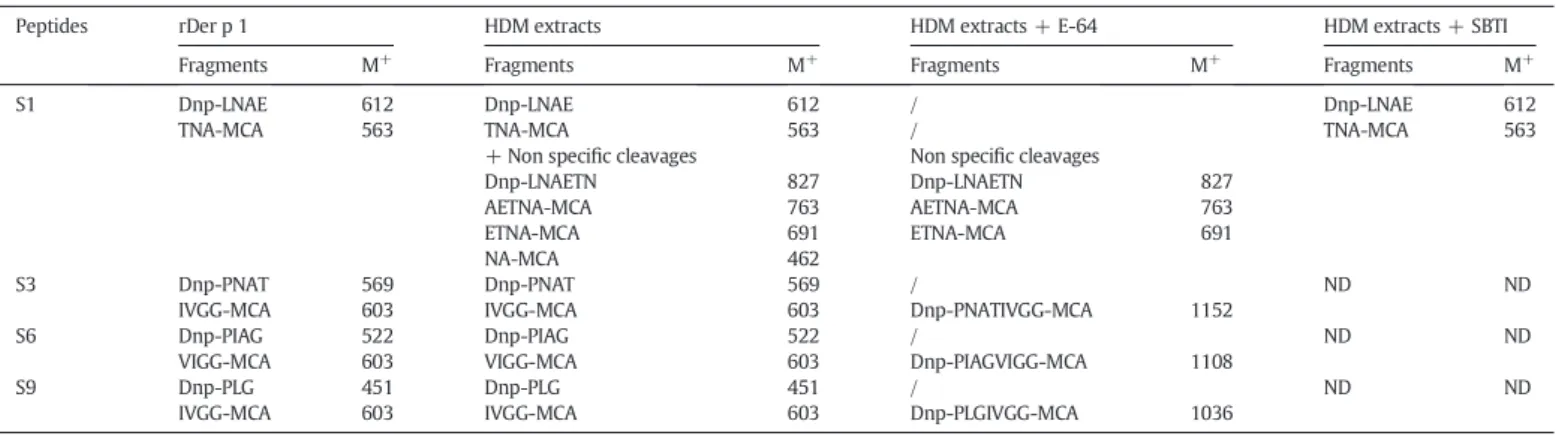
We showed that Der p 1 (either recombinant or in HDM extracts) cleaved the presented peptides at the expected sites, i.e., between the P1 and P′1 positions of the activation sites of the zymogens (Table 2
and Fig. S2, supplemental data). This can be explained by the presence of similar residues at the C-terminal end of the zymogen prosequences, which correspond to the specificity of Der p 1[13]. These observations validated our methodology by confirming the role of Der p 1 as an auto-catalytic enzyme and in proDer p 3 maturation. More interestingly, this study broadens the established activating role of the major allergen Der p 1 to include all mite protease zymogens. Indeed, the addition of E-64 inhibited the specific cleavage of all of the peptides at the predicted processing sites.
Nevertheless, a small amount of non-specific hydrolysis of S1 was observed when using HDM extracts (Table 2). We showed that the presence of additional peptide fragments was due to the activity of serine proteases, as this phenomenon was totally abolished in the pres-ence of SBTI. These data suggest that whereas Der p 1 is involved in the activation of zymogens, the serine proteases are likely to be responsible for non-specific protease degradations.
3.3. Expression, purification and activation of recombinant proDer p 6 We determined the activation mechanism for the recombinant proDer p 6 zymogen. ProDer p 6 was expressed in the culture superna-tant of P. pastoris after 36 h of methanol induction, and 1 mg of the zymogen was then purified from 1 l of culture. SDS-PAGE analysis (Fig. 2A, lane pDp6) indicated that proDer p 6 was present in two pre-dominant forms with apparent molecular masses of 30 and ~ 28 kDa. A sequence analysis indicated that these forms corresponded to the entire zymogen (molecular mass of 28 834 Da) and to proDer p 6 with the 25 N-terminal residues deleted (proDer p 6′: 25872 Da), respectively. Recombinant proDer p 6 was then incubated in the presence of either rDer p 1 or HDM extracts. SDS-PAGE analysis was performed
(Fig. 2A and B), and the time course of chymotrypsin activity was analysed using AAPF-AMC (Fig. 2G). The two bands (~30 and 28 kDa) corresponding to proDer p 6 and proDer p 6′ disappeared over time after the addition of rDer p 1, and a new band corresponding to rDer p 6 appeared (~ 25 kDa) (Fig. 2B). The expected N-terminal sequence (V35IGGDQ) of rDer p 6 was confirmed. The incubation of proDer p 6 with rDer p 1 or HDM extracts for 1 h led to fully activated Der p 6. The loss of the propeptide was related to an increase in chymotrypsin activity. The maximum activity was reached after 60 min (Fig. 2G).
The same results were obtained following the addition of HDM extracts previously incubated with SBTI (~20 kDa) to proDer p 6, sug-gesting that serine proteases do not participate in the activation of the zymogen (Fig. 2C). In contrast, the addition of the cysteine protease
Table 2
Identification of cleavage sites of the S1–S9 peptides after hydrolysis by rDer p 1 or HDM extracts. Analytical HPLC analysis of peptides after their incubation with rDer p 1 or HDM extracts in presence or absence of inhibitors was performed followed by the characterization of the different peaks obtained by ESI-MS. The serine protease inhibitor is the Soybean-Trypsin Inhibitor (SBTI) and the cysteine protease inhibitor is the E-64. M+indicates the molecular mass of the peptide. ND is not determined.
Peptides rDer p 1 HDM extracts HDM extracts + E-64 HDM extracts + SBTI Fragments M+
Fragments M+
Fragments M+
Fragments M+
S1 Dnp-LNAE 612 Dnp-LNAE 612 / Dnp-LNAE 612
TNA-MCA 563 TNA-MCA 563 / TNA-MCA 563
+ Non specific cleavages Non specific cleavages
Dnp-LNAETN 827 Dnp-LNAETN 827
AETNA-MCA 763 AETNA-MCA 763
ETNA-MCA 691 ETNA-MCA 691
NA-MCA 462
S3 Dnp-PNAT 569 Dnp-PNAT 569 / ND ND
IVGG-MCA 603 IVGG-MCA 603 Dnp-PNATIVGG-MCA 1152
S6 Dnp-PIAG 522 Dnp-PIAG 522 / ND ND
VIGG-MCA 603 VIGG-MCA 603 Dnp-PIAGVIGG-MCA 1108
S9 Dnp-PLG 451 Dnp-PLG 451 / ND ND
IVGG-MCA 603 IVGG-MCA 603 Dnp-PLGIVGG-MCA 1036
Fig. 1. Cleavage of S1, S3, S6 and S9 substrates. Enzymatic activity of rDer p 1 (0.04μM or 2.32 μM) or HDM extracts (1 mg/ml) measured with 8 μM (A) S1, (B) S2, (C) S3 and (D) S9 in absence (black) or presence of 100μM E-64 (grey) or 10 μM SBTI (white).
inhibitor (E-64) to rDer p 1 or HDM extracts totally abolished rDer p 6 production (data not shown andFig. 2D, respectively) and no chymo-trypsin activity was detected (Fig. 2G). Moreover, in the absence of DTT, which enhances the activity of cysteine proteases, very weak acti-vation of proDer p 6 by rDer p 1 or HDM extracts was observed (Fig. S3, supplemental data).
To further confirm the role of cysteine protease Der p 1 in the activa-tion mechanism of proDer p 6, HDM extracts were pre-incubated with the highly specific inhibitory propeptide of Der p 1 (KD7 nM[10]). The rate of activation of proDer p 6 in the presence of such HDM extracts was drastically delayed (Fig. 2E and G). It is noteworthy that the gain of proDer p 6 activation after 30 min is due to the degradation of the propeptide of Der p 1 by serine proteases of HDM extracts[27]. Indeed, the addition of SBTI abolished this phenomenon and no activation could be observed (Fig. 2F), whereas SBTI alone was not able to inhibit the proDer p 6 activation by HDM extracts (Fig. 2C). Therefore, in the absence of Der p 1 activity, no auto-activation of proDer p 6 could be observed. The intermolecular activation of proDer p 6 confirmed the results of the FRET experiments and the crucial role of the cysteine pro-tease Der p 1.
After activation of proDer p 6 with rDer p 1, rDer p 6 was purified. The catalytic parameters of natural and recombinant Der p 6 were measured using AAPF-AMC and were found to be similar. Indeed, the KMvalues were (120 ± 10)μM and (40 ± 10) μM and the kcatvalues were (0.38 ± 0.01) s−1and (0.38 ± 0.01) s−1for nDer p 6 and rDer
p 6, respectively. The optimum pH was determined to be 7, although the protease displayed high activity (more than 90%) between pH 6 and 10 (Fig. S4, supplemental data).
3.4. Determination of the role of the propeptide
The mechanism underlying the maturation of the inactive zymogen proDer p 6 mediated by Der p 1 corresponds to loss of the propeptide and an increase in chymotrypsin activity, suggesting that the propeptide covalently linked to the protease acts as an inhibitor of Der p 6. To explore the role of this propeptide in the inhibition of Der p 6 when it is free in solution, the prosequence was synthesised. We measured the inhibition constant of the propeptide for rDer p 6 using AAPF-AMC as a reporter substrate in either the absence or presence of increasing con-centrations of the peptide. The Hanes plot (Fig. S5 supplemental data) obtained using different concentrations of inhibitor shows that the propeptide acts as a competitive inhibitor, with an inhibition constant of (11 ± 2)μM.
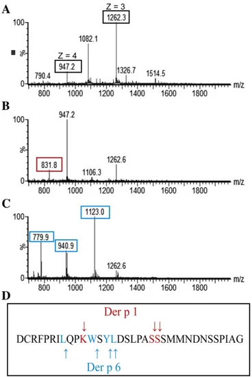
Nevertheless, after a 1 minute incubation of the propeptide with rDer p 6, an increase of chymotrypsin activity was observed, indicating that the prosequence could be processed by the protease. To confirm this hypothesis, a mass spectrometry analysis of the propeptide in the presence of rDer p 6 was performed.Fig. 3shows the results of ESI-MS analysis of the propeptide alone (Fig. 3A) and after a 5 minute incuba-tion with rDer p 6 (Fig. 3C). This analysis was also performed in the
Fig. 2. Inter-molecular activation of proDer p 6 by rDer p 1 or HDM extracts in presence or absence of SBTI, E-64 and/or Der p 1 propeptide. SDS-PAGE analysis: proDer p 6 (pDp6) was incubated at 37 °C in polybuffer with 1 mM DTT/EDTA pH 7.4 with (A) HDMe; (B) rDer p 1; (C) HDMe with SBTI, (D) with E64, (E) with propeptide of Der p 1 or (F) with propeptide and SBTI.(E) rDer p 6 activity for each activation time (0 to 90 min) in presence of rDer p 1 (blue diamond shapes), HDM extracts (black circles), HDMe with propeptide (dark grey circles) and HDM extracts with E-64 (light grey circles).
presence of rDer p 1 (Fig. 3B). The peptide was found to be cleaved in several places by the two proteases (Fig. 3D). However, the sites of hydrolysis are different for the two proteases and correspond to their respective cleavage specificities. Indeed, rDer p 6 and rDer p 1 cleaved the peptide after aromatic (tryptophan and leucine) or positively charged (arginine, lysine and serine) residues, respectively.
Here, we demonstrated that the propeptide inhibits the proteolytic activity of Der p 6. However, whereas this inhibition is very stable when the propeptide is covalently linked to the protease, the free propeptide in solution is quickly cleaved by Der p 6 as well as Der p 1, rendering it unable to inhibit the neo-formed Der p 6.
3.5. Immunolocalisation of Der p 1 and Der p 6 in D. pteronyssinus We determined the localisation of Der p 6 and Der p 1 in D. pteronyssinus. Examination of coronal sections under a confocal microscope revealed that Der p 6 immunoreactivity is only present in the digestive tract and, more specifically, in the hindgut (Hg) following the terminology of Brody et al.[28]. Indeed, Der p 6 was observed in cells in the absence of ingested material and in the gut lumen (red fluorescence,Fig. 4A and B). Similar localisation patterns have previously been observed for Der f 3, the Der p 3 homologous found in D. farinae
[9].
Protease Der p 1 was localised in lining cells of the anterior midgut (AMg, yellowfluorescence,Fig. 4C) and in the lumen of the posterior midgut (PMg,Fig. 4D), as previously reported by Thomas et al.[7].
Fig. 4C and D shows that Der p 1 and Der p 6 are co-localised in the hindgut (Hg, orangefluorescence).
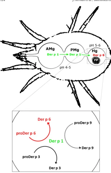
Previous observations of the digestive tract of the mite showed that cells lining the anterior midgut, where pH is comprised between 4 and 5, would be gradually detached in the gut lumen and would then be taken in the faecal pellets in the posterior midgut and the hindgut, where pH reaches 6[28–30]. It is thus likely that the auto-activation of proDer p 1 occurring at acidic pH proceeds in the anterior midgut, thereby providing active protease Der p 1. As the Der p 1 displays a high activity at pH 6[10], it may activate the proDer p 3 and proDer p 6 zymogens, and most probably proDer p 9, in the hindgut where the serine proteases and Der p 1 were co-localised (Fig. 5)[7,9].
Fig. 3. Mass spectrometry analysis. ESI-QTOF analysis of (A) the peptide corresponding to the propeptide of proDer p 6 (3μM) and the peptide after 5 minute incubation with 50 nM (B) rDer p 1 or (C) rDer p 6; (D) represents the propeptide sequence and the arrow indicates the cleavage sites of rDer p 1 (red) and rDer p 6 (blue).
Fig. 4. Immunolocalisation of Der p 1 and Der p 6 in longitudinal sections of Dermatophagoides pteronyssinus body. (A–B) Localisation of Der p 6 (red fluorescence) in the posterior midgut and (C–D) co-localisation of Der p 1 (green fluorescence) in the anterior to posterior gut with Der p 6 only detected in posterior part. Mite bodies are in blue fluorescence with DAPI. The scale bar is 50μM. AMg, anterior midgut; Hg, hindgut; PMg, posterior midgut.
4. Conclusion
Ourfindings demonstrate that Der p 1 is essential for the activation of the mite protease zymogens proDer p 1, proDer p 3, proDer p 6 and probably proDer p 9 (Fig. 5). Moreover, a similar proteolytic cascade might also occur in D. farinae and Euroglyphus maynei, as the propeptides of the corresponding zymogens in these species exhibit a high degree of similarity with those of D. pteronyssinus (Table S2, supplemental data).
According to the critical role of proteolytically active HDM protease allergens in the initiation and in the chronicity of the allergic response, the elucidation of the proDer p 6 maturation process will provide a source of recombinant highly pure and fully active Der p 6 allergen offering new opportunities to deeply characterise its proteolytic speci-ficity and to better define its roles in the activation of innate immune pathways.
Supplementary data to this article can be found online athttp://dx. doi.org/10.1016/j.bbagen.2013.11.017.
Acknowledgements
We thank Professor Jean-Marie Frère and Professor André Matagne for helpful comments and critical reading of the manuscript, Philippe
[5] L. Gough, et al., Proteolytic activity of the house dust mite allergen Der p 1 enhances allergenicity in a mouse inhalation model, Clin. Exp. Allergy 33 (8) (2003) 1159–1163.
[6] H. Wan, et al., The transmembrane protein occludin of epithelial tight junctions is a functional target for serine peptidases from faecal pellets of Dermatophagoides pteronyssinus, Clin. Exp. Allergy 31 (2) (2001) 279–294.
[7] B. Thomas, P. Heap, F. Carswell, Ultrastructural localization of the allergen Der p I in the gut of the house dust mite Dermatophagoides pteronyssinus, Int. Arch. Allergy Appl. Immunol. 94 (1–4) (1991) 365–367.
[8] E.R. Tovey, B.A. Baldo, Localization of antigens and allergens in thin sections of the house dust mite, Dermatophagoides pteronyssinus (Acari: Pyroglyphidae), J. Med. Entomol. 27 (3) (1990) 368–376.
[9] Z.K. Zhan, et al., Monoclonal antibodies against recombinant Der f 3 reveal localiza-tion of Der f 3 in the gut and faecal pellets of Dermatophagoides farinae, Exp. Appl. Acarol. 52 (1) (2010) 63–71.
[10]A. Chevigne, et al., Relationship between propeptide pH unfolding and inhibitory ability during ProDer p 1 activation mechanism, J. Mol. Biol. 374 (1) (2007) 170–185.
[11] M.E. Dumez, et al., Activation mechanism of recombinant Der p 3 allergen zymogen: contribution of cysteine protease Der p 1 and effect of propeptide glycosylation, J. Biol. Chem. 283 (45) (2008) 30606–30617.
[12] B.J. Bennett, W.R. Thomas, Cloning and sequencing of the group 6 allergen of Dermatophagoides pteronyssinus, Clin. Exp. Allergy 26 (10) (1996) 1150–1154.
[13] J. Harris, et al., Activity profile of dust mite allergen extract using substrate libraries and functional proteomic microarrays, Chem. Biol. 11 (10) (2004) 1361–1372.
[14] H. Yasueda, et al., Allergens from Dermatophagoides mites with chymotryptic activity, Clin. Exp. Allergy 23 (5) (1993) 384–390.
[15]B.J. Hales, et al., IgE and IgG anti-house dust mite specificities in allergic disease, J. Allergy Clin. Immunol. 118 (2) (2006) 361–367.
[16] A.C. Angus, S.T. Ong, F.T. Chew, Sequence tag catalogs of dust mite-expressed genomes: utility in allergen and acarologic studies, Am. J. Pharmacogenomics 4 (6) (2004) 357–369.
[17] Y. Cui, et al., Molecular cloning, expression, sequence analyses of dust mite allergen Der f 6 and its IgE-binding reactivity with mite allergic asthma patients in southeast China, Mol. Biol. Rep. 39 (2) (2012) 961–968.
[18]S. Kawamoto, et al., Cloning and expression of Der f 6, a serine protease allergen from the house dust mite, Dermatophagoides farinae, Biochim. Biophys. Acta 1454 (2) (1999) 201–207.
[19]C. King, et al., The isolation and characterization of a novel collagenolytic serine protease allergen (Der p 9) from the dust mite Dermatophagoides pteronyssinus, J. Allergy Clin. Immunol. 98 (4) (1996) 739–747.
[20] G.A. Stewart, et al., The group III allergen from the house dust mite Dermatophagoides pteronyssinus is a trypsin-like enzyme, Immunology 75 (1) (1992) 29–35.
[21]W.R. Thomas, The advent of recombinant allergens and allergen cloning, J. Allergy Clin. Immunol. 127 (4) (2011) 855–859.
[22] T. Takai, et al., Recombinant Der p 1 and Der f 1 exhibit cysteine protease activity but no serine protease activity, Biochem. Biophys. Res. Commun. 328 (4) (2005) 944–952.
[23] A.-C. Mailleux, et al., Collective migration in house dust mites, Ethology 116 (2010) 11.
[24] S. Grahn, D. Ullmann, H. Jakubke, Design and synthesis offluorogenic trypsin peptide substrates based on resonance energy transfer, Anal. Biochem. 265 (2) (1998) 225–231.
[25]M. Egger, et al., Development of recombinant allergens for diagnosis and therapy, Front. Biosci. (Elite Ed.) 1 (2009) 77–90.
[26] I. Schechter, A. Berger, On the size of the active site in proteases. I. Papain, Biochem. Biophys. Res. Commun. 27 (2) (1967) 157–162.
[27] A. Chevigne, et al., Comparative study of mature and zymogen mite cysteine protease stability and pH unfolding, Biochim. Biophys. Acta 1800 (9) (2010) 937–945.
[28] M.J. Colloff, Dust Mites, Springer and CSIRO, 2009. 583.
[29]Y.Y. Zhang, X. Sun, Z.G. Liu, Morphology and three-dimensional reconstruction of the digestive system of Dermatophagoides farinae, Int. Arch. Allergy Immunol. 146 (3) (2008) 219–226.
[30] T. Erban, J. Hubert, Determination of pH in regions of the midguts of acaridid mites, J. Insect Sci. 10 (2010) 42.
Fig. 5. Proteases activation cascade in the digestive tract of the mite Dermatophagoides pteronyssinus. The box represents the activation cascade in the hindgut. AMg, anterior midgut; FP, faecal pellet; Hg, hindgut; PMg, posterior midgut.