i
Université de Montréal
Functional Analysis of Nuclear β-Adrenergic Receptors in the Myocardium
par George Vaniotis Département de Biochimie
Faculté de Médecine
Thèse présentée à la Faculté des études supérieures en vue de l’obtention du grade de
Philosophiae Doctor (Ph.D.) en biochimie
30 Septembre 2012
ii
Université de Montréal Faculté des etudes supérieures
Cette these intitulée:
Functional Analysis of Nuclear β-Adrenergic Receptors in the Myocardium
présentée par: George Vaniotis
a été évalué par un jury compose des personnes suivantes:
Dr. Nikolaus Heveker President-rapporteur
Dr. Bruce Gordon Allen Directeur de recherche Dr. Terence E. Hébert Co-directeur de recherché Dr. Denis deBlois Membre du jury Dr. Timothy O’Connell Examinateur externe Dr. Nathalie Arbour Représentant du doyen
iii
ABSTRACT
Recently several G protein-coupled receptors (GPCRs) have been shown to localize to intracellular membranes, in particular the nuclear membrane. As such, we sought to determine if the β-adrenergic receptor (βAR) subtypes and their associated signalling machinery are functionally localized to nuclear membranes. We demonstrated the presence of β1AR and the β3AR, but not the β2AR, in adult ventricular myocyte nuclei by western blotting, confocal microscopy and functional assays. Downstream signalling partners such as GαS, Gαi and adenylyl cyclase II and V/VI were also present. Nuclear-localized βARs were functional with respect to ligand binding and effector activation. In isolated nuclei, the non-selective βAR agonist isoproterenol (ISO) and the β3AR-selective ligand BRL37344, but not the β1AR-selective xamoterol, stimulated transcription initiation in a pertussis toxin (PTX)-sensitive manner. In contrast, stimulation of type B endothelin receptors (ETB), another GPCR family shown to be present on the nuclear membrane, decreased de novo RNA synthesis. To investigate the signalling pathway(s) involved in GPCR-mediated regulation of RNA synthesis, nuclei were isolated from intact adult rat hearts and treated with receptor agonists in the presence or absence of inhibitors of the PI3K/PKB and mitogen-activated protein kinase (MAPK) pathways. Components of p38, JNK, and ERK1/2 MAPK cascades as well as PKB were detected in nuclear preparations. Inhibition of PKB with triciribine converted the activation of the βAR from stimulatory to inhibitory with regards to transcription initiation. Analysis by qPCR indicated isoproterenol treatment increased 18S rRNA but decreased NFκB mRNA. In contrast, ET-1 had no effect on 18S rRNA expression. Further investigation using pathway-specific PCR arrays revealed that isoproterenol treatment also reduced the expression of several other genes involved in the activation of NFκB and that ERK1/2 and PKB inhibitors attenuated this effect. Subsequent genome-wide microarray analysis has revealed that nuclear βAR and ETB regulated a host of genes in an overlapping but distinct manner. Moreover, both ET-1 and ISO produced an L-NAME-sensitive increase in NO production in isolated cardiac nuclei. These observations were confirmed in intact cardiomyocytes using novel caged
iv
analogues of ISO and ET-1 and the cell-permable NO-sensitive fluorescent dye, DAF-2 DA. Briefly, both ET-1 and isoproterenol increased NO production, and this increase was prevented upon preincubation with L-NAME. Moreover, the ability of isoproterenol to increase transcription initiation in isolated nuclei was blocked by L-NAME or the PKG inhibitor KT5823, indicating the NO-GC-PKG pathway is involved in the regulation of gene expression by nuclear βARs. Hence, we have shown that βARs and ETRs in the nuclear membrane activate distinct signalling pathways, resulting in different effects on gene transcription and thus represent potentially important targets for drug development.
Keywords: β-adrenergic receptors, endothelin receptors, G protein coupled receptors (GPCRs), heart, nuclear membrane, transcription, nitric oxide.
v
RÉSUMÉ
Récemment plusieurs récepteurs couplés aux protéines G (RCPGs) ont été caractérisés au niveau des membranes intracellulaires, dont la membrane nucléaire. Notre objectif était de déterminer si les sous-types de récepteurs β-adrénergiques (βAR) et leurs machineries de signalisation étaient fonctionnels et localisés à la membrane nucléaire des cardiomyocytes. Nous avons démontré la présence des β1AR et β3AR, mais pas du β2AR à la membrane nucléaire de myocytes ventriculaires adultes par immunobuvardage, par microscopie confocale, et par des essais fonctionnels. De plus, certains partenaires de signalisation comme les protéines GαS, Gαi, l’adénylate cyclase II, et V/VI y étaient également localisés. Les sous-types de βAR nucléaires étaient fonctionnels puisqu'ils pouvaient lier leurs ligands et activer leurs effecteurs. En utilisant des noyaux isolés, nous avons observé que l'agoniste non-sélectif isoprotérénol (ISO), et que le BRL37344, un ligand sélectif du β3AR, stimulaient l'initiation de la synthèse de l’ARN, contrairement à l'agoniste sélectif du β1AR, le xamotérol. Cette synthèse était abolie par la toxine pertussique (PTX). Cependant, la stimulation des récepteurs nucléaires de type B de l’endothéline (ETB) causaient une réduction de l'initiation de la synthèse d’ARN. Les voies de signalisations impliquées dans la régulation de la synthèse d’ARN par les RCPGs ont ensuite été étudiées en utilisant des noyaux isolés stimulés par des agonistes en présence ou absence de différents inhibiteurs des voies MAP Kinases (proteines kinases activées par mitogènes) et de la voie PI3K/PKB. Les protéines impliquées dans les voies de signalisation de p38, JNK, ERK MAP Kinase et PKB étaient présents dans les noyaux isolés. L'inhibition de PKB par la triciribine, inhibait la synthèse d’ARN. Nous avons ensuite pu mettre en évidence par qPCR que la stimulation par l’ISO entrainait une augmentation du niveau d'ARNr 18S ainsi qu’une diminution de l'expression d’ARNm de NFκB. En contraste, l’ET-1 n’avait aucun effet sur le niveau d’expression de l’ARNr 18S. Nous avons ensuite montré que la stimulation par l’ISO réduisait l’expression de plusieurs gènes impliqués dans l'activation de NFκB, tandis que l’inhibition de ERK1/2 et PKB renversait cet effet. Un microarray global nous a ensuite permis de démontrer que les βARs et les ETRs nucléaires régulaient un grand nombre de gènes distincts. Finalement, les βARs et ETRs nucléaires augmentaient
vi
aussi une production de NO de noyaux isolés, ce qui pouvait être inhibée par le L-NAME. Ces résultats ont été confirmés dans des cardiomyocytes intacts en utilisant des analogues cagés et perméables d’ISO et de l'ET-1: l'augmentation de NO nucléaire détectée par DAF2-DA, causée par l'ET-1 et l'ISO, pouvait être prévenue par le L-NAME. Finalement, l’augmentation de l’initiation de la transcription induite par l'ISO était aussi bloquée par le L-NAME ou par un inbitheur de PKG, le KT5823, suggérant que la voie NO-GC-PKG est impliquée dans la régulation de la transcription par les βAR. En conclusion, les βARs et les ETRs nucléaires utilisent des voies de signalisation différentes et exercent ainsi des effets distincts sur l’expression des gènes cardiaques. Ils représentent donc une avenue intéressante pour le développement de drogues pharmacologiques.
Mots Clés: récepteur β-adrénergiques, récepteurs aux endothelin, récepteurs couplé aux protéines G (RCPG), coeur, membrane nucléaire, transcription, oxide nitrique.
vii
TABLE OF CONTENTS
Section Page
Abstract ... iii
Résumé ... v
Table of Contents ... vii
List of Tables ... x
List of Figures ... xi
List of Abbreviations ... xiii
Acknowledgements ……...………...………. xvi
Chapter 1: Introduction ... 1
I) The Cardiovascular System ………...…...… 1
II) G Protein-Coupled Receptors ... 3
a) GPCR Classes ... 3 b) Heterotrimeric G Proteins ... 5 c) GPCR Ligands ... 10 d) GPCR Regulation ... 12 e) GPCR Oligomerization …………...……….… 14 f) Intracellular GPCRs ... 15
III) Adrenergic Receptors ... 17
IV) Endothelin Receptors ... 23
V) Nitric Oxide ... 25
VI) Gene Transcription ... 27
Figure Legends ... 31
Figures …... 33
Tables ... 38
Hypothesis and Objectives ... 42
viii
Chapter 3: Functional β-adrenergic receptor signalling on nuclear membranes in adult
rat and mouse ventricular cardiomyocytes ... 50
Abstract ... 52
1. Introduction ... 53
2. Materials and Methods ... 54
3. Results ... 58
4. Discussion ... 62
Figure Legends ... 66
References ... 68
Figures ... 75
Chapter 4: Nuclear β-adrenergic receptors modulate gene expression in adult rat heart 82 Abstract ... 84
1. Introduction ... 85
2. Materials and Methods ... 88
3. Results ... 92 4. Discussion ... 98 5. Conclusions ... 101 Figure Legends ... 103 References ... 106 Figures ... 111 Tables ... 121
Chapter 5: Regulation of cardiac nitric oxide signalling by nuclear β-adrenergic and endothelin receptors... 122
Abstract ... 125
1. Introduction ... 127
2. Materials and Methods ... 129
3. Results ... 137
ix Conclusions ... 151 Figure Legends ... 154 References ... 161 Figures ... 168 Tables ... 179
Chapter 6: General Discussion ..………..………...……… 180
Conclusions ……….………...……….. 199
x
LIST OF TABLES
Table Page Chapter 1
1.1 Nuclear GPCRs in various organ systems ... 38 1.2 Intracellular ligands involved in cardiac physiology ... 41 Chapter 4
4.1 Primers used during real-time qPCR …………...……….……...….… 121 4.2 RT2 profiler PCR array analysis of the NFκB signalling pathway in isolated cardiac nuclei ... 121 Chapter 5
5.1 Primers used during real-time qPCR ... 179 General Discussion
6.1 Genes regulated by ETR or βAR classified according to biological function 192 6.2 Predicted upstream regulators ... 193 6.3 Pathology …... 194
xi
LIST OF FIGURES
Figure Page Chapter 1
1.1 Cross section of the heart ... 33
1.2 Cardiac action potential ... 34
1.3 Diversity of G protein-coupled receptor signalling ……...… 35
1.4 Possible orientation of nuclear βAR signalling complexes ... 36
1.5 Subtype-specific signalling pathways of cardiac βAR isoforms ... 37
Chapter 3 3.1 125I-CYP binding in enriched nuclear fractions …………...….………. 75
3.2 Detection of βAR isoforms in crude and enriched nuclei using western blotting ………. 76
3.3 Detection of βAR isoforms in isolated adult rat ventricular cardiomyocytes using immunocytochemistry ………....……..………..………..………...…. 77
3.4 Colocalization of βAR isoforms in isolated adult mouse ventricular cardiomyocytes with a marker of the nuclear membrane ……… 78
3.5 Detection of βAR signalling partners in isolated adult rat left ventricular cardiomyocytes using immunocytochemistry ………..……..……....…….. 79
3.6 Functional coupling of β1AR but not β3AR to adenylyl cyclase stimulation in enriched nuclear preparations ………..……….…………...… 80
3.7 Functional coupling of β3AR but not β1AR to initiation of de novo transcription in enriched nuclear preparations ……..…………...……...…….……..…………... 81
Chapter 4 4.1 Effect of MAP kinase inhibition on transcription initiation …...………. 111
4.2 Effect of PI3K/PKB inhibition on transcription initiation ……..…….…..….. 112
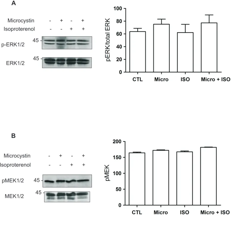
4.3 Effect of microcystin on transcription initiation ………..………….... 113
4.4 Phosphorylation of ERK1/2 and MEK1/2 in isolated nuclei following treatment with isoproterenol ………..………..……..………. 114
xii
4.6 Phosphorylation of PKB following treatment with isoproterenol ………..…. 116
4.7 βAR stimulation alters the abundance of 18S rRNA and NFκB mRNA ……. 117
4.8 Effects of α-amanitin on isoproterenol-mediated transcription initiation …... 118
4.9 Detection of NFκB following treatment with isoproterenol …..….………... 119
4.10 Signalling diagram of post-nuclear βAR and ETR activation ………... 120
Chapter 5 5.1 Synthesis of a caged isoproterenol analog …... 168
5.2 Effect of various agonists on NO production …... 169
5.3 Effect of pertussis toxin and triciribine on NO production …... 170
5.4 Effect of NOS pathway inhibition on isoproterenol and ET-1 induced transcription initiation …... 171
5.5 βARs regulate rRNA and mRNA transcription in intact myocytes …... 172
5.6 Activation of βAR signalling by a caged isoproterenol analogue, ZCS-1-67, in HEK 293 cells ... 173
5.7 Measurement of NO production in live cardiomyocytes by confocal microscopy …... 174
5.S1 Regulation of NO production ... 175
5.S2 Identification of NOS isoforms in isolated nuclei ... 176
5.S3 Effect of KT5823 on NO production ... 177
xiii
LIST OF ABBREVIATIONS
αAR α-adrenergic receptor βAR β-adrenergic receptor AC adenylyl cyclase
AKAP A kinase anchoring protein AMP adenosine monophosphate ATF-2 activating transcription factor 2 ATP adenosine triphosphate
BAD Bcl-2 associated death promoter
CAMKII Ca2+/calmodulin-dependent protein kinase II cAMP cyclic adenosine monophosphate
cGMP cyclic guanosine monophosphate CRE cAMP response elements
CREB cAMP response element binding DAG diacylglycerol
DNA deoxyribonucleic acid
ECE endothelin converting enzyme eNOS endothelial NOS
EPAC exchange proteins activated by cAMP ER endoplasmic reticulum
ERK1/2 extracellular signal-regulated kinase 1/2 ET-1 endothelin 1
ETA endothelin receptor A ETB endothelin receptor B ETR endothelin receptor
GAP GTPase activating protein GC guanylyl cyclase
GDP guanosine diphosphate
GEF guanosine nucleotide exchange factor
xiv GMP guanosine monophosphate GPCR G protein-coupled receptor GRK G protein-coupled receptor kinase GSK-3 glycogen synthase kinase-3 GTP guanosine triphosphate HAT histone acetyltransferase HDAC histone deacetylase IκB inhibitor of κB IKK Iκ-B kinase iNOS inducible NOS IP3 inositol trisphosphate ISO isoproterenol
JNK c-Jun N-terminal kinases
MAPK mitogen-activated protein kinase mGLU metabotropic glutamate
mRNA messenger RNA
mTOR mammalian target of rapamycin
NFκB nuclear factor kappa-light-chain-enhancer of activated B-cells NO nitric oxide
NOS nitric oxide synthase nNOS neuronal NOS PDE phosphodiesterase
PDK1 phosphoinositide-dependent protein kinase 1 PI3K phosphoinositide 3-kinase
PIP2 phosphotidylinositol 4, 5-bisphosphate PKA protein kinase A
PKB protein kinase B PKC protein kinase C PKG protein kinase G PLA phospholipase A PLC phospholipase C
xv POL I RNA polymerase I
POL II RNA polymerase II POL III RNA polymerase III PP2 protein phosphatase 2 PTX pertussis toxin
qPCR quantitative real-time polymerase chain reaction RGS regulator of G protein signalling
RNA ribonucleic acid
ROCK Rho-associated protein kinase ROS reactive oxygen species rRNA ribosomal RNA
SERCA sarco/endoplasmic reticulum calcium ATPase sGC soluble guanylate cyclase
xvi
ACKNOWLEDGEMENTS
I would like to thank both of my PhD supervisors, Dr. Bruce G. Allen and Dr. Terry Hébert, without whom this work would not have been possible. Their guidance and knowledge was a great asset during my training, and thanks in no small part to their seemingly endless patience I was able to learn a great deal about signalling in heart, and about research as a whole.
I would also like to thank Dr. Nikolaus Heveker and Dr. André Tremblay, who both served on my thesis committee, for their time and advice.
I wish to also thank the funding agencies that made this work possible, in particular the CIHR and the Montreal Heart Institute.
Finally, I would like to thank all the members of both Dr. Allen’s and Dr. Hébert’s labs for all their help during my PhD training, in particular Dr. Clemence Merlen and Dr. Dharmendra Dingar whose friendship and support made it a true pleasure to go to the lab every day.
1
CHAPTER 1: INTRODUCTION
I) The Cardiovascular System
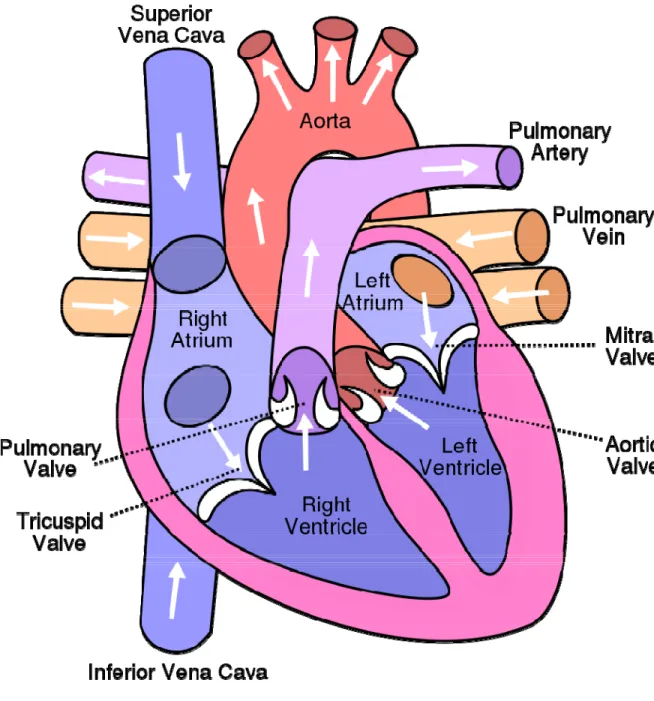
The heart is responsible for pumping blood throughout the body to ensure adequate delivery of oxygen and nutrients as wells as removal of metabolic waste products. The myocardium is composed primarily of cardiac muscle cells, endothelial cells and fibroblasts. The mammalian heart is divided into four chambers, consisting of a right and left ventricle and atrium (Figure 1). The right atrium and ventricle are responsible for pumping blood to the respiratory system, while the left atrium and ventricle pump blood to the remainder of the body. The heart acts as a dual pump with the atria contracting first followed in quick succession by the ventricles. The contraction of the atria ensures the proper filling of the ventricle, termed the preload. This contraction is achieved by a change in the membrane potential from its resting negative state to a positive one. This depolarization causes the contraction of the cardiomyocyte and is the basis for the electrical activity that controls the heart. The original depolarization, which controls the basic automaticity of the heart occurs in the sinoatrial node, located in the right atrium, and quickly spreads throughout the atria and into the ventricles. The discharge rate of the sinoatrial node determines the heart rate. Depolarization is a result of the opening of voltage-gated sodium channels and generates an action potential in the plasma membrane of muscle cells. This in turn increases the cell’s cytosolic calcium concentration, causing a contraction. The cardiac action potential consists of five phases (Figure 2). The primary phase is the resting membrane potential of the cardiomyocyte, prior to stimulation. Upon stimulation, the action potential enters the rapid depolarization phase, due to the opening of the fast sodium channels. The following phase involves the inactivation of the sodium channels, followed by the plateau phase, resulting from a sustained balance between inward movement of calcium through L-type calcium channels and outward movement of potassium through slow delayed rectifier potassium channels. The final phase is the rapid repolarization phase, where the calcium channels close, and other potassium channels open, mainly the rapid delayed rectifier potassium channels. This final phase allows for a return to the resting membrane potential. During the depolarization of the
2
cardiomyocyte, voltage-gated calcium channels located in the T tubules open, allowing calcium to enter the cell. This small increase in calcium facilitates calcium binding to ryanodine-sensitive calcium channels (RyR) on the sarcoplasmic reticulum (SR) and this leads to opening of the RyR allowing a large efflux of calcium from the SR into the cytosol. This release of calcium from the SR is termed calcium-induced calcium release and is responsible for cardiac contraction. Contraction occurs as a result of binding of calcium to the regulatory protein troponin, which initiates cross-bridge formation between actin and myosin. Following cardiomyocyte contraction is relaxation, which allows the cardiomyocytes to return to their basal state, where they can undergo another contraction/relaxation cycle. Cardiomyocyte relaxation involves the reuptake of calcium into the sarcoplasmic reticulum, and is mediated primarily by sarco/endoplasmic reticulum calcium ATPase (SERCA). Calsequestrin, a calcium-binding protein located within the lumen of the SR, facilitates SERCA function by lowering the free calcium concentration within the SR. The rate of calcium reuptake by SERCA is regulated by phospholamaban, which is in turn under the control of PKA phosphorylation. Phospholamban normally inhibits SERCA, though phosphorylation of phospholamban by PKA relieves this inhibition, resulting in an increase in the rate of calcium movement.
Cardiac output is the product of heart rate (HR) and stroke volume (SV), the blood volume ejected by the left ventricle with each beat. Changes in either of these two parameters can alter cardiac output, and increase or decrease the amount of blood pumped by each ventricle. HR and SV are primarily controlled by the sympathetic (increase) and parasympathetic (decrease) nervous systems. The main target of the sympathetic nervous system, our focus here, are the adrenergic receptors, which respond to the hormones epinephrine and norepinephrine. Under baseline conditions cardiac output should remain fairly stable, while it will increase in response to stress or strenuous activity.
Control of arterial pressure by the sympathetic system is normally acute, and subsides as soon as the stimulus passes, commonly referred to as the “fight-or-flight”
3
response. However, sustained changes in arterial pressure can also lead to alterations of cardiac output and eventually to the development of various pathological conditions. One such instance is the case of chronically increased arterial blood pressure, which leads to the development of hypertension. Many factors can contribute to the development of hypertension, though the main underlying cause remains unclear. Hypertension causes a variety of problems throughout the cardiovascular system, and is a primary cause of left ventricular hypertrophy. Hypertrophy is at first an adaptive increase in muscle mass that helps to compensate for the increased pressure. However, this adaptive response causes changes in the organization and function of the cells within the myocardium, and eventually leads to the development of maladaptive hypertrophy. This in turn can lead to the development of heart failure. The development of heart failure can lead to pulmonary edema, arrhythmias, myocardial infarction and ultimately, mortality.
Cardiovascular diseases are among the largest causes of death worldwide, and as such their treatment is of paramount importance. In fact, 77% of all Canadians over the age of 60 are either hypertensive or prehypertensive. Currently one of the most common treatment options for both hypertension and end-stage heart failure is administration of β-adrenergic receptor antagonists (β-blockers). However, while effective, these drugs are not without side effects, which have required the development of newer more selective and specific β-blockers, in terms of both receptor subtype (discussed below) and signalling pathways modulated. As such, developing a better understanding of the signalling pathways the β-adrenergic receptors target in the heart, which genes they might regulate, as well as how they are regulated, is of critical importance in the development of the next generation of β-blockers.
II) G Protein-Coupled Receptors a) GPCR Classes
G protein-coupled receptors (GPCRs) comprise the largest family of receptors encoded by the human genome with roughly 900 members, and represent the largest class of proteins currently targeted by therapeutic agents (1-3). Drugs targeting GPCRs
4
are used to treat a wide range of human pathologies including neurodegenerative, immune, cardiovascular and renal diseases, and cancer (3). There are 5 recognized families of GPCRs, each consisting of several sub-families.
The class A, or rhodopsin-like receptors, is the largest class of GPCRs. This class contains 672 family members, divided into 19 separate sub-families, representing almost 75% of all GPCRs, making it the most diverse of all the classes (4). Almost half of these receptors are predicted to be olfactory receptors. Resulting from its large diversity, class A receptors have been shown to respond to a variety of different ligands, including hormones and neurotransmitters. This class contains the adrenergic, endothelin, angiotensin and chemokine receptors sub-families, to name a few. This class also comprises of a large number of receptors having no known physiologic ligand.
Class B GPCRs consist primarily of hormone receptors, including secretin, glucagon and parathyroid hormone (5). This receptor class contains three main sub-families, the B1 sub-family of secretin and glucagon receptors, the B2 sub-family receptors which are differentiated by large N-terminal extensions that mediate ligand binding (6) and the B3 sub-family which includes methuselah and methuselah-related
Drosophila receptors (7). There are also several as yet unclassified receptors that make
up this class.
Class C includes receptors that play a particular role in the central and peripheral nervous systems. This includes the metabotropic glutamate (mGlu) as well as the taste receptors and the GABA-B receptors (8, 9). The defining characteristic of the class C receptors is their large N-terminal domain, which is necessary for ligand binding (6).
The final two GPCR classes are the adhesion and frizzled/taste receptor families, which encompass the rest of the GPCRs (4). The frizzled/taste family is an important regulator of the Wnt signalling pathway, through the activation of the protein dishevelled.
5
b) Heterotrimeric G Proteins
The best known property of GPCRs is the way in which they activate intracellular signalling pathways through their association with and activation of heterotrimeric G protein complexes, consisting of an α, β and γ subunit. The classic GPCR signalling paradigm as written in textbooks holds that upon ligand binding, the receptor undergoes a conformational change leading to the activation of the G protein α (Gα). In their inactive state, Gα subunits are bound to guanosine diphosphate (GDP) (10). However, once activated by receptors, Gα exchanges GDP for guanosine triphosphate (GTP) and dissociates from the receptor, forming the Gα-GTP monomer as well as a Gβγ dimer, which are now free to modulate the activity of other intracellular proteins (11). Gα inactivation is mediated via its intrinsic GTPase activity which hydrolyses GTP to GDP (12). This process is facilitated by regulator of G protein signalling (RGS) proteins, a type of GTPase-activating protein (GAP) that have the ability to stimulate the GTPase activity of Gα. Following the activation of the effector, GTP is hydrolyzed, resulting in the inactivation of Gα, and its reassociation with Gβγ to re-form the G protein heterotrimer, which can again bind the GPCR. However, recent evidence indicates that in certain cases the heterotrimeric G protein complex will simply undergo a conformational change, rather than dissociating, as a result of ligand binding. This conformational change would then allow the G ptotein to interact with its effector either by revealing the binding site, or by bringing them into close proximity, this in turn leads to the activation of the effector (13). Furthermore, there is also recent evidence indicating that GPCRs can also signal independently of their G proteins, leading to the suggestion that they be renamed seven transmembrane receptors instead of GPCRs (4).
There are four sub-classes of Gα subunit which differ in their sensitivity to different bacterial toxins such as cholera toxin or pertussis toxin. The Gα that is associated with the receptor determines, in large measure, which downstream effectors are activated, leading to the production or release of different second messengers and, as a result, the activation of diverse signalling pathways (Figure 3). Certain GPCRs can bind more than one subclass of Gα subunit and can even switch between them
6
depending on how they are regulated. One of the main subclasses is GαS which also includes Golf and Ggust. GαS activation primarily leads to the activation of adenylyl cyclase (AC) located at the plasma membrane. There are 10 known types of AC, nine of which are membrane bound and one which is soluble (sAC): AC5 and AC6 are the major isoforms present in myocytes, though all except AC8 are expressed in the heart (14). Once activated, AC catalyzes the conversion of its substrate ATP to 3’,5’-cyclic AMP (cAMP) (14). The second messenger cAMP, then binds and activates a protein serine/threonine kinase, protein kinase A (PKA), also known as cAMP-dependent protein kinase. PKA is normally inactive, in the form of a tetrameric holoenzyme, consisting of two catalytic and two regulatory subunits (15). cAMP binds to the regulatory subunits, causing their dissociation from the catalytic subunits and thereby allowing the catalytic subunits to bind to and phosphorylate their specific substrates. cAMP has also been shown to activate exchange proteins activated by cAMP (EPAC) in a similar manner, leading to the activation of small Ras-like GTPase proteins (16). There are two isoforms of EPAC, and recent data has revealed that they can play critical roles in both physiologic and pathologic processes (16). cAMP is subsequently degraded to 5’-AMP by cyclic nucleotide phosphodiesterases (PDEs) (14). The cyclic nucleotide phosphodiesterases can either be specific for cAMP (PDE4, 7, 8), cGMP (PDE5, 6, 9) or can hydrolyze both (PDE1, 2, 3, 10, 11).
The effects of PKA actvation can vary greatly depending on the cell type. In the cardiomyocyte, PKA has been shown to regulate calcium uptake into the sarcoplasmic reticulum (SR) through the phosphorylation of phospholamban (17). Phospholamban normally inhibits the sarco/endoplasmic reticulum calcium ATPase (SERCA). This inhibition is relieved upon phosphorylation of phospholamban by PKA, thus increasing the rate of muscle relaxation as well as contractility (17). Furthermore, PKA can also regulate voltage-gated L-type calcium channels directly through phosphorylation (18). PKA is also able to directly activate cAMP response element binding (CREB), a transcription factor that is able to bind cAMP response elements on DNA and thus affect transcription (19).
7
The accessibility of PKA to its downstream substrates is also tightly regulated by the localization of PKA within the cell. This localization is in turn regulated by the scaffolding proteins A-kinase anchor proteins (AKAPs). AKAPs interact with the regulatory domains of PKA, and usually, also assemble other signalling elements into a complex, thus facilitating PKA function. AKAPs often bring together opposing regulatory molecules, thereby setting up localized temporal regulation of signal transduction pathways. There are over 50 different AKAPs with AKAP6, also termed mAKAP, being a predominant member in cardiac muscle, localized predominantly to the SR and the nucleus (20). In fact, mAKAP has been shown to determine the subcellular localization of several proteins that are thought to be involved in cardiac hypertrophy, including protein phosphatases and MAPKs (14).
Gαi/o subunits work in the exact opposite manner to GαS. Gαi/o inhibits AC function, and in so doing decreases cellular levels of cAMP and thus the activity of PKA. Hence the activity of AC is tightly controlled by these two Gα proteins, and the balance between the two in conjunction with cAMP-selective phosphodiesterases, determines the cytosolic levels and subcellular distribution of the second messenger cAMP.
Another sub-class of Gα protein is Gαq/11 which activates phospholipase C (PLC). There are 6 PLC sub-families that differ in mode of activation, regulation and cellular localization (21). PLC-δ is considered the prototypical PLC isoform, and is the best understood, although PLC-β is the one most commonly associated with GPCR signalling. Nevertheless, all isoforms of PLC catalyze the same reaction, the hydrolysis of phosphotidylinositol 4,5-bisphosphate (PIP2) into inositol trisphosphate (IP3) and 1,2-diacylglycerol (DAG) (21). Unlike DAG, IP3 is soluble and can more freely diffuse through the cytoplasm, where it can bind IP3-sensitive calcium channels (IP3R) on the endoplasmic reticulum (ER), or SR in muscle, and cause the release of calcium from the ER into the cytoplasm. In the heart, the entry of calcium from the voltage-sensitive calcium channels can also lead to the activation of the RyR on the SR, triggering a further increase in cytosolic calcium. This process is termed calcium-induced calcium
8
release. On the other hand, DAG remains in the plasma membrane as a result of its hydrophobic nature. DAG is able to recruit the serine/threonine protein kinase C (PKC) to the plasma membrane, and thus facilitate its activation. There are three main sub-families of PKC, the conventional, the novel and the atypical. The conventional family of PKC requires DAG as well as calcium in order to be activated, while the novel PKC family requires DAG but not calcium. The atypical PKC family however requires neither DAG nor calcium for its activation (22). Similar to PKA, the effects of PKC vary greatly depending on the cell type in question. PKC has been implicated in a multitude of cellular functions including receptor desensitization, and regulating cell growth and transcription. In the heart, PKC is primarily involved in the regulation of ion channel-linked receptor, or ionotropic receptor, function, and has been implicated in the devolpment of heart failure (23). One method by which PKC alters contractility is by phosphorylating G protein receptor kinase 2 (GRK2), which is known to regulate the βΑRs (23).
The fourth and final type of Gα subunit is Gα12/13. Gα12/13 regulates cellular processes through the modulation of guanosine nucleotide exchange factors (GEF) known as RhoGEFs (12). When bound to Gα12/13, these RhoGEFs can allosterically activate the small GTPase Rho, and are typically implicate in the regulation of the cellular cytoskeleton (12). The Rho family of GTPases is part of the Ras superfamily, and contains 20 known members. However, only three members have been seriously studied to date; cdc42, Rac1 and RhoA. Once Rho binds GTP it can activate a host of proteins, including Rho-associated protein kinase (ROCK), a serine/threonine kinase and a major downstream effector of RhoA (24). There are two ROCK isoforms, with ROCK2 found predominantly in the brain and heart, where the RhoA/ROCK pathway contributes to remodeling following ischemic injury or persistent hypertrophic stress (24).
In addition, the ability of Gβγ subunits to modulate signalling should not be overlooked. While Gβγ was long believed to primarily be important for the inhibition of Gα, subsequent work indicates that Gβγ can activate signalling pathways independently
9
of Gα. There are five Gβ isoforms and twelve Gγ isoforms, resulting in the formation of a host of different Gβγ dimers depending on the cell type. Gβγ has been shown to regulate ion channels, particularly the G protein-coupled inwardly rectifying potassium channel (GIRK or Kir3 channels), phospholipase A and C, and of particular interest to the work presented here, phosphoinositide 3-kinase (PI3K) (12).
The PI3K family is subdivided into three classes, with class I shown to be activated by GPCRs. Class I PI3K are composed of a regulatory and catalytic subunit, with at least seven known variants of the regulatory subunit, and three of the catalytic subunit (25). Upon activation, PI3K produces various 3-phosphorylated phosphoinositides including PtdIns(3,4,5)P3 and PtdIns(3,4)P2 (25). These two phosphoinositides bind the pleckstrin homology domain of serine/threonine protein kinase B (PKB, also known as Akt), and hence activation of PI3K can result in the recruitment of PKB to the plasma membrane. The phosphoinositide-dependent protein kinase 1 (PDK1) is similarly recruited to the plasma membrane following the activation of PI3K. The colocalization of PKB with activated PDK1 allows PDK1 to phosphorylate PKB at threonine 308, leading to the partially activation of PKB (26). PKB is then phosphorylated at serine 473; this double phosphorylation fully activates the enzyme. The phosphorylation of PKB at serine 473 is mediated by either the PI3K related family mammalian target of rapamycin complex 2 (mTORC2) or DNA-PK, depending on the original stimulus as well as cellular context (26). There are three PKB family members, each the product of a different gene. PKB has a variety of different downstream targets, including but not limited to GSK3β, mTOR, Bcl-2-associated death promoter (BAD), Iκ-B kinase (IΚΚ) and Pim-1, and is involved in cell proliferation, differentiation and survival (25). Glycogen synthase kinase 3 (GSK3β) and BAD are involved in the regulation of apoptosis, and both are inhibited by PKB phosphorylation. On the other hand, mTOR and nuclear factor kappa-light-chain-enhancer of activated B-cells (NFκB) are activated by PKB, thus regulating protein synthesis as well as the pro-survival pathway (27). In the case of NFκB, PKB leads to its activation by phosphorylating IKK, causing its dissociation from NFκB, which allows it to freely migrate to the nucleus, where it can modulate transcription. Finally, Pim-1 expression
10
has been linked to many of the cardioprotective effects normally associated with PKB, as well as the regulation of c-Myc (28, 29). Like many of the other protein kinases discussed, PKB is deactivated by a protein phosphatase (PP2). This serine/threonine phosphatase has broad substrate specificity and is fairly ubiquitously expressed. PP2 consists of a dimeric core enzyme composed of the structural A and catalytic C subunits, and a regulatory B subunit.
More recent studies have begun to assign a role in cellular signalling to the Gβγ dimer, independent of its association to GPCRs. In fact, there appears to be a role for G proteins in other receptor-mediated signalling pathways, as well as modulation of other proteins, like GEFs and protein phosphatases. The Gβγ dimer has also been implicated in the direct regulation of transcription through potential regulation of transcription factors like the AP-1 complex, or even direct interaction with histone deacetylase 5 (HDAC5) (12, 30).
c) GPCR Ligands
Given the variety of GPCRs, the multiple G protein subtypes, and the wide array of downstream effectors, it comes as no surprise that there is a wide diversity of ligands for GPCRs. In fact GPCRs have been shown to be activated by everything from small amines, to peptides, and lipids (31). Ligands are usually classified as agonists, antagonists or inverse agonists. Agonists would thus result in the activation of a given signalling pathway. Inverse agonist however would have the opposing effect of the agonist, resulting in the inhibition of constitutive signalling in the absence of agonist. Antagonists block the effect of either agonists or inverse agonists. In the case where all three types of ligands are present, the affinity of the receptor for the individual ligands, termed receptor specificity, will determine which ligand binds and as a result the level of activation of the downstream signalling pathway. Full agonists or inverse agonists, bind the receptor and display full efficacy, producing a maximal functional response. This efficacy is determined relative to the endogenous ligand. Partial agonists on the other hand have only partial efficacy, and result in a reduced activation compared to the endogenous ligand.
11
In the case of the antagonists, there are both competitive and non-competitive antagonists. Competitive antagonists bind reversibly to the receptor, usually at the same binding site as the agonist, blocking agonist or inverse agonist binding. As such, these ligands compete for the same binding site, and the level of receptor activity will be determined by the receptor’s relative affinity for each ligand as well as the concentration of each ligand. In this case, simply increasing the concentration of the agonist can overcome the effect of the antagonist and will result in a greater activation of the receptor. Non-competitive antagonists however bind the receptor irreversibly, or nearly so, and at either the active site or an allosteric site. Non-competitive antagonists that bind to an alternative alosteric site usually exert their function by preventing conformational changes in the receptor required for activation (32). In both cases, this results in a decrease in the magnitude of the maximal possible response, regardless of the amount of agonist present.
Complicating the issue of ligand diversity is the matter of ligand specificity versus selectivity. Recent evidence has begun to mount indicating that a single receptor binding one distinct ligand, may result in the differential activation of particular signalling pathways, or even the activation of completely distinct pathways, termed ligand-biased signalling (4). The standard ligand nomenclature has arisen as a way to define the action of each receptor on a specific signalling pathway following ligand binding. Adding yet another level of complexity to GPCR signalling dynamics is the fact that certain individual ligands can also bind more than just one receptor. As a result, a particular ligand can affect a specific signalling pathway in a completely different way depending on the receptor to which it has bound. This phenomenon, termed binding selectivity, results in ligands potentially acting as either agonists or inverse-agonists towards a single signalling pathway, depending on the receptor being activated (33). Hence, the classification of ligands is much more complicated than originally believed, and necessitates the precision of a receptor and signalling pathway of interest. This likely plays a factor in why many currently available drugs that target specific GPCRs usually have unwanted side effects.
12
d) GPCR Regulation
GPCR activation is also a tightly regulated process, as they become desensitized, as well as internalized, as a result of sustained activation. There are two types of desensitization; homologous, where the activated receptor type is desensitized upon chronic stimulation and heterologous, where repeated stimulation leads to general desensitization towards multiple agonists. One way in which GPCRs are desensitized is by PKA phosphorylation. As a sort of feedback mechanism, PKA which is activated by cAMP, can then in turn phosphorylate the receptor and desensitize it. In the case of the β2AR, this phosphorylation can actually switch its coupling from GαS to Gαi (34). There are also protein kinases which specifically regulate GPCRs. These proteins, termed G protein-coupled receptor kinases (GRKs) are serine/threonine protein kinases that phosphorylate only activated GPCRs. There are seven known GRKs with different types associated to the regulation of different GPCRs. Upon phosphorylation by GRKs, the GPCR is endocytosed, dephosphorylated within endosomal vesicles and then recycled to the plasma membrane (35). This allows for the resensitization of the receptor. Alternatively, the GPCR can also be degraded by being targeted to the lysozome. The phosphorylation of GPCRs by GRKs also has another effect, the recruitment of arrestin proteins. The phosphorylated receptor can recruit arrestin, which reduces the receptor’s ability to productively activate G proteins.
There are 4 arrestin subtypes, termed simply arrestins 1-4. The subtype most commonly found in the heart is arrestin-2, also known as β-arrestin. Once β-arrestin has bound to the GPCR it undergoes a conformational change that allows it to act as a scaffolding protein for the adaptor complex AP-2, which in turn recruits clathrin (36). This complex, which also includes PI3K, facilitates internalization of the GPCR via clathrin-coated pits (37). PI3K also aids in endocytosis by phosphorylating non-muscle tropomyosin, thereby enhancing the endocytotic process (37). β-arrestin also has the ability to recruit other proteins, and has been shown to mediate post G protein signalling. β-arrestin can mediate these signalling cascades by selectively scaffolding certain components, including the small GTPases like Ras, and members of the mitogen-activated protein kinase (MAPK) cascade (36).
13
The MAPKs are serine/threonine protein kinases activated in response to extracellular stimuli, regulating signalling cascades involved in cellular proliferation, gene expression, cell survival and apoptosis. The MAPK family consists of three main groups, the classical extracellular signal-regulated kinases (ERK1/2), and the stress activated kinases which consist of c-Jun N-terminal kinases (JNKs) and p38. As with all MAPKs, ERK1/2 is activated by a cascade that involves a MAPK kinase (MAPKK) as well as a MAPK kinase kinase (MAPKKK). The activation of ERK1/2 involves Ras GTPases, leading to the activation of Raf-1, a MAPKKK. Raf-1 can directly bind the Ras family members, leading to its activation (38). Once activated, Raf-1 phosphorylates the MAPKKs 1/2 also known as MEK1/2. MEK1/2 will then in turn phosphorylate, and hence activate, ERK1/2. ERK1/2 regulates cell proliferation and differentiation, and has been shown to activate various transcription factors, like Elk-1 (39). Furthermore, ERK1/2 is implicated in cardiac hypertrophy, though it does not appear to be absolutely necessary, it does appear to provide critical protective effects (40). In addition, a role for ERK1/2 in the nucleus has also been shown, under the apparent control of casein kinase 2, as well as a novel auto-phosphorylation site that directs ERK1/2 to the nucleus leading to enhanced phosphorylation of nuclear targets (41, 42). ERK5 is also a member of this family, and is activated by MEK5. The MEK5/ERK5 pathway is less studied, although it has been shown to be implicated in survival, and anti-apoptotic signalling (43). In addition, the C-terminus of ERK5 is significantly larger than most MAPKs, and contains both a auto-inhibitory domain and nuclear-shuttling functions (43). There are also two atypical MAPKs, ERK3 and ERK4 (44). These MAPKs only require a single phosphorylation to be activated, unlike the classical MAPKs that require dual phosphorylation, and display major differences in their C-terminal tail. ERK3 is unstable, unlike ERK4, and both are primarily found in the cytoplasm (44). Otherwise, not much is known about their regulation, possible substrates and function in the cell.
There are three main JNK genes (JNK1, JNK2 and JNK3), each with multiple isoforms. Activation of JNK is mediated by MAPKK4 and MAPKK7, and requires dual phosphorylation of threonine and tyrosine residues within a thr-pro-tyr motif (45). As
14
previously stated, JNKs are activated by stress stimuli, such as ultraviolet radiation, cytokines, and/or heat or osmotic shock. Once activated, JNKs have been shown to be involved in the regulation of differentiation, survival and apoptosis, through the activation of various proteins, including c-Jun, ATF2 and p53 (45).
Four p38 MAPKs have been identified (α, β, γ and δ), with p38α and p38β more widely expressed, as compared to the other two isoforms (46). Similar to JNKs, the p38 MAPKs are activated by MAPKK3 and MAPKK6 by dual phosphorylation of threonine and tyrosine residues within a thr-gly-tyr motif. p38 MAPK is activated by similar stress stimuli as JNK, and also regulates transcription factors involved in survival and apoptosis, like ATF2 and MEF2 (47).
e) GPCR Oligomerization
GPCR oligomerization has begun to be studied in a wide range of different GPCR subfamilies, and appears to play a role predominantly in class C GPCRs. However, the notion of GPCR oligomerization remains controversial, and opinions tend to vary according to which class, and even subfamily is being discussed (6). While the question of oligomerization of class C receptors is now more widely accepted, whether this is also the case for class A remains debated. The study of stable class C receptor dimers has however produced a wealth of knowledge on the subject, which could then potentially be applied to the other classes of GPCRs. Recent evidence has indicated that while these receptors might form stable dimers, one receptor is sufficient for everything from ligand binding, to G protein activation and even β-arrestin binding (6). Although dimerization can alter which G protein associates with a given receptor (4). Oligomerization of GPCRs can also play a role in receptor trafficking, as is the case for the GABA receptors, where heterodimerization of GABAB1 with GABAB2 is necessary for proper trafficking to the plasma membrane (48). Moreover, the GABA-B receptors are not the only GPCRs that appear to be able to heterodimerize, as other GPCRs have recently also been shown to have the ability to heterodimerize, as is the case of the β-adrenergic receptors and the endothelin receptors (49, 50).
15
Moreover, the notion of oligomerization is further complicated by recent evidence that indicates that, contrary to the classical view of GPCR signalling, GPCRs might in fact form stable complexes with their heterotrimeric G proteins as well as downstream effectors and even regulatory proteins (51). These GPCR complexes would undergo a conformational change as a result of ligand binding, rather than the classical separation of the G protein heterotrimer (52). These complexes could potentially form as early as in the ER, where the β2AR has been shown to interact with the Gβγ dimer, and would thus be exported to the plasma membrane as an intact complex (51). GPCRs have been shown to associate with AC as well as with Kir3.1 potassium channels, indicating that these two effectors might form stable complexes with the receptors and their G proteins (53).
f) Intracellular GPCRs
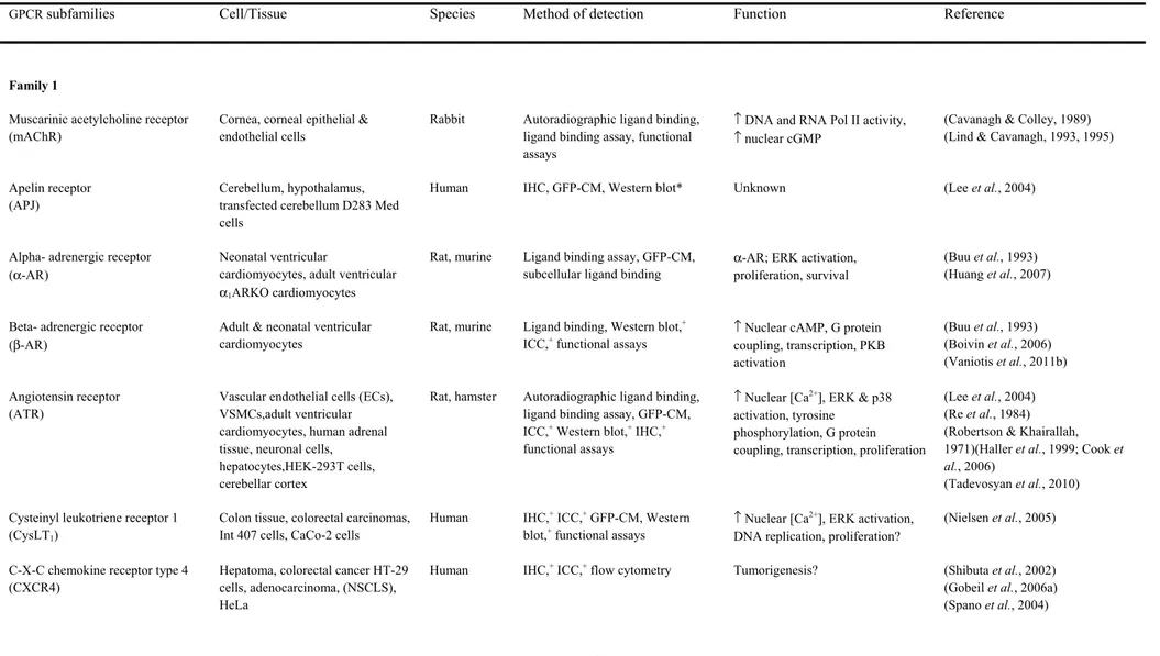
Recently, GPCRs have also been shown to localize to intracellular membranes, in particular the nuclear membrane (Table 1). GPCRs have also been shown to signal from other intracellular locations, with the angiotensin receptor localized to mitochondria (54). The discovery of nuclear GPCRs is particularly relevant for class A GPCRs, as many sub-families have been found to localize to the nucleus including but not limited to the endothelin receptor (ETR), angiotensin receptor (ATR) and adrenergic receptor families (55-57). Class B and C GPCRs have also been shown to localize to the nucleus, with the parathyroid receptor being discovered in the nucleus of osteoclast cells (58), and metabotropic glutamate receptors (mGluR) have been demonstrated on the nuclear membrane of neurons (9, 59). In addition, all four types of Gα proteins and various Gβγ subtypes have also been detected at the level of the nucleus (60-62). Furthermore, many of the downstream effectors associated to GPCRs have also been shown to localize to the plasma membrane, including PLA, PLC and AC (63-65), and several ion channels, including Ca2+ and K+ channels (66-68). Moreover, the various second messengers generated by these effectors have also been observed at the level of the nucleus, including cAMP (69), IP3 and DAG, as well as cGMP (70). Additionally, these nuclear GPCRs have been shown to interact with their downstream effectors. Nuclear bradykinin type 2, ETB, mGlu5 and AT1 receptors, to name a few, have been
16
demonstrated to be able to regulate nuclear calcium, with mGlu5 and AT1 receptors also shown to generate nuclear IP3 (61, 70-75). On the other hand, nuclear β1AR appears to activate AC and leads to the increase of nuclear cAMP (57). Furthermore, several of the GPCR regulatory proteins are also located at the nucleus. This includes the RGS proteins (76), β-arrestin 1 (77), as well as the GRKs (78, 79). This implies that nuclear GPCRs are also under tight regulation, not unlike GPCRs located at the plasma membrane, and further supports the notion that these receptors are in fact functionally relevant. In addition, other downstream effectors, associated with both G protein-mediated and G protein-independent signalling have also been shown at the nucleus, and appear to be activatable (30, 80, 81). Further, some GPCRs, such as the GABA-B receptors, have also been shown to interact directly with certain transcription factors, as have certain G proteins (82-84), while others appear to exert their effects by way of histone acetylation, as is the case of the gonadotropin and bradykinin receptors (74, 85).
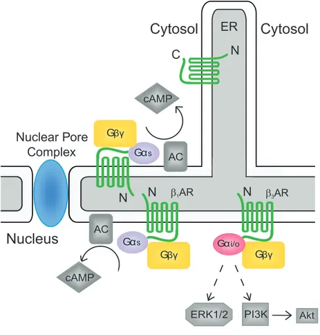
The unique structure of the nuclear envelope however does make the organization of nuclear GPCR signalling more complex than at the plasma membrane. The nuclear envelope consists of a dual membrane with an intermembrane space, the perinuclear space or lumen, which is contiguous with the ER. Additionally, at regular intervals the two membranes are joined together, forming nuclear pores. As such nuclear GPCRs can potentially be present on either the inner or outer nuclear membrane, or perhaps both (Figure 4). However, we believe that the N-terminal end of the receptor, containing the ligand binding sites, will never the less be situated in the lumen. This would mean that the ligand would have to be transported into the lumen of the nuclear membrane in order to bind its receptor and effect signalling. Furthermore, depending on its orientation nuclear GPCRs would either be directing signalling toward the nucleus or the cytosol. To date no study has conclusively shown the orientation of any nuclear-localized GPCR.
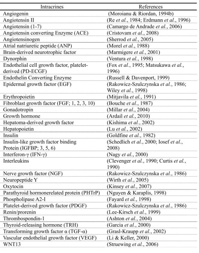
The presence of GPCRs at the level of the nuclear membrane means there must be a pool of intracrine ligands that can reach and bind to these nuclear receptors (Table 2). These ligands can be either synthesized within the cell, or internalized from the extracellular space. Intracrine ligands synthesized within the cell can be targeted to the
17
Golgi apparatus for secretion, though they may act intracellularly before or following secretion, by way of reuptake. Intracrine ligands that are internalized from the extracellular space may do so in a variety of ways. These ligands might be internalized in conjunction with their native receptor located at the plasma membrane, or can potentially enter the cell through passive diffusion. Internalized intracrine ligands can also be internalized by way of transmembrane transporters, or pass through channels or pores in the membrane.
III) Adrenergic Receptors
One GPCR sub-family of particular interest is the adrenergic receptor family. The adrenergic receptor family is part of the class A GPCRs, with the ability to bind catecholamines, especially epinephrine (adrenaline) as well as norepinephrine (noradrenaline) endogenously. Upon binding their ligand, these receptors will generally mediate sympathetic nervous system responses; this is normally referred to as the “fight-or-flight” response. Catecholamines are synthesized from DOPA, with dopamine being the first catecholamine synthesized, followed by norepinephrine and epinephrine. Norepinephrine and epinephrine are produced in certain neurons of the sympathetic nervous system, the latter more predominantly in the chromaffin cells of the adrenal medulla. Both catecholamines can act as either a hormone or a neurotransmitter, depending on their site of action, and have half-lives of only a few minutes in the circulation. Their release throughout the entire body is triggered by a variety of stresses, including a physical threat, excitement and loud noises. Catecholamines have been shown to directly increase heart and respiratory rate, trigger the release of glucose from energy stores, as well as lipolysis and increase blood flow and muscle contraction.
There are two primary types of adrenergic receptors, the α-adrenergic receptors (αAR) and the β-adrenergic receptors (βAR). These two groups can be further subdivide into 5 subtypes; the α1AR and α2AR, and the β1AR, β2AR and β3AR. The α and βARs have differential cellular localization and usually play opposing roles in the tissues where they are both present. In fact, in the circulation, the αARs are less sensitive to
18
epinephrine than the βARs, however when activated they override the vasodilatory effects of the βARs. This will only occur at higher levels of circulating epinephrine.
There are three α1AR subtypes, α(1A), α(1B) and α(1D), and all display a higher affinity for norepinephrine than epinephrine. All three subtypes generally activate Gαq. The α1AR is found predominantly in smooth muscle, where it mediates vasoconstriction. Activation of the receptor/G protein complex leads to the activation of PLC. PLC in turn causes an increase in DAG and IP3, triggering further effects, including calcium release and the activation of PKC (86). The α1AR is also present in other tissues, regulating glycogen metabolism in adipose tissue and liver, sodium reabsorption in the kidney, and a variety of other effects in the nervous system. In the heart, α1AR activation tends to have a positive inotropic effect, though to a lesser extent than the βARs, with α(1B) being the predominant isoform (86, 87). The physiologic function of the α1AR in the heart is still not completely clear, though they have been shown to play a role in hypertrophy and stimulation of transcription (87). Under pathologic conditions, it is believed that the αAR may play a compensatory role to counterbalance the desensitization of the βAR. Furthermore, the α1AR is believed to be important in developmental cardiac growth, as well as pathologic hypertrophy (86). The α1AR also appears to produce pro-survival effects in the cardiomyocyte, while also protecting it from decompensated heart failure. Moreover, the α1AR has been shown to localize to the nucleus in adult ventricular myocytes, where it is capable of activating the ERK1/2 pathway (88, 89).
Similarly to α1AR, the α2AR also has three subtypes, α(2A), α(2B) and α(2C). However, α2AR displays higher affinity for epinephrine, and primarily activate Gαi (90). Upon receptor activation, Gαi is able to associate with AC, and thereby inhibit its activity. This in turn causes a reduction in the levels of cAMP and a decrease in the activity of PKA. The α2AR is predominantly expressed in the nervous system and in smooth muscle. The α2AR can cause either vasoconstriction or vasodilation, depending on the tissue of interest (91). In the nervous system, α2AR can act as either an
19
autoreceptor, inhibiting the exocytosis of its own neurotransmitters, or as a heteroreceptor, inhibiting the release of many other neurotransmitters (90). The α2AR has also been shown to inhibit insulin release from the pancreas, to inhibit the norepinephrine system in the brain and increase thrombocyte aggregation (92). In fact, during the development of hypertrophy, the α2AR are desensitized by GRK2 in adrenergic neurons, causing an increase in circulating adrenaline levels, likely contributing to the eventually development of heart failure (90). In the heart, α2AR appear to play a cardioprotective role, as α2AR agonists are a common perioperative treatment, though this remains a contentious and controversial area of medicine.
The β1AR displays equal affinity for both epinephrine and norepinephrine and signals mainly through GαS (34). Upon activation of GαS, the G protein associates with AC, and promotes the conversion of ATP to cAMP, increasing its levels, and leading to the activation of PKA. This activation of PKA can be a fairly diffuse cellular response, leading to effects throughout the cell. The β1AR is the predominant βAR expressed in the heart, where it represents roughly 70% of the βAR density (93). The β1AR has also been shown to be expressed in adipose tissue and the cerebral cortex. In the heart, activation of the β1AR leads to an increase in cardiac output, via increases in heart rate as well as contractility, and while the β2AR can perform a similar role, knock-out studies have shown that the β1AR is sufficient (93). β1AR-mediated cAMP signalling has also been shown to regulate L-type calcium channels as well as phospholamban (93). During the development of hypertrophy, and eventually heart failure, there is an increase in circulating catecholamines, resulting in chronic stimulation of the β1AR which is thought to play a role in the further development of pathologic hypertrophy, and heart failure. During the progression towards heart failure, several changes occur within the cardiomyocyte, which include alterations in β1AR signalling. There is a downregulation of the β1AR subtype by around 50%. Such changes are not seen in the other subtypes, although the β2AR does undergo desensitization (94). Similar to other GPCRs, the βAR also undergo desensitization following phosphorylation by GRKs. The principal GRK responsible for desensitizing the βARs is GRK2, also termed
β-20
adrenergic receptor kinase 1 (βARK) (34). Moreover, there is an increase in the expression of GRK2, which is responsible for the pronounced desensitization of the βARs. This downregulation of βAR signalling, coupled with desensitization, is initially cardioprotective but eventually contributes to the onset of heart failure. This has lead to the use of βAR antagonists, or β-blockers to treat heart failure. However, the method by which they improve cardiac function is still somewhat contested. While some believe that β-blockers work by antagonizing the βARs, there is also evidence that they may work by resensitizing the failing myocardium to adrenergic stimulation. Another change in β1AR signalling is the observation that persistent activation augments contractility through calcium/calmodulin-dependent kinase II (CAMKII) signalling, and independently of the cAMP/PKA signalling system (93). This was shown through the use of inhibitors and a dominant-negative mutant of CAMKII. In conditions of chronic β1AR stimulation, inhibition of PKA is unable to block the sustained increases in myocyte contractility and calcium transients, while CAMKII inhibition does (95). Furthermore, studies have shown that following chronic β1AR stimulation, along with a progressive activation of CAMKII, there is also a concomitant desensitization of the PKA pathway, indicating that in response to chronic stimulation there is a time dependent switch from PKA to CAMKII (95).
CAMKII is a serine/threonine protein kinase regulated by the calcium/calmodulin complex. CAMKII consists of four structural domains, a catalytic domain, an autoinhibitory domain, a variable segment and a self-association domain (96). The sensitivity of CAMKII to calcium and calmodulin is regulated by the variable and self-association domains. As greater amounts of calcium and calmodulin become present, CAMKII autophosphorylates, and together with a partially reversible oxidation of a pair of methionines in the CAMKII regulatory domain, leads to persistent activation for a short period of time (96). CAMKII has also been linked to the apoptotic effects of sustained β1AR stimulation, as inhibition of CAMKII can block the apoptotic effects, and overexpression of CAMKII-δC, a cardiac CAMKII isoform, markedly aggravates this apoptotic effect (97). As such, the switch from PKA to CAMKII has also been linked to the catecholamine-induced hypertrophic response in cardiomyocytes (98). This
21
has been further demonstrated by treatment with a CAMKII peptide inhibitor. Overexpression of this inhibitor in transgenic mice prevents the maladaptive cardiac remodeling and contractile dysfunstion associated with chronic βAR stimulation and myocardial infarction (99). Also involved in this PKA independent hypertrophic response is EPAC, which has been shown to lead to the activation of CAMKII through the release of calcium by ryanodine receptors located on the SR (16). Given that EPAC plays a role as a GEF for small GTPases like Rap1 and Rap2, this would suggest that this switch to CAMKII activity involves the activation of small GTPases rather than cAMP production.
The β2AR displays higher affinity for epinephrine over norepinephrine, and signals through both GαS and Gαi. Similarly to the β1AR, upon activation the β2AR leads to the activation of AC, catalyzing the formation of cAMP, and leading to the activation of PKA. However, unlike the β1AR, this activation of PKA remains highly localized. The mechanism responsible for this localized response is likely its association with AKAPs, although coupling of the β2AR to Gαi is suggested to also play a role (100). The β2AR is primarily expressed in smooth muscle and the heart, where it represents roughly 30% of the βAR density (93). In the heart, the β2AR has been linked to positive inotropic and chronotropic effects, and has been shown to regulate the class C L-type calcium channels. A key feature of the β2AR is an ability to undergo spontaneous activation which has not been observed in the β1AR (101). As such, the β2AR can signal in the absence of its endogenous ligand, and this effect is dependent on receptor density. Moreover, in contrast to the β1AR, chronic activation of the β2AR is actually believed to play a cardioprotective effect (102). This cardioprotective effect is largely believed to be mediated through Gαi, and involves Gβγ, and the activation of the PI3K/PKB survival pathway. This pathway is thought to be responsible due to the fact that the β1AR cannot interact with Gαi, and that blocking any of these elements results in the loss of the protective effect (93). In addition, one of the main recognized changes in βAR signalling during the development of heart failure is the switch of β2AR coupling from GαS to Gαi in cardiomyocytes (34). This potential cardioprotective effect
22
of β2AR signalling has lead, in part, to the development of a new generation of β-blockers that are β1AR selective agents. Furthermore, the development of β-blockers that would antagonize the β1AR while acting as partial agonists towards the β2AR has also been explored. Furthermore, recent evidence indicates that the β2AR is able to form a heterodimer with the β1AR (103), this heterodimerization might be important in the proper localization of the receptors, as well as cause the heterodimer to have a unique ligand binding profile.
The β3AR displays higher affinity for norepinephrine than epinephrine and, similarly to the β2AR, signals through both GαS and Gαi. The β3AR is found predominantly in adipose tissue, where it is involved in the regulation of lipolysis and thermogenesis (104). However, the β3AR has also been localized to the brain, gut, liver and myocardium (105). A key distinguishing feature of the β3AR is the relatively fewer serine and threonine sites in its C-terminal tail, and the lack of a consensus PKA phosphorylation site, of which there are two in the β2AR and one in the β1AR (105, 106). These differences affect its propensity to be desensitized, and in fact in remains to be determinded conclusively whether or not the β3AR can desensitize. In adipose tissue, where the β3AR can interact with both GαS and Gαi, it has been shown to activate two parallel signalling pathways, the cAMP/PKA pathway through GαS, and the ERK MAPK pathway through Gαi and recruitment of the tyrosine kinase c-Src (105). The activation of PKA activates the protein kinase cascade leading to p38 activation. This in turn activates a subset of transcription factors, including ATF2. The β3AR is also present in the heart, though it represents a negligible percentage of the total βAR density in comparison to the other two βAR subtypes (104). As in adipose tissue, it has been linked to both GαS and Gαi, although the signalling effectors downstream of either G protein are less well understood than in the adipose tissue. Interestingly, stimulation of the β3AR actually results in a decrease in cardiac contractility, which is in clear contrast to the other βARs. Also of note, in cardiac tissue, β3AR activation of Gαi appears to lead to the production of nitric oxide (NO) by a constitutively expressed isoform of NO synthase (NOS), probably endothelial NOS (eNOS) (104). The production of NO is
23
likely responsible for some of the negative inotropic effects observed following β3AR activation, through the activation of sGC (107). As in adipose tissue, in the heart, the β3AR is believed to activate various MAPK cascades, which potentially contribute to the observed negative inotropic effects. Another unique feature of the β3AR, is that its expression is upregulated in the failing heart, in contrast to the β1AR and β2AR (104). Given the negative inotropic effect of the β3AR, this upregulation might prove beneficial at first, but likely contributes to the eventual development of heart failure. Interestingly, the β3AR has also been linked to various cardioprotective effects, principally through its activation of the PI3K/PKB pathway, demonstrating the complexity of β3AR signalling, and the necessity for further study (93).
IV) Endothelin Receptors
The endothelin receptors (ETR) also belong to the class A family of GPCRs. The ligands for ETR are endothelins, vasoconstricting peptides consisting of 21 amino acids, primarily produced in the endothelium, which play a role in vascular homeostasis. There are three isoforms of endothelin, derived from different genes, termed endothelin 1 (ET-1), ET-2 and ET-3. All three endothelins are synthesized as larger precursor proteins, pre-proETs, and subsequently cleaved to 37-41 amino acid proforms, termed big endothelins. Endothelin converting enzymes (ECEs) are responsible for converting big endothelins to their mature forms (108). In the myocardium, ET-1 is believed to play a paracrine/autocrine role, with ET-1 synthesized, stored and secreted by adult cardiomyocytes under basal conditions and in response to various stimuli (109, 110). Moreover, extracellular ET-1 has been shown to regulate ET-1 mRNA levels, thereby regulating its own expression (111). In addition, there are two receptor subtypes, ETA and ETB, each with its own expression and signalling profile. Another subtype, ETC, has been cloned in Xenopus laevis, although a mammalian homolog has yet to be discovered (112).
ETA is equally selective for ET-1 and ET-2, over ET-3, couples predominantly to Gαq, although it has been shown to couple to GαS as well, and is implicated in the regulation of intracellular calcium. ETA is located primarily in the smooth muscle of