Role and Modulation of Maternal Transcripts During the
First Cleavage Divisions in Bovine Embryos
Thèse
Ernesto Orozco-Lucero
Doctorat en sciences animales
Philosophiæ doctor (Ph.D.)
Québec, Canada
Résumé
Ce travail porte sur l’identification, la fonction et la régulation des molécules maternelles d’ARNm qui dirigent la compétence développementale juste après la fécondation chez les bovins.
Tout d’abord, en utilisant le modèle du temps écoulé jusqu’au premier clivage zygotique et à travers l’évaluation du transcriptome des embryons à 2-cellules, il fut possible de déterminer la signature moléculaire des niveaux extrêmes de compétence au développement et sélectionner des molécules candidates pour des études postérieures. Les résultats ont montré que les embryons de capacité développementale variable diffèrent dans certaines fonctions comme la réparation de l’ADN, le traitement de l’ARN, la synthèse de protéines et l'expression génique définies par des ARNm synthétisés par l’ovocyte.
Pour obtenir une confirmation fonctionnelle, une paire de transcrits maternels (l’un détecté dans notre sondage précédent et l’autre étant une molécule reliée) ont été inhibés par « knock-down » dans des ovocytes. Les effets du knock-down de ces facteurs de transcription sont apparus avant la formation des blastocystes dû à une diminution de la capacité au clivage et celle à progresser après le stage de 8-cellules. L’analyse moléculaire des embryons knock-down survivants suggère qu’un de ces facteurs de transcription est un contrôleur crucial de l’activation du génome embryonnaire, qui représente une fenêtre développementale dans l’embryogenèse précoce.
Dans la dernièr étude, nous avons testé si les facteurs de transcription d'intérêt sont modulés au niveau traductionnel. Des ARNm rapporteurs couplés à la GFP (Protéine fluorescente) contenant soit la version courte ou la version longue de la séquence 3’-UTR des deux molécules furent injectées dans des zygotes pour évaluer leur dynamique traductionnelle. Les résultats ont montré que les éléments cis-régulateurs localisés dans les 3’-UTRs contrôlent leur synchronisation traductionnelle et suggèrent une association entre la compétence développementale et la capacité de synthèse de ces protéines. Ceci conduit à
l’idée que ces facteurs de transcription cruciaux sont aussi contrôlés au niveau traductionnel chez les embryons précoces.
Les connaissances acquises ont joué un rôle essentiel pour définir le contrôle potentiel des molécules maternelles sur les embryons au début de leur développement. Cette étude nous montre aussi une utilisation potentielle de cette information ainsi que les nouveaux défis présents dans le secteur des technologies reproductives.
Abstract
This work explores the identity, the function, and the regulation of maternal mRNA molecules that drive developmental competence shortly after fertilization in cattle.
First of all, by using the model of the time of first zygotic cleavage and assessing the transcriptome of 2-cell embryos, it was possible to determine the molecular fingerprint of extreme levels of developmental competence and select candidate molecules for further monitoring. Data implied that early embryos of variable developmental capacity differ in functions including DNA repair, RNA processing, protein synthesis, and gene expression that are dictated by oocyte-synthesized mRNA.
To obtain a functional confirmation, a pair of maternal transcripts (one detected in our previous survey and other related molecule) were knocked-down in oocytes that were further cultured. The effects of ablating these transcription factors were evident before blastocyst formation due to a decrease in cleavage capacity, as well as progression past the 8-cell stage. The molecular analysis of surviving knocked-down embryos suggested that one of these transcription factors is a pivotal orchestrator of the activation of the embryonic genome, a critical developmental window in early embryogenesis.
In the last survey, we asked whether the transcription factors of interest are modulated at the translational level. Reporter mRNAs containing either short or long versions of the 3’-UTR sequences of both molecules were injected in zygotes to look at their translational dynamics. Results showed that cis-acting elements located in the 3’-UTRs govern their timely translation and suggested an association between developmental competence and protein synthesis capacity. This led to the notion that these crucial transcription factors are also controlled at the translational level in early embryos.
The acquired knowledge was instrumental to define the possible control operated by maternal molecules on embryos at the onset of their development, as well as some of the challenges and potential use of this information in the field of reproductive technologies.
Contents
Résumé ... III Abstract ... V Contents ... VII List of Tables ... XIII List of Figures ... XV List of Abbreviations and Symbols... XVII Acknowledgements... XXV Foreword ... XXVII
1. Introduction to Early Development ... 1
1.1. The Oocyte and Its Origin ... 1
1.2. Oocyte Growth and Folliculogenesis ... 4
1.2.1. Pre-antral follicular development ... 4
1.2.2. Antral follicular phase ... 7
1.3. Oocyte Maturation ... 9
1.3.1. Nuclear maturation ... 9
1.3.2. Cytoplasmic maturation ... 11
1.3.3. Molecular maturation ... 13
1.4. Molecular Regulation of the Maternal Stockpile ... 14
1.4.1. Oocyte transcription activation and silence ... 14
1.4.2. Oocyte global transcriptome profile during maturation ... 15
1.4.3. Polyadenylation and deadenylation during maturation ... 17
1.4.5. Protein synthesis during maturation ... 21
1.5. Ovulation and Fertilization ... 22
1.6. Embryonic Cleavage and First Cell Cycles ... 24
1.7. Maternal-Embryonic Transition ... 30
1.7.1. Embryonic genome activation ... 31
1.7.2. Potential existence of minor embryonic genome activation ... 35
1.7.3. Nuclear organization ... 37
1.7.4. DNA damage repair ... 40
1.7.5. Cell cycle and the first transcription products ... 42
1.7.6. Post-transcriptional and post-translational processing of maternal gene products ... 45
1.7.6.1. Protein degradation and posttranslational modifications ... 46
1.7.6.2. Turnover of maternal transcripts ... 48
1.7.6.3. Recruitment for translation ... 54
1.7.7. Transcriptional machinery ... 59
1.8. Activating Transcription Factors ... 61
1.9. Knock-Down of Specific Transcripts ... 66
1.9.1. Different types of systems for knock-down assays ... 67
1.9.2. Knock-down technology in early developmental studies ... 70
1.10. Hypothesis and Objectives ... 75
1.11. References ... 77
2. Rapidly Cleaving Bovine Two-cell Embryos Have Better Developmental Potential and a Distinctive mRNA Pattern ... 111
2.2. Abstract ... 113
2.3. Introduction ... 114
2.4. Results ... 115
2.4.1. Relationship between zygotic cleavage timing and blastocyst rate ... 115
2.4.2. Large-scale transcriptome analysis ... 116
2.4.3. Reverse transcriptase-quantitative PCR validation ... 116
2.4.4. Functional characterization ... 116
2.5. Discussion ... 117
2.5.1. Accuracy of the zygotic cleavage timing model ... 117
2.5.2. Transcriptome analysis ... 118
2.5.2.1. Cell cycle ... 118
2.5.2.2. Transcription control ... 119
2.5.2.3. Upstream regulators ... 120
2.6. Materials and Methods ... 121
2.6.1. In vitro production of bovine blastocysts ... 121
2.6.2. Separation of fast- and slow-cleaving 2-cell embryos and survey of the developmental outcome ... 122
2.6.2.1. Developmental competence estimation ... 122
2.6.3. Determination of differential transcript levels in 2-cell embryos ... 122
2.6.3.1. RT-qPCR ... 123
2.6.4. Functional analysis of differential mRNA levels profile in 2-cell embryos ... 123
2.7. Declaration of Interest ... 124
2.8. Acknowledgements ... 124
2.9. References ... 125
3. The Knockdown of ATF1 and ATF2 Transcripts in Germinal Vesicle Oocytes
Reveals their Crucial Roles in Bovine Early Development ... 143
3.1. Résumé ... 144
3.2. Abstract ... 145
3.3. Introduction ... 146
3.4. Materials and Methods ... 147
3.4.1. In vitro production of bovine blastocysts ... 147
3.4.2. Microinjection of oocytes ... 148
3.4.3. Estimation of the effect on phenotype ... 149
3.4.4. Western blot ... 149
3.4.5. Immunofluorescence ... 150
3.4.6. Determination of differential transcript levels in 8-cell embryos ... 150
3.4.7. Transcription factor binding sites prediction ... 150
3.5. Results ... 151
3.5.1. ATF1 and ATF2 protein expression through different stages in bovines ... 151
3.5.2. Effects of morpholino oligonucleotides against ATF1 and ATF2 on development ... 151
3.5.3. Western blot of microinjected 8-cell embryos ... 152
3.5.4. Immunofluorescence of microinjected 8-cell embryos ... 152
3.5.5. Transcription factor binding sites prediction ... 152
3.5.6. RT-qPCR analysis of microinjected 8-cell embryos ... 152
3.6. Discussion ... 153
3.7. Declaration of interest ... 161
3.8. Funding ... 161
3.10. References ... 163
3.11. Tables and Figures ... 171
4. Regulation of ATF1 and ATF2 Transcripts by Sequences in their 3’-Untranslated Region in Cleavage-stage Cattle Embryos ... 181
4.1. Résumé ... 182
4.2. Abstract ... 183
4.3. Introduction ... 184
4.4. Results ... 186
4.4.1. Effect of poly(A) tail presence and time on reporter expression ... 186
4.4.2. Impact of cleaving capacity on GFP expression ... 187
4.4.3. 3’-Untranslated region sequences ... 188
4.5. Discussion ... 188
4.5.1. Presence of poly(A) tail or 3’-untranslated region is necessary for GFP translation ... 189
4.5.2. Elapsed time from fertilization affects GFP expression ... 189
4.5.3. Increased cleaving capacity improves translation of long constructs ... 190
4.5.4. Potential Motifs in the 3’-untranslated region and possible regulatory upstream factors ... 190
4.6. Materials and Methods ... 194
4.6.1. Preparation of mRNAs ... 194
4.6.2. In vitro production of bovine embryos ... 195
4.6.3. Microinjection of presumptive zygotes ... 195
4.6.4. Fluorescence assessment ... 196
4.7. Acknowledgements ... 196
4.8. References ... 197
4.9. Tables and Figures ... 203
Supplemental File 4-1 ... 214
Supplemental File 4-2 ... 219
5. General Conclusion ... 223
5.1. References ... 229
Appendix 1: 6. Molecular Markers of Fertility in Cattle Oocytes and Embryos – Progress and Challenges ... 231
6.1. Résumé ... 232
6.2. Abstract ... 233
6.3. Introduction ... 234
6.4. Fertility status of oocytes and embryos ... 235
6.5. Molecular Markers of Quality Prior to Fertilization ... 236
6.5.1. Immature oocytes ... 236
6.5.2. Mature oocytes ... 240
6.6. Molecular Markers of Quality in Embryos ... 240
6.6.1. From fertilization to embryonic genome activation ... 240
6.6.2. Morulae and blastocysts ... 243
6.7. Conclusion ... 246
6.8. Acknowledgements ... 247
List of Tables
Table 1-1 Reports of gene knockdown in cattle pre-implantation development ... 72
Table 2-1 Symmetrical raw fold changes and p-values of microarray analysis and RT-qPCR validation of fast compared to slow 2-cell embryos ... 132
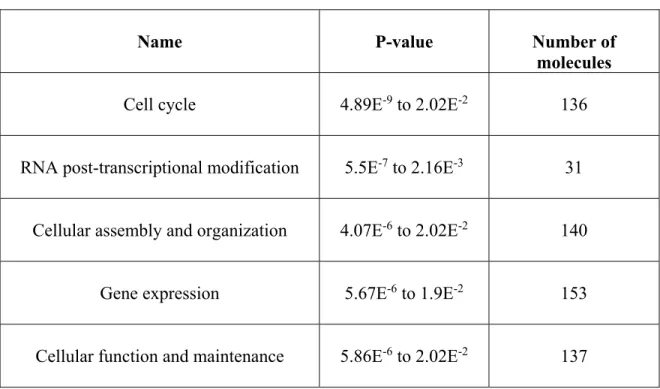
Table 2-2 Top molecular and cellular functions (biological functions) ... 132
Table 2-3 Top canonical pathways ... 133
Table 2-4 Details of RT-qPCR in bovine 2-cell embryos ... 134
Table 3-1 Details of RT-qPCR in 8-cell embryos ... 171
Table 3-2 Transcription factor binding sites prediction ... 173
Table 4-1 Number of 3’-UTR motifs localized in bovine ATF1 and ATF2 mRNA ... 203
Supplemental Table 4-2 Details of PCRs for mRNA preparation ... 220
Supplemental Table 4-3 Primer sequences ... 221
Table 6-1 Molecular markers of developmental competence in oocytes ... 257
Table 6-2 Molecular markers of developmental competence in follicles ... 258
Table 6-3 Molecular markers of developmental competence in embryos ... 259
List of Figures
Figure 1-1 Folliculogenesis (Orisaka et al. 2009) ... 5
Figure 1-2 Follicular dynamics (Aerts and Bols 2010b) ... 8
Figure 1-3 Nuclear maturation (Ferreira et al. 2009) ... 11
Figure 1-4 Cytoplasmic maturation (Ferreira et al. 2009) ... 13
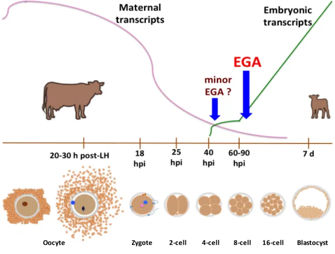
Figure 1-5 Dynamics of early development in cattle and embryonic genome activation ... 26
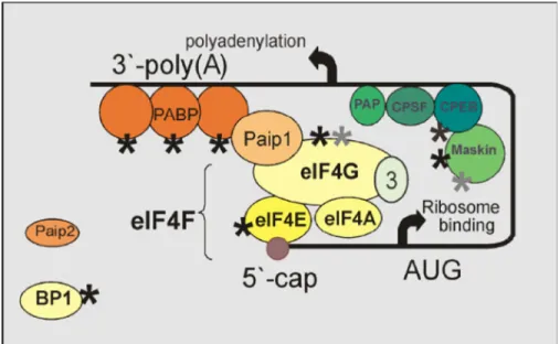
Figure 1-6 Closed-loop model of mRNA recruitment (Tomek and Wollenhaupt 2012) ... 56
Figure 2-1 Impact of cleavage timing on blastocyst production ... 135
Figure 2-2 Overall comparison by microarray of gene transcript targets present at the 2-cell stage ... 136
Figure 2-3 RT-qPCR validation of transcript abundance in fast- (29.5 hpi) and slow-cleaving (46 hpi) 2-cell embryos ... 137
Figure 2-4 ATM-related pathway ... 139
Figure 2-5 Upstream regulator prediction ... 140
Figure 3-1 Microinjection process ... 174
Figure 3-2 ATF1 and ATF2 protein expression through different stages in bovines ... 174
Figure 3-3 Effects of morpholino oligonucleotides (MO) on development ... 175
Figure 3-4 Embryos produced after morpholino oligonucleotides (MO) microinjection ... 176
Figure 3-5 Western blot of microinjected 8-cell embryos ... 177
Figure 3-6 Immunofluorescence of microinjected 8-cell embryos ... 178
Figure 3-7 Determination of differential transcript levels in 8-cell embryos by RT - qPCR ... 179
Figure 3-8 Proposed model of ATF1 and ATF2 regulation at time of embryonic genome activation (EGA) ... 180
Figure 4-1 Epifluorescence micrographies of presumptive zygotes/cleaved embryos microinjected with GFP mRNA ... 204
Figure 4-2 Effects of time on GFP expression of different constructs microinjected in presumptive zygotes ... 205 Figure 4-3 Comparison of results of the same mRNA with distinct polyadenylation status upon microinjection in presumptive zygotes used in this study ... 206 Figure 4-4 Epifluorescence micrographies of presumptive zygotes/cleaved embryos microinjected with either ATF2sA- or ATF2LA- mRNA ... 207 Figure 4-5 Effects of cleavage on GFP expression of different GFP mRNAs microinjected in presumptive zygotes ... 208 Figure 4-6 Effects of cleavage on GFP expression of different GFP-ATF1_UTR3 constructs microinjected in presumptive zygotes ... 209 Figure 4-7 Effects of cleavage on GFP expression of different GFP-ATF2_UTR3 constructs microinjected in presumptive zygotes ... 210 Figure 4-8 Constructs used in this study ... 211 Figure 4-9 Proposed model of ATF1 and ATF2 translational regulation through sequence motifs in their 3’-UTR ... 212 Figure 4-10 Zygote microinjection ... 213
List of Abbreviations and Symbols
(E) Embryonic day
[Ca2+]i Intracellular calcium % Percentage A- Deadenylated A+ Polyadenylated AARE Amino acid response element
AAUAAA Hexanucleotide; nuclear polyadenylation signal ACTB Beta-actin
ANOVA Analysis of variance ANXA Annexin
APC Adenomatous Polyposis Coli APC/C Anaphase-promoter complex
ARE A/U-rich element
ARNT Aryl hydrocarbon receptor nuclear translocator ART Applied reproductive technology ATF Activating Transcription Factor ATF1L GFP-Long ATF1_UTR3 construct ATF1mo ATF1 morpholino
ATF1s GFP-short ATF1_UTR3 construct ATF2L GFP-Long ATF2_UTR3 construct ATF2mo ATF2 morpholino
ATF2s GFP-short ATF2_UTR3 construct ATM Ataxia Telangiectasia Mutated
ATP1A1 ATPase, Na+/K+ Transporting, Alpha 1 Polypeptide
ATR Ataxia-telangiectasia and Rad3-related
AUG Start codon
AURKAIP1 Aurora kinase A-interacting protein 1 BCB Brilliant cresyl blue
bp Pair of bases
BRCA Breast cancer early onset
BTAF1 BTFIID transcription factor-associated, 170-kDa Mot1 hom. (S. cerevisiae) BTC Betacellulin
bZIP Basic leucine zipper domain
cAMP AMP, cyclic
CBP CREB-binding protein
CCNA Cyclin A
CCND2 Cyclin D2
CDKN1A Cyclin-dependent kinase inhibitor 1A (p21, Cip1) CENPF Centromere protein E
CHUK Conserved helix-loop-helix ubiquitous kinase CHX Cycloheximide
CKS1B CDC28 Protein Kinase Regulatory Subunit 1B
COC Cumulus-oocyte complex
CPEB Cytoplasmic polyadenylation element-binding protein CPSF Cleavage and polyadenylation specific factor
CRE cAMP-response element
CREB cAMP-response element-binding protein CREBBP CREB-binding protein
CREM cAMP-response element modulator CTNNB1 Beta-catenin-1
d Day
DAZL Deleted in azoospermia-like DBF4 DBF4 homolog (S. cerevisiae)
DNAJA1 DnaJ (Hsp40) Homolog, Subfamily A, Member 1
DRE DAZL-recognition element
DSB DNA double-strand break
dsRNA Double-stranded RNA
dTXr Dextran Texas red
DUSP6 Dual Specificity Phosphatase 6
DYNC1I1 Dynein, Cytoplasmic 1, Intermediate Chain 1 eCPE Embryonic cytoplasmic polyadenylation element
ED External diameter
EDEN Embryo deadenylation element
EEF1A1 Eukaryotic Translation Elongation Factor 1 Alpha 1 EGA Embryonic genome activation
EGFP Enhanced Green Fluorescent Protein EGFR Epidermal Growth Factor Receptor EIF Eukaryotic Translation Initiation Factor ELAVL1 ELAV Like RNA Binding Protein 1
ENY2 Enhancer Of Yellow 2 Homolog (Drosophila) EPAB Embryonic poly(A)-binding protein
EXOSC10 Exosome component
FC Fold change
FIGLA Factor in the germline, alpha
G6PDH Glucose-6-phosphate dehydrogenase GFP Green Fluorescent Protein
GFPA- GFP mRNA deadenylated GFPA+ GFP mRNA polyadenylated GFPA30 GFP mRNA 30 adenine residues
GO Gene ontology
GRB2 Growth factor receptor-bound protein 2
GV Germinal vesicle
GVBD Germinal vesicle-breakdown h Hours
HAS Hyaluronan synthase
HEX Hexanucleotide, nuclear polyadenylation signal
HKG Housekeeping gene
hpi Hours post-insemination
HSP70 Heat shock 70kDa protein
IF Immunofluorescence IFN Interferon
IGF2 Insulin-like growth factor 2
INHB Inhibin, beta
IVC In vitro culture
IVD In vitro-derived
IVM In vitro maturation
IVP In vitro produced / in vitro embryo production
KCNJ8 Potassium Channel, Inwardly Rectifying Subfamily J, Member 8 KD Knockdown
kDa Kilodaltons KLF4 Kruppel-like factor 4 (gut) KO Knockout L Long
ldsRNA Long double-stranded RNA
MAPS Motif Associated with Polyadenylation Signal MATER Maternal Antigen That Embryos Require Mb
MBE
Mega-bases
Musashi-binding element
MET Maternal-embryonic transition
MII Metaphase II
miRNA Micro-RNA
MO Morpholino oligonucleotides
MRE11A Meiotic recombination 11 homologA (S. cerevisiae)
mRNA Messenger RNA
MSH6 MutS homolog 6 (Escherichia coli) MSX1 Msh Homeobox 1
MSY2 (YBX2, Y Box Binding Protein 2) MuERV-L Murine endogenous retrovirus
MW Molecular weight
MYC V-Myc Avian Myelocytomatosis Viral Oncogene Homolog MZT Maternal-zygotic transition
NASP Nuclear Autoantigenic Sperm Protein (Histone-Binding) NDFIP1 Nedd4 Family Interacting Protein 1
NDUFS NADH Dehydrogenase (Ubiquinone) Fe-S Protein NPS Nuclear polyadenylation signal
nt Nucleotides
NUSAP1 Nucleolar And Spindle Associated Protein 1 OCT4 Octamer-Binding Protein 4
p-Zy Presumptive zygote
P38-MAPK P38 Mitogen-Activated Kinase (MAPK14) PABP Poly(A)-binding protein
PABPC1 Poly(A)-binding protein, cytoplasmic 1 PABPN1 Poly(A)-binding protein, nuclear 1 PABPNL1 Poly(A)-binding protein, nuclear like-1
Paf 1-o-alkyl-2-acetyl-sn-gylcero-3- phosphocholine PAIP1 Poly(A)-interacting protein 1
PAP Poly(A)-polymerase
PARN Poly(A)-Specific Ribonuclease
PB Polar body
PBE Pumilio-binding element
PCBP1 Poly(RC) Binding Protein 1 PCNA Proliferating cell nuclear antigen PKA Protein kinase, cAMP-dependent pL Picoliters
PN Pronucleus poly(A) Poly-adenine
POU5F1 POU Class 5 Homeobox 1 PRDX1 Peroxiredoxin 1
PRE Polyadenylation response element
PSMB2 Proteasome (Prosome, Macropain) Subunit, Beta Type, 2 PTTG1 Pituitary Tumor-Transforming 1 (securin)
Pum Pumilio
RAD51 RAD51 Recombinase
Rb Retinoblastoma protein
RGS16 Regulator Of G-Protein Signaling 16 RISC RNA-induced silencing complex RNAi RNA-interference RPL Ribosomal protein L
rRNA Ribosomal RNA
RT-qPCR Reverse Transcriptase-quantitative PCR s Short
SDS-PAGE SDS-Polyacrylamide gel electrophoresis SIAH2 Siah E3 Ubiquitin Protein Ligase 2 siRNA Small-interference RNA
SKIIP SKI-Interacting Protein (SNW1, SNW Domain Containing 1) SKP1 S-Phase Kinase-Associated Protein 1
SLBP Stem-loop binding protein
SOD Superoxide dismutase
SP1 Sp1 transcription factor
SRFS3 Serine/Arginine-Rich Splicing Factor 3 (SRSF3) STDmo Standard control morpholino
SUPT4H1 Suppressor of Ty 4 homolog 1 (S. cerevisiae)
TACC3 Transforming, Acidic Coiled-Coil Containing Protein 3 TAE Translation activating element
TAF RNApolymerase II, TATA box binding protein associated factor, 150 kDa.
TF Transcription factor
TFBS Transcription factor-binding site
TNFAIP6 Tumor Necrosis Factor alpha–Induced Protein 6 TP53 Tumor protein P53
TRAPPC3 Trafficking Protein Particle Complex 3 TRC Transcription-requiring complex TRE Translation repressing element UBE2K Ubiquitin-conjugating enzyme E2K
uninj Uninjected
UPP Ubiquitin-proteasome pathway
UTR Untranslated region
WB Western blot
YWHAG Tyr. 3-monooxygenase/tryptophan 5-monooxygenase activat. prot. gamma
YY1 Ying-yang 1
ZGA Zygotic genome activation
ZP Zona pellucida
… al amor, con amor
“Heard melodies are sweet, but those unheard are sweeter” (J.K). “All we need to do is make sure we keep talking” (S.H).
Acknowledgements
Through the years in Québec I had the opportunity to meet wonderful people. Now, is time to express how grateful I am for their unique help and the experiences we shared. It is not possible to mention all these nice people, but they should be sure I always remember them. First of all, I would want to express my deepest gratitude to my advisor Marc-André Sirard. After having the honor of being accepted as his student, I received not only an opportunity of education within a top-class work group, but also to learn from his discipline, responsibility, unbeatable energy, problem-solving mind, innovation, as well as passion for science. Thanks, so much for your patience and daily encouragement to improve. You’re with no doubt one of the main role models in my life.
The benefits of my stay at ULaval would have been far from complete without my co-supervisor Claude Robert. Thanks for your example and critical spirit, your indispensable support to my project and to the group, in addition to the incomparable quality of your classes and molecular expertise, as well as your sharp reasoning. I’m also in profound debt with my other professors and members of the former CRBR, particularly Janice Bailey, Pierre Leclerc, Jacques Tremblay, and François Richard for their support even before my arrival to ULaval, for sharing their expertise and contributing to the consolidation of an amazing research center. As for François R and Jacques, together with Jon LaMarre from Guelph, besides their indispensable support during the years and now the final feedback by serving on my PhD committee, they have been exceptional examples in research.
I’m also thankful to other professors and members of SAN and FSAA for their crucial help: Dany Cinq-Mars, Robert Chartrand, Jean-Paul Laforest, Lyne Fortier, Janny Bérubé, and Jean-Claude Dufour. On the lab bench and next to it, a research student requires the irreplaceable assistance of our technicians, professionals, and managers: Isabelle Dufort, Isabelle Laflamme, Christine G, Alexandre Bastien, Dominic G, Eric Fournier, Isabelle Gilbert, Julie N, Nancy B, Micheline G, Serge Daigle, and Richard Prince. Thanks a lot for teaching me all the necessary, driving me through the labs, and of course, your analysis and for making our projects possible. I was also lucky to meet and learn from post doc fellows,
former students, and visitor researchers, as Daulat, Nicolas Guillemin, Maëlla G, Béatrice de M, Dessie Salilew-Wondim, Morteza, Julieta, Christian Vigneault, and Annick Bergeron. I show my appreciation for your unique example, teaching, company, and support. In addition, last year I had the chance to stay at UOGuelph for a pivotal part of my project at the lab of Allan King, Elizabeth St-John, Pavneesh Madan, Jon LaMarre, Monica Antenos, Ed, Fazl, Gyu-Jin and N-Y Rho, Ryoung-Hoon, Tamas, and Leslie. I’m quite grateful for your welcoming, support, and friendship, as well as that of Nihad, Cristina, Sharon, and Harold during that unforgettable summer. Moreover, I extend my gratitude to all singular students I was lucky to met at CRBR, including Sara, Angus (both that starting me to show what is living in Québec and Canada, and who survived to me at my arrival), Habib, Akintayo, O. D’Amours, Luis, Audrey Bunel, Florence, Annie Girard, Gaël, Rémi L, Marie C, and Anne-Laure… « et aussi à la nouvelle gang composée de » Léoni, David, Patricia, Simon, Rachèle et Chloé. Thanks, so much for your friendship and help.
Besides the lab mates, I met a bunch of nice people with whom to share some free time: “los compadres” Arturo Duarte y Maria Garcia, Aude Popek, Mahder, Vishal, Rafael IB, Ana Sofia, Emperador, Yhosvanni, Mylene A, Patrick M, Luc Pelletier, Alma D, Dr. Pinedo, Pascal D, Donatien, Analisse, José Luis M, and Stéphanie Robert. I always remember those who helped me in Mexico shortly before my initial and last departures: Alejandro Martinez (my other great role model), Dora F and Ángel D, Moisés B and Karla, F. Plenge, N. Lobo, Pepe Nuñez, Boni, as well as Beto Grado. In my very personal circle in Quebec I’m indebted to all the community of Saint-Thomas-D’Aquin, especially Alexandre Julien, Martin Lagacé, and Marie-Josée G: You don’t know how much you helped me! Of course, I incur everything to my loving parents, as well as to my siblings and their families, to whom I owe my life and my whole current being. They were always available in spite of any circumstance… in addition to the little lady that has recast all my life, together with her family: Lupita Férnandez. She brought me back and took incomparable care of me. I have no words to thank my beloved ones for everything: All the honor and love that you deserve! Now, I’m going back to my México, which I missed. I’m quite sure I’ll miss Québec so much: My life completely changed here… Thanks!
Foreword
Chapters 2 and 6 consist on published papers. Chapters 3 and 4 will be soon submitted for publication.
Chapter 2:
Orozco-Lucero E, Dufort I, Robert C, Sirard MA. 2014. Rapidly cleaving bovine two-cell embryos have better developmental potential and a distinctive mRNA pattern. Mol Reprod
Dev 81: 31-41.
In this chapter Ernesto Orozco-Lucero is responsible of all experiments, data analysis, and writing the paper. Isabelle Dufort designed and collaborated in the experiments, as well as data analysis, and reviewed the paper. Claude Robert participated in the project elaboration, interpretation of results, and paper revision. Marc-André Sirard conceived the project, interpreted the analysis, and reviewed the manuscript. All coauthors approved the final version of the manuscript.
Chapter 3:
The knockdown of ATF1 and ATF2 transcripts in germinal vesicle oocytes reveals their crucial roles in bovine early development.
Chapter 4:
Regulation of ATF1 and ATF2 transcripts by sequences in their 3’-untranslated region in cleavage-stage cattle embryos.
In these two chapters Ernesto Orozco-Lucero collaborated in all experiments, data analysis, and writing the paper. Isabelle Dufort designed and collaborated in the experiments, as well as data analysis, and reviewed the paper. Claude Robert participated in the project elaboration, interpretation of results, and paper revision. Marc-André Sirard conceived the project, interpreted the analysis, and reviewed the manuscript.
Chapter 6:
Orozco-Lucero E, Sirard MA. 2014. Molecular markers of fertility in cattle oocytes and embryos: Progress and challenges. Anim Reprod 11: 183-94.
In this chapter Ernesto Orozco-Lucero is responsible of obtaining data and writing the manuscript. Marc-André Sirard obtained information, conceptualized the paper, and reviewed the manuscript. The coauthors approved the final version of the manuscript.
1. Introduction to Early Development
1.1. The Oocyte and Its Origin
The oocyte is a highly specialized cell capable of supporting fertilization and early embryogenesis in metazoans once it completes its differentiation. It provides half of the genetic material (haploid complement, 1N) to the embryo by the moment of fertilization, while the sperm collaborates with the other part, to finally restore the diploid chromosomal complement (2N). In addition, the female gamete contributes with transcripts and proteins to allow developmental progression throughout the first stages of embryogenesis during a transcriptionally silenced period, also termed the maternal or embryonic program, before the activation of the genome from embryonic source. The egg is a totipotent cell as it possesses the capacity to generate all the cell types of the future embryo and the adult organism. Furthermore, the oocyte is able to reprogram the genetic material of a somatic and differentiated cell following the transfer of the nucleus of the later to a recipient ooplasm and then constitute a viable embryo (rev. Song and Wessel 2005; Macaulay et al. 2011; Von Stetina and Orr-Weaver 2011), in an analogous manner to that of the remodeling of the sperm nuclear material by the oocyte cytoplasm upon fertilization (McLay and Clarke 2003). To better understand the complex biology of the egg, it is important to focus on the origin of the germ cell lineage during early embryonic life and the molecular and cellular events that will lead to the rise of female gametes.
Germ cells (GCs) in both male and female embryos are derived from undifferentiated precursors called primordial germ cells (PGCs) in early development. During their lifespan, PGCs pass through the phases of establishment of the germ cell lineage, migration, colonization of the primordial gonad, and proliferation (Vanderhyden 2002). In turn, the ancestors of the PGCs are located in the proximal (posterior) epiblast, structure originated from the inner cell mass (ICM) of the blastocyst. In such place these ancestor cells are induced to become GCs by BMP4 and BMP8b signalization produced by the adjacent extra-embryonic ectoderm. This precursor cell population expresses Pou5f1, Dppa3 (Stella) and Ifitm3 (Fragilis) markers of GCs (Saitou et al. 2002; Saitou et al. 2005). However, these three factors are not exclusive of the germinal lineage, since they are also expressed in early embryos. In addition, these ancestor cells can produce both companion somatic cells and actually specified GCs. Consequently, it is not until the expression of Blimp1 (Prdm1)
in a small subpopulation of around six cells by embryonic day (E) 6.25 in the mouse epiblast (Ohinata et al. 2005) that the precursor cells are first committed to a germinal fate, given the repression of somatic cell-specific Hox genes by Blimp1 (McLaren and Lawson 2005). Subsequently, the founder population of approximately 45 PGCs moves to the base of the allantois by (E)7.25 in mice. PGCs show tissue-non-specific alkaline phosphatase (TNAP) activity, which has been used to help in localization of this kind of cells, but is not exclusive of GCs (De Felici et al. 2004). On the other side, Wrobel and Suss (1998) reported that in the bovine embryo the putative PGCs population is first detected by (E)18, and later incorporated close to the mesonephros by (E)23-25. As it can be noticed, PGCs are originally located in extragonadal compartments in distinct species.
In order to colonize the developing gonad, the PGCs (which in the cow have a diameter of 30 μm) start a migration from their place of origin next to the primitive ectoderm, passing through the embryonic posterior gut by using amoeboid movements and pseudopodia (Aerts and Bols 2010a). Durcova-Hills et al. (2003) reported that in the mouse this migration takes place from (E)9.5 to (E)11. By contrast, in bovines it occurs from (E)30 to (E)64 when all PGCs finally reach the developing gonad. It is strongly suggested that throughout the migratory pathway, the PGCs are driven to the gonad by chemotactic signals produced by the genital ridge. These molecules are apparently also important for PGCs survival and proliferation during the displacement period and include Kit Ligand (KL, SCF), bFGF, TNF-alpha, LIF, CNTF, oncostatin-M, SDF-1, BMP4, TGFβ1, activin, Gas6, neuregulin- β , and PACAP (Donovan et al. 2001; De Felici et al. 2004). For example, KL is expressed in the surface of the somatic cells in the migratory pathway, while its receptor, KIT, is produced by PGCs (Vanderhyden 2002) creating a regulation system kept later on until the oocyte-cumulus cells interaction. Moreover, KL possibly assures PGCs survival by inhibiting the apoptotic molecular system that can be induced by BAX (Krysko et al. 2008). Concerning attachment, the adhesion properties offered by the substrate to the PGCs during their migration appear fundamental, as it has been observed by the interaction of this group of cells with collagen IV, fibronectin, and laminin, to which these cells connect through integrins (Garcia-Castro et al. 1997). It must be noted that as PGCs progress towards the gonadal territory their population experiences an important
initial proliferation. In mice the founder population of around 40-45 PGCs increases to more than 3,000 cells by (E)11, moment when they reach the gonadal crests in this species (Morrish and Sinclair 2002).
During migration, PGCs behavior and phenotype are identical in embryos of both sexes (McLaren 2001). Nevertheless, the molecular events that determine the phenotypic sex of the GCs start by the end of the PGCs displacement, and in such processes the gonadal somatic cells are directly involved. In female embryos, the triggering factor of GCs sex specification is Dax1 (Morrish and Sinclair 2002). This gene is expressed in the bipotential gonad but diminishes its transcript levels in male embryos by (E)12.5, the time of gonad differentiation in the mouse, while it remains highly expressed by somatic cells in the future ovary. Thus, it constitutes a possible antagonist of the Sry masculinizing factor (Morrish and Sinclair 2002). In the bovine, the differentiation of the gonad occurs by (E)40 (Wrobel and Suss 1998), implying that the first PGCs arriving to the developing gonads (still genital crests) in this species by (E)30-35 do so when such structure is about to exhibit sexual dimorphism. When PGCs are located in the already differentiated gonad, the female GCs are now termed oogonia. This cellular population is mitotically very active and continues into the embryonic ovary the proliferation that had shown during their previous displacement. In the cow, the maximum number of these cells is estimated in 2.1 x 10⁶
(Aerts and Bols 2010a). At this moment the GCs will be prepared to switch from mitotic divisions to meiosis before forming primordial follicles.
The induction of the first meiotic division occurs in the cow by (E)70 (Magre and Vigier 2001) and there is evidence that the timing of this process is dictated by the surrounding somatic cells. The triggering mechanism for meiosis seems to involve the induction of retinoic acid (RA) of the expression of the meiotic markers Stra8, Sycp3, and Dmc1, given the high levels of RA in the embryonic female gonad in comparison to the testis, in which the male GCs do not enter meiosis until puberty (Swain 2006; Bowles and Koopman 2007). Once the oogonia start meiotic divisions they are called oocytes. In all mammals meiosis stops at prophase I and this occurs in the cow by (E)90, a concomitant event with the first complete assembly of primordial follicles (Aerts and Bols 2010a), which according to the
observations of Nilsson and Skinner (2009) start to form by (E)80. This initial blockage of meiosis can persist during months or years, depending on the species, and such period is termed dictyate. According to Zheng and Dean (2007), mouse oocytes stop their first meiotic division specifically at diplotene, while there is no clear consensus of the exact time point at which it occurs at the end of prophase I in the bovine, but it is considered to take place in a moment between pachytene and diplotene stages. The oocytes meiotically arrested in prophase I contain a nucleus called germinal vesicle (GV). Meiosis restarts and rupture of GV will take place later during development induced by the gonadotropin surge (Edwards 1965).
1.2. Oocyte Growth and Folliculogenesis
The growth of the oocyte in the follicle takes place from the establishment of primordial follicles up to just before the moment of final maturation. In contrast, the follicle grows until the moment of ovulation and passes through the stages of primary, secondary (pre-antral), tertiary, and pre-ovulatory follicle (antral).
1.2.1. Pre-antral follicular development
Contrary to rodents, which do not form primordial follicles until birth, folliculogenesis in bovines starts by (E)90 with the presence of fully enclosed oocytes in primordial follicles. These follicles are located in the ovary cortex and their gametes (primary oocytes) measure around 30 μm in diameter (Braw-Tal and Yossefi 1997). Nilsson and Skinner (2009) have proved that progesterone regulates primordial follicle assembly since decreasing levels of this hormone are correlated with high primordial follicle formation at the end of gestation in cattle. Previously, the mitotically active oogonia form GCs clusters, which establish a syncytium, surrounded by somatic cells derived from sex cords. After inception of meiosis, the nurse cells extend cytoplasmic projections into the interconnected oogonia to divide the clusters and start surrounding individual oocytes, constituting a single layer of flat pre-granulosa cells around each gamete (Fig. 1-1). In turn, this cellular monolayer is enclosed by a basal lamina (van Wezel and Rodgers 1996; Aerts and Bols 2010a).
Figure 1-1 Folliculogenesis (Orisaka et al. 2009)
The main features of the disctint developmental stages throughout folliculogenesis are illustrated. Copyright-free scheme reproduced and adapted from BioMed Central © 2009.
A first selection of oocytes occurs by the moment of meiosis entry since many of these gametes suffer attrition, basically occurring through apoptotic mechanisms (Krysko et al. 2008). This is reflected by the number of primordial follicles at the moment of birth, which in a calf is approximately 1.1 x 10⁶, in contrast to the peak of GCs during embryogenesis.
However, bovine fetuses show an average of 130,000 primordial follicles in such moment, while in most domestic species the number of this type of follicles ranges from 100,000 to 400,000 per newborn (Senbon et al. 2003; Aerts and Bols 2010a). These primordial follicles constitute the ovary complement from which oocytes will be recruited for ovulation during all the female reproductive lifespan without de novo replenishment (Senbon et al. 2003; Aerts and Bols 2010a). In recent years, the reports of the laboratory of J.L. Tilly (Johnson et al. 2004; Johnson et al. 2005) opened the discussion concerning the possible renewal of GCs stock during mammalian female adulthood from undifferentiated cells, which could be generated from bone marrow stem cell populations. Nevertheless, this presumptive biological mechanism is still highly controversial and needs further demonstration (Telfer et al. 2005; Vogel et al. 2005). As it has been mentioned, folliculogenesis begins with primordial follicles and these will enter the growing pools of follicles that will be recruited
Primordial Primary Secondary Preantral Antral Flat granulosa Cubic
granulosa granulosaCubic
Cumulus Mural granulosa Antrum Expanded cumulus Ovula on Oocyte
until they enlarge and form primary and secondary follicles. This phase is called follicle
growth initiation or follicle activation, and it is an irreversible process. To get into the
growing wave, primordial follicles must leave the resting pool where quiescent follicles remain and account for the vast majority of the follicular population in the ovary. This is probably induced by KL signaling from the neighbor granulosa cells (Aerts and Bols 2010a). Moreover, Yang and Fortune (2008) have demonstrated that in the cow the meiotic arrest must be completed in prophase I, with high ovarian expression of YBX2 (MSY2) mRNA before activation of primordial follicles.
Although there is no tangible difference in oocyte diameter between primordial and primary follicles, the later ones are already committed to growth, and the developmental progress to the primary follicular stage is marked by one of the first major changes during folliculogenesis: The transformation of flat pre-granulosa cells to cuboidal granulosa cells, as well as the proliferation of the same (Aerts and Bols 2010a). In spite that most of the growth waves occur after birth in cattle, a few primordial follicles are activated during fetal life and the earlier primary and secondary follicles are found by (E)140 and (E)210, respectively (Yang and Fortune 2008). Besides the KIT/KL communication system (also present in primordial follicles) between the gamete and its nurturing follicular cells, modulation control exists between both cell types through gap junctions (heterologous channels), as well as among the granulosa cell population (homologous channels). The integrity of the heterologous channels is kept even when the Zona Pellucida (ZP) is synthetised at a later stage. Furthermore, the oocyte-secreted factors, GDF9 and BMP15, have been shown to be crucial for follicular development as they are mitogens for granulosa cells (de Matos et al. 1997; Carabatsos et al. 2000; Knight and Glister 2003; Senbon et al. 2003; Knight and Glister 2006). By all these molecular interchange mechanisms the distinct types of cells inside a follicle are interconnected and a bidirectional communication is established between the oocyte and granulosa cells. Through this system the granulosa cells provide nutrients, maturation and meiotic regulating molecules (ribonucleosides), and elements used upon fertilization (e.g. cysteine, precursor of glutathione) to the gamete, while the oocyte orchestrates the proliferation, differentiation, and some functions (glycolysis, steroidogenesis) of the granulosa cells, and later the cumulus expansion (de
Matos et al. 1997; Carabatsos et al. 2000; Knight and Glister 2003; Senbon et al. 2003; Knight and Glister 2006).
In agreement with the data of Braw-Tal and Yossefi (1997) the initial deposition of the glycoprotein-containing ZP matrix, fundamental for fertilization, over the bovine oocyte’s plasma membrane takes place in the early secondary follicular stage, and the first appearance of fully-enclosed oocytes by ZP occurs by the late secondary stage. Similarly, another important developmental change in the secondary follicle is the conformation of multiple layers of granulosa cells around the oocyte, in addition to an important growth of the gamete, reaching a diameter of around 70 μm by the late secondary stage (Braw-Tal and Yossefi 1997). Finally, the same group reported that the inner theca begins to form around the granulosa cells by the early secondary follicular stage in cattle, and subsequently the basal lamina is totally surrounded by theca cells in the late secondary follicle. The theca accounts for the vascularization of the ovarian follicle, contrary to the avascular internal portion beyond the basal lamina, constituted by granulosa cells and the oocyte (Braw-Tal and Yossefi 1997).
1.2.2. Antral follicular phase
The tertiary follicle (also called antral or Graafian) is characterized by the presence of a fluid-filled cavity (antrum) in the middle of the granulosa cell population, ensuing in this way the differentiation of such cells in two subpopulations: 1) The most peripheral one includes those cells termed mural granulosa; 2) those in direct contact with the gamete (corona radiata) together with the ones comprising the cell layers separating it from the antral fluid and the mural granulosa cells constitute the cumulus. The antral space contains a complex liquid mixture of proteins, hormones, and ions involved in endocrine/paracrine regulation (Senbon et al. 2003; Huang et al. 2006; Adams et al. 2008). It must be remarked that in contrast to rodents, whose oocytes reach their maximum size by the moment of follicular antrum appearance, oocytes of antral follicles in domestic species continue with a remarkable growth during this developmental stage (Motlik et al. 1984). Hence, the early (small) antral follicle in the cow that attains a size of 250-500 μm harbours an oocyte with an average diameter of 93 μm and on whose plasma membrane the deposition of the ZP still
increases (Braw-Tal and Yossefi 1997). Thereafter, the preovulatory (mature Graafian) includes an oocyte with the biggest size: Its diameter reaches or can slightly surpass 130 μm in cattle (Otoi et al. 1997; Fair 2003). If this gamete has restarted meiosis after the prophase-I arrest it is then termed secondary oocyte. Nonetheless, in monovulatory species typically only a gamete is ovulated at the end of each estrous cycle. Before that, the ovulating follicle must become dominant (Fig. 1-2).
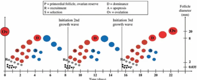
Figure 1-2 Follicular dynamics (Aerts and Bols 2010b)
Representation of the distinct follicular phases and their timing during a three-follicular wave estrous in the cow. Scheme reproduced with permission of Wiley © 2010.
As in most domesticated animals and humans, the estrous cycle in cattle presents follicular growth waves, and in this last species it commonly manifests in 2-3 waves per cycle. In general, only one follicle per wave becomes dominant. Only the dominant follicle (DF) of the last wave in the estrus cycle ovulates. The length of the estrous cycle in bovines has an average of 21 or 23 days for cycles with two or three waves, respectively (Adams et al. 2008; Aerts and Bols 2010b). In comparison with the pre-antral stage, the antral follicular phase represents the shortest period of the follicular development. In cows, the antral growth has an average length of 42 out of the 180 days of the total folliculogenesis. Therefore, a complete antral phase typically needs the extent of two estrous cycles to be finished. Each growth wave consists of recruitment, selection, and dominance periods (Adams et al. 2008; Aerts and Bols 2010b). In the first phase a cohort of 5-10 antral
follicles avoid atresia due to the presence of high levels of circulating FSH, which performs a recruitment function for the new cohort of tertiary growing follicles at the beginning of each wave. The selection takes place when the largest follicle reaches around 8.5 mm in diameter. By this moment, a follicle commits to become the DF (generally the largest and/or the most developed one), while the rest turn into subordinate follicles (SF) and start regression: The dominance stage starts. The DF then becomes the major suppressor (by synthesizing inhibin and estradiol) of FSH secretion (thus, of SFs growth and emergence of new follicular cohorts), since this follicle is by this moment capable to survive and continue its growth even in basal levels of FSH (Adams et al. 2008; Aerts and Bols 2010b). This is achieved by the DF by switching from FSH to LH dependence. If the DF rises when a
corpus luteum (CL) is still present, the progesterone secreted will inhibit LH pulse
frequency, preventing the DF from obtaining sufficient amounts of this hormone to complete its growth and thus the DF will enter atresia (Fig. 1-2). On the contrary, when the DF emerges during follicular phase (absence of CL) the increased LH pulsatility will allow it to finish growth until ovulation (Adams et al. 2008; Aerts and Bols 2010b). However, in order to accomplish developmental viability, the to-be-ovulated gamete must attain maturation during the final stages of folliculogenesis.
1.3. Oocyte Maturation
Although the oocyte experiences maturation to some extent and an extensive growth during the earlier phases of oogenesis, a unique form of final maturation is provoked in the in vivo environment by the gonadotrophic discharge of LH at the last stages of the peri-ovulatory follicle evolution. Altogether with the previous development, this oocyte terminal maturation is crucial to confer the developing egg with the capacity to be successfully fertilized and produce a viable embryo and a healthy offspring. It can be considered that this process has three different subtypes of maturation: Nuclear (meiotic), cytoplasmic, and
molecular (Sirard et al. 2006; Mermillod et al. 2008).
1.3.1. Nuclear maturation
Meiosis is a cellular division exclusive to GCs of both genders in which, by contrast to mitosis, GCs first duplicate their nuclear DNA and subsequently are subjected to a double
cell division with the same number of chromosomal complement partition. In this way, both oocyte and sperm assure that their genetic material is haploid by the moment of their final differentiation in order to restore the diploid chromosomal state upon fertilization (Brunet and Maro 2005; Richard 2007). Moreover, the meiotic division results in a high genetic recombination following chromosomal synapses. In the oocyte, meiotic or nuclear
maturation refers to the meiotic re-activation after nuclear arrest at the late prophase-I stage
in mammals. As other maturation events, meiotic resumption is prompted in vivo by the LH surge, or by the oocyte retrieval from its follicle (Brunet and Maro 2005; Richard 2007). The ability of the female gamete to reinitiate meiosis is associated with its size in a progressively acquired process. Fair et al. (1995) demonstrated in cattle that although the oocytes with a diameter slightly smaller than 100 μm are able to progress to MI-phase in
vitro, the number of those reaching MII is low, while only oocytes surpassing a 110 μm size
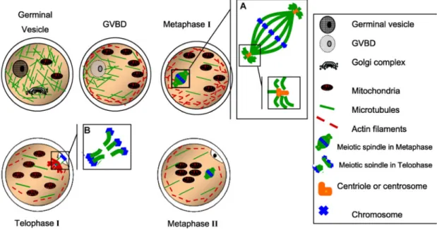
are able to achieve the final steps of meiosis. The later diameter is accomplished in bovine oocytes from follicles of 2-<3 mm (Fair et al. 1995). Under the microscope the first mark of meiotic restart is the disappearance of the GV membrane, or germinal vesicle breakdown (GVBD), followed by chromosomal condensation and alignment into the first metaphase plate and extrusion of the first polar body (PB) to eliminate the duplicated genetic material and keep a transitory 2N chromosomal number. Meiosis then progresses until MII stage when a new blockage takes place (Fig. 1-3). Finally, the meiotic division is again resumed upon sperm activation with the consequent second PB expulsion (Massicotte 2006; Ferreira et al. 2009).
At the molecular level, the activity of the maturation promoting factor (MPF) controls the entry into and exit from M-phase during meiotic progression. MPF that is in turn regulated by the cytostatic factor (CSF), in which Mos (c-Mos) is a component, exerts a kinase activity rendered by p34/cdc2 (CDK1), which forms a complex with the regulatory subunit cyclin B1 (Brunet and Maro 2005; Malcuit and Fissore 2007). As in the mouse (Brandeis et al. 1998), CycB2 (CCNB2) exists in the bovine, although only CycB1 (CCNB1) is the limiting factor for meiosis restart in the latter species: CycB1 is one (Levesque and Sirard 1996; Sirard et al. 1998; Sirard 2001) of the newly synthesized proteins during the GV-GVBD transition and is required for such developmental progression (Coenen et al. 2004;
Massicotte et al. 2006). It must be noted that CycB1 in cattle has two alternative isoforms whose translation is regulated through polyadenylation (Tremblay et al. 2005). Concerning the molecular dynamics of CycB1 and MPF activity through bovine oocyte maturation, it has been observed that CycB1 protein appears after 3 h of initiated IVM (Levesque and Sirard 1996) and 4 h in advance to GVBD (Coenen et al. 2004). Subsequently, as demonstrated by the studies of Wu et al. (1997) a peak of MPF activity allows chromosome condensation (MI) by 6-12 h of maturation, while MPF activity level decreases at the AI/TI transition after 16-18 h of culture (the Bos taurus protein bears the classic biphasic activity pattern of CycB1), then leading to 1st PB extrusion by 18-20 h with an increase in MPF
activity with a plateau by 20-24 h IVM (MII). Finally, MPF in conjunction with CSF brings the oocyte into an arrested state at MII (Russo et al. 2009).
Figure 1-3 Nuclear maturation (Ferreira et al. 2009)
A) and B) insets of the meiotic spindle. GVBD, germinal vesicle breakdown. Scheme reproduced with permission of Elsevier © 2009.
1.3.2. Cytoplasmic maturation
This series of events occurs in the ooplasm and although cytoplasmic maturation begins before LH surge, it is simultaneous at some points with meiosis resumption since cytoplasmic modifications are not finished until the final capacitation of the egg. According to Sirard (2001) and Ferreira et al. (2009), cytoplasmic maturation consists in both the
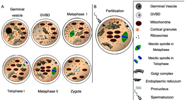
repositioning of organelles and cortical granules (CGs) developed during oocyte growth, as well as cytoskeletal dynamic changes (Fig. 1-4). Frequently, molecular maturation is considered as part of the cytoplasmic process, but the first will be discussed later.
Organelle redistribution directly depends on the appropriate function of cytoskeletal components like microtubules and microfilaments. According to the cell’s requirements mitochondria are transferred to subcellular compartments of high- energy demand during oocyte maturation. Hence, such organelles migrate from a more peripheral distribution before LH discharge in vivo (GV-oocyte) to a more homogeneous allocation by 15 h after LH stimulation (MI), and to a centrally clustered localization closely before or after ovulation, around 24 h following the LH peak (MII). The same reallocation occurs for lipid droplets that increase in number and size. The movement of mitochondria and lipid droplets follows a similar pattern during IVM (Wang et al. 2009). Simultaneously, Golgi complexes gradually decrease in number. Other structures are subjected to dynamic changes towards the end of maturation: The GCs notably proliferate in comparison to their first appearance by the secondary follicle stage, and are placed directly below the plasma membrane in preparation to block polyspermy through exocytosis of their content (Soloy et al. 1997; Ferreira et al. 2009). Such redistribution of CGs (Fig. 1-4) is apparently associated with oocyte size in the bovine since Otoi et al. (1997) observed that those gametes reaching a diameter of 115-120 μm are less susceptible to polyspermy. In addition, ribosomes regroup around chromosomes. All these changes reflect a basal metabolic level in the overall cell and the aim of the oocyte to save energy in preparation for fertilization, and the readiness of energy sources (lipid compartments associated to mitochondria) for the earlier embryonic cleavages (Hyttel et al. 1997). Alternatively, it is known that the oocyte loses its attachments to the cumulus cells through gap junctions by the end of maturation as a pre-requisite for cumulus expansion. This communication has been demonstrated as indispensable during maturation as the gamete utilizes lactate, pyruvate, and even alanine, malate, aspartate, and oxalacetate as energy molecules taken from the cumulus cells and then processed in the ooplasm (Cetica et al. 2003), in addition to their participation in meiotic modulation (Ali and Sirard 2005; Atef et al. 2005).
Figure 1-4 Cytoplasmic maturation (Ferreira et al. 2009)
A) Stages from germinal vesicle to zygote; B) intracellular calcium (Ca2+) release upon
sperm entry. GVBD, germinal vesicle breakdown. Scheme reproduced with permission of Elsevier © 2009.
1.3.3. Molecular maturation
Typically, molecular maturation has been considered as part of the cytoplasmic maturation, since the first occurs in the cytoplasm of the developing egg (or at least the vast majority of such process does). However, the complexity and the undeniable importance of the molecular maturation for oocyte development has frequently prompted discussion as a separate biological event. According to Sirard et al. (2006) and Ferreira et al. (2009), the molecular maturation consists of the transcription, stocking, and processing of transcripts in the oocyte during the growth of the gamete before the transcriptionally silenced period that marks the final maturation of the oocyte up to the activation of the embryonic genome. These events are concomitant at some points with the nuclear and cytoplasmic maturation, and also include the translation and storage of some of the codified proteins by the transcribed/stored mRNAs. Conversely, a wide diversity and amount of the molecular messages will only be translated at the appropriate moment when their codified proteins become necessary for the female gamete. For this reason, an adequate mechanism for the deposition of stabilized transcripts must be accomplished in the ooplasm. Moreover, such a system must also guarantee the safety of the mRNAs to avoid their degradation (Sirard et
al. 2006; Ferreira et al. 2009). Some details of the mechanisms involved in molecular maturation are discussed next.
1.4. Molecular Regulation of the Maternal Stockpile
1.4.1. Oocyte transcription activation and silence
In comparison to the oocyte in mice, the female gamete in cattle is transcriptionally inactive both during the primordial and primary follicle stages (granular nucleoli lacking of fibrillar centers), until the formation of the secondary follicle. At such moment, transcription is then instigated in the bovine oocyte, accompanied by the transformation of the nucleolus to a conformation capable of producing ribosomes (migration of fibrillar centers towards nucleoli). Subsequently, synthesis of mRNAs importantly increases up to a maximum level at the early antral phase, when the oocyte reaches approximately 110 μm in diameter closely before ceasing to grow up (Fair et al. 1997; Hyttel et al. 1997; Hyttel et al. 2001). Since that moment the oocyte decreases mRNA production rates. When it measures 120 μm in diameter (still at GV-phase, just before the onset of the final maturation), the cow’s gamete is practically transcriptionally quiescent, as remarked by Lodde et al. (2007) and Lodde et al. (2008), who observed a correlation of this condition with an electron-dense fibrillar nucleolus and condensed chromatin (non-permissive conformation for transcriptional machinery). This transcriptionally inert form is common to distinct species and will be kept in the oocyte during all maturation steps, as well as throughout the first embryonic cleavages until embryonic genome activation, EGA (rev. De Sousa et al. 1998; Sirard 2010; Macaulay et al. 2011; Clarke 2012). Of note is the fact that Mamo et al. (2011) determined by microarray analysis that the abundance level of 589 transcripts (~28% of the evaluated pool) increased upon IVM in bovine oocytes and this trend was verified by RT-qPCR of selected candidates, where seven transcripts corroborated such behavior even when random primers were employed. Such findings could be explained by the very low transcription rate existent at the very end of the growth phase, just prior transcription it is completely suppressed at the onset of meiosis resumption (rev. Labrecque and Sirard 2014). Thus, the female gamete must produce and store enough molecular messages and proteins during the growth phase in order to support development in the course of transcriptional inactivity (rev. De Sousa et al. 1998; Sirard 2010; Macaulay et al. 2011; Clarke 2012).
1.4.2. Oocyte global transcriptome profile during maturation
Several studies on the analysis of the differences in mRNA levels during maturation of cattle oocytes will be described in this part of the review. There are strong evidences suggesting that a crucial part of the processes conferring the bovine oocyte with the molecular machinery to be developmentally competent take place during oocyte growth (Blondin et al. 1997a; Blondin et al. 1997b; Sirard et al. 1999; Blondin et al. 2002), before the final evolution from GV to MII. As a brief note, Sirard et al. (2006) defined
developmental competence as the capacity of an oocyte to produce a healthy offspring. A
widely accepted simplified measure of developmental competence is the ability of an egg to produce a blastocyst. For a thorough review of molecular markers of competence in oocytes from mouse, human, and cow see Wrenzycki et al. (2007) and Labrecque and Sirard (2014). A number of models used to discern the developmental competence level of oocytes in cattle include: Follicle size (Robert et al. 2000; Donnison and Pfeffer 2004; Mourot et al. 2006; Racedo et al. 2008; Caixeta et al. 2009), in vivo or in vitro maturation (Lonergan et al. 2003; Katz-Jaffe et al. 2009), age of donor (Patel et al. 2007; Dorji et al. 2012), follicular stage (Ghanem et al. 2007; Lingenfelter et al. 2007), electrophoretic migration (Dessie et al. 2007), FSH coasting duration (Labrecque et al. 2013), BCB staining (Ghanem et al. 2007; Opiela et al. 2008; Torner et al. 2008; Opiela et al. 2010), and chromatin configuration (Lodde et al. 2007; Lodde et al. 2008; Labrecque et al. 2015).
Oocyte maturation represents the first developmental phase following the start of the transcription blockage subsequent to the female gamete growth. Therefore, the study of the transcriptome at this stage can help understanding both the molecular regulation aimed at favoring an adequate maturation, as well as the subsequent modulation of the progressing steps through the first embryonic cleavages until EGA. In agreement with Fair et al. (2007), there is a difference in the mRNA levels of several genes at distinct moments during maturation of cattle oocytes, even considering the transcriptional quiescent period encompassing GV-MII. Discrepancies in transcripts levels pre- and post-maturation have also been reported in mouse (Cui et al. 2007) and human oocytes (Assou et al. 2006). Nevertheless, the variation of the amount of specific mRNA species is likely influenced by their poly(A) elongation and/or shortening, and selective degradation or translation, rather
than transcription occurrence (Fair et al. 2007). Overall, it is considered that throughout the transition from MI to MII about 30% of mRNA stockpiles are selectively degraded (Conti 2011).
Dalbies-Tran and Mermillod (2003) compared the transcript profile of cattle GV- and MII oocytes and found differential abundance in the mRNA of 70 genes during nuclear maturation, which were ontogenetically grouped in cell cycle control, apoptosis, and DNA transcription. Later on Fair et al. (2007) indicated that transcripts like GDF9, ZP2, ZP3, and
NALP5 (MATER) are overrepresented in GV- in comparison to MII- bovine oocytes. In
such studies it was concluded that the major presence in immature gametes of mRNAs with preferential-, GDF9 and NALP5 (Pennetier et al. 2004), and specific presence, ZP2 and ZP3 (Topper et al. 1997), in cattle oocytes might indicate an important and time-specific action of such genes prior to nuclear maturation (Fair et al. 2007). In the last survey more than 800 transcripts displayed variable levels during meiotic maturation. By comparing these results with the findings of Misirlioglu et al. (2006), Fair’s team found that common transcripts with higher abundance at MII mainly belonged to gene ontology (GO) categories as metabolism, transport, and cell death regulation. Thus, it was concluded that mRNAs involved in DNA regulation, metabolism, and internal/external signaling appear to be overrepresented at MII in bovine oocytes (Fair et al. 2007). This could appear contradictory due to the general regard of oocyte maturation as a period when mRNA synthesis is practically null. However, in agreement with Memili et al. (1998) and Pennetier et al. (2005) transcriptional activity is present in bovine oocytes during a short time window at the beginning of IVM (before GVBD). In a more recent survey, Mamo et al. (2011) detected that the top networks enriched in transcripts with differential abundance levels between GV and MII cattle oocytes corresponded to cellular assembly and organization, protein trafficking, translation, post-translational modification, and cell to cell signaling. On the other side, special care should be taken when considering a given transcript as preferentially represented in MII- in comparison to GV-oocytes as such difference might rather obey a major detection of the mRNA species due to poly(A) tail length variation and not to an increase in the presence of the transcript (Thelie et al. 2009). Such mechanism will be further discussed in section 1.4.3. Other possible source for the increase in the levels of
specific transcripts during maturation could be a proposed novel model of RNA transfer in the follicle, given that it was determined migration of mRNA and non-coding RNA from cumulus cells to the oocyte during maturation in the cow (Macaulay et al. 2014; Macaulay et al. 2015).
By using cross hybridization it has been possible to identify oocyte-specific (or preferential) genes conserved in distinct species. Vallee et al. (2005) and Vallee et al. (2006) confirmed that the transcripts GDF9, BMP15, ZP, and Spindlin with a widely known presence in oocytes, together with several with no reported role, as MLF1IP, BTG4, and xPTB are located only in the oocyte of mice (GV-), cattle (GV-), and X. laevis (immature, stage IV-V). Interestingly, a total of 208 mRNAs, including ZAR1, were found as maternal transcripts shared in all the species involved, suggesting a wide conservation of regulatory molecular mechanisms in oocytes. Furthermore, gene ontology (GO) analyses revealed that the top biological processes were RNA metabolism and cell cycle, while the main cellular component and the major molecular function were RNP complex and RNA binding, respectively (Vallee et al. 2008). Even if not a complete picture of the evolutionarily conserved oocyte maturation mechanisms was obtained since only immature oocytes were processed, results provided relevant insights of common molecular networks conserved in three phylogenetically distant species at the onset of female gamete maturation (Vallee et al. 2008).
1.4.3. Polyadenylation and deadenylation during maturation
In addition to the oocyte mRNA control by the masking system described for mRNP bodies, the 3’-polyadenylation status of the transcripts is a basic system used by the egg to modulate the recruitment of a given mRNA for translation (to suppress or to induce it). In somatic cells most nascent transcripts receive a poly(A) tail in the nucleus and bind to ribosomes for protein synthesis shortly after they are exported to the cytoplasm. Conversely, some mRNAs produced during oocyte growth (and several of them remaining up to embryonic cleavage stage) are translationally repressed by cytoplasmic deadenylation (Bachvarova et al. 1985; Paynton et al. 1988). At this point deadenylation precedes masking and storage. Thus, Eichenlaub-Ritter and Peschke (2002) remark that transcripts with a