Effect of exercise training on preeclampsia superimposed on chronic hypertension in a mouse model
par
Suzanne Dominique Genest
Département de Physiologie Faculté de Médecine
Mémoire présenté à la Faculté de Médecine en vue de l’obtention du grade de maîtrise en Physiologie moléculaire, cellulaire et intégrative
Août 2013
Université de Montréal
Faculté des études supérieures et postdoctorales
Ce mémoire intitulé:
Effect of exercise training on preeclampsia superimposed on chronic hypertension in a mouse model
Présenté par :
Suzanne Dominique Genest
a été évalué par un jury composé des personnes suivantes :
Guy Rousseau, président-rapporteur Julie L. Lavoie, directrice de recherche
Jolanta Gutkowska, co-directrice Kristi Adamo, membre du jury
Résumé
La prééclampsie est l’une des causes primaires de mortalité et morbidité périnatales, touchant 2-7% des grossesses. Sa prévalence augmente à 10-25% chez les femmes hypertendues. Jusqu’à maintenant, aucun traitement, mis à part l’accouchement précoce, n’est connu. Néanmoins, plusieurs études épidémiologiques suggèrent une diminution de l’incidence de la prééclampsie chez les femmes entraînées quoique, ces études sont considérées insuffisantes. Ainsi, le but de cette étude est de déterminer si l’entraînement avant et pendant la grossesse prévient la maladie dans un modèle animal de prééclampsie superposée à de l’hypertension chronique (SPE).
Nous avons utilisé des souris double transgéniques, surexpirmant la rénine et l’angiotensinogène humaines (R+A+), puisqu'elles sont hypertensives à la base, et développent plusieurs symptômes de la prééclampsie. Pour l'entraînement, les souris ont été mises dans des cages d’exercice 4 semaines avant leur grossesse et y sont restées jusqu’au sacrifice.
L'entraînement physique a prévenu la hausse de pression artérielle en fin de gestation présente chez les souris R+A+ sédentaires, possiblement via l’axe de l’angiotensine-(1-7). Le rapport entre l’albumine: créatinine a également été réduit avec l’entraînement. Les altérations placentaires ont été prévenues chez les souris entraînées, améliorant le développement placentaire et fœtal. Ceci était accompagné d'une normalisation de sFlt-1 circulant et placentaire. De plus, l’augmentation du récepteur à l’angiotensine II de type 1 et la diminution du récepteur Mas dans le placenta étaient renversées.
L’entraînement semble prévenir plusieurs symptômes de la SPE dans un modèle animal suggérant qu'il pourrait être d'une grande utilité dans la prévention de la maladie chez la femme.
Mots-clés : Prééclampsie superposée à de l’hypertension chronique, entraînement physique, modèle transgénique, système rénine-angiotensine, placenta.
Abstract
Preeclampsia is among the leading causes of perinatal mortality and morbidity, affecting 2-7% of pregnancies. Its incidence increases to 10-25% in already hypertensive women. To date, no treatment, aside from delivery, is known. Interestingly, several studies have reported that exercise training (ExT) can reduce preeclampsia prevalence although the available studies are considered insufficient. Therefore, the aim of this study is to determine the impact of ExT when practiced before and during gestation on pregnancy outcome in a mouse model of preeclampsia superimposed on chronic hypertension (SPE).
To do so, mice overexpressing both human angiotensinogen and renin (R+A+) were used because they are hypertensive at baseline and they develop many hallmark features of SPE. Mice were trained by placing them in a cage with access to a running wheel 4 weeks before and during gestation.
ExT in this study prevented the rise in blood pressure at term observed in the sedentary transgenic mothers. This may be realized through an increased activity of the angiotensin-(1-7) axis in the aorta. In addition, ExT prevented the increase in albumin/creatinine ratio. Moreover, placental alterations were prevented with training in transgenic mice, leading to improvements in placental and fetal development. Placental mRNA and circulating levels of sFlt-1 were normalized with training. Additionally, the increase in angiotensin II type I receptor and the decrease in Mas receptor protein were reversed with training.
ExT appears to prevent many SPE-like features that develop in this animal model and may be of use in the prevention of preeclampsia in women.
Keywords: Superimposed preeclampsia on chronic hypertension, exercise training, transgenic model, renin-angiotensin system, placenta.
TABLE OF CONTENTS
Résumé ... i
Abstract ... iii
List of tables ... vii
List of figures ... viii
List of abbreviations ... ix Acknowledgements... xv Chapter 1 - Introduction ... 1 Chapter 2 - Pregnancy ... 1 2.1 The placenta ... 2 2.1.1 Placentation ... 2
2.1.2 Role of oxygen tension ... 4
2.2 Cardiovascular and hemodynamic adaptations ... 7
Chapter 3 - Preeclampsia ... 15
3.1 Symptoms ... 15
3.2 Risk factors of PE ... 19
3.2.1 Epidemiology ... 19
* Hydrops fetalis: Fetal disorder characterized by an abnormal accumulation of fluide in a minimum of two (2) fetal compartments. ... 22
3.2.2 Genetic factors ... 22
3.2.3 Dietary supplementation, stress, smoking and exercise training ... 24
3.3 Etiologies of PE ... 26
3.3.1 Abnormal placentation ... 26
3.3.2 Endothelial dysfunction ... 30
3.3.3 Oxidative stress ... 34
3.3.4 Renin-angiotensin system ... 35
Chapter 4 - Exercise training ... 40
4.1 Exercise training benefits for non-pregnant individuals ... 40
4.2 Exercise training and pregnancy ... 44
4.3 Exercise training and PE ... 47
4.3.1 Potential mechanisms involved in the beneficial impact of exercise of PE54 Chapter 5: Objectives and Hypotheses... 59
Chapter 6: Novel role of the renin-angiotensin system in the beneficial effects of exercise on a preeclampsia model. ... 60
Chapter 7: Discussion ... 117
List of tables
Table 1: PE risk factors ... 22
Table 2 - Circulating factors in pregnancy and preeclampsia ... 34
Table 31: Effect of ExT and SPE on ratio of the whole heart and its compartments to tibia length at the end of pregnancy. ... 91
Table 42: Characterization of placental pathology. ... 92
Table 53. Fetal consequences of SPE-like phenotype and ExT. ... 93
Table 6S1: Primer sequences used for real-time PCR. ... 111
Table 7S2: Maternal characteristics. ... 112
Table 8S3: Cardiac parameters calculated following echocardiography. ... 113
List of figures
Figure 1: The RAS and the effects of the different components via their respective receptors . 9 Figure 2 : Regulators of gestational blood pressure control ... 14
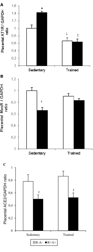
Figure 31. Effect of training and genotype on mean arterial pressure (MAP) and proteinuria. ... 94 Figure 42. Left ventricular gene expression of Nab1 and BNP. ... 95 Figure 53. Effect of ExT and SPE on angiogenic balance. ... 96 Figure 64. Modulation of placental AT1R, MasR and ACE2 protein expression by a SPE and
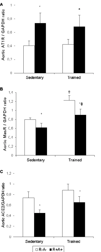
ExT. ... 97 Figure 75. Effect of SPE-like phenotype and exercise on aortic AT1R, MasR and ACE2 protein
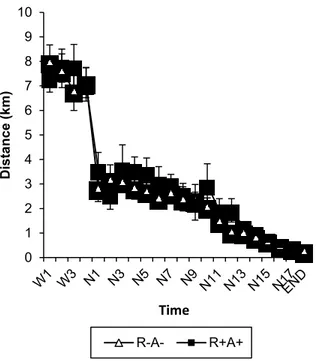
expression, respectively. ... 98 Figure 8S1: Distance travelled prior to and throughout gestation. ... 107 Figure 9S2: Effects of exercise training and SPE on placental cytokeratin immunostaining. 108 Figure 1S3: Effects of exercise training and SPE on placental histone H3 immunostaining. . 109 Figure 1S4: Changes in circulating glucose levels with ExT and SPE-like phenotype. ... 110
List of abbreviations (P)RR: Pro-renin receptor
ACE: Angiotensin-converting enzyme ACE2: Angiotensin-converting enzyme 2
ACEI: Angiotensin-converting enzyme inhibitors
ACOG: American College of Obstetricians and Gynecologists ACR: Albumin/creatinine ratio
ADH: antidiuretic hormone AGT: Angiotensinogen AMPA: Aminopeptidase A AMPM: Aminopeptidase M AngI: Angiotensin I Ang-(1-7): Angiotensin-(1-7) AngII: Angiotensin II AngIV: Angiotensin IV AP: Arterial pressure
ARB: AT1 receptor blockers
AT1R: Angiotensin II type 1 receptor AT2R: Angiotensin II type 2 receptor AT1-AA: AT1R auto-antibodies BNP: Brain natriuretic peptide CaCl2: Calcium chloride
cDNA: complementary DNA
cGMP: cyclic guanosine monophosphate CO: Cardiac output
CVD: Cardiovascular diseases DNA: Deoxyribonucleic acid E-cadherin: Epithelial cadherin
EDTA: Ethylenediaminotetraacetic acid EF: Ejection fraction
ELISA: Enzyme-linked immunosorbent assay eNOS: endothelial nitric oxide synthase ExT: Exercise training
FS: Fractional shortening
GAPDH: Glyceraldehyde 3-phosphate dehydrogenase GFR: Glomerular filtration rate
GPx: Glutathione peroxidase hANG: Human angiotensinogen HR: Heart rate
HELLP: Hemolysis elevated liver enzymes low platelet HIF-1: Hypoxia-inducible factor-1
HLA: Human leukocyte antigen HPS: Hematoxylin phloxine saffron hREN: human renin
HSP: Heat shock protein
ICAM-1: Intercellular adhesion molecule-1 IL: interleukin
IFN-: Interferon-
iNOS: inducible nitric oxide synthase
IRAP: Insulin-regulated aminopeptidase – Angiotensin IV receptor IUGR: intra-uterine growth restriction
IVS: Interventricular septum
KIR: Killer-cell immunoglobulin-like receptors LV: Left ventricle
LVD: Left ventricular diameter
LVEDD: Left ventricular end-diastolic diameter LVEDV: Left ventricular end-diastolic volume LVESD: Left ventricular end-systolic diameter LVESV: Left ventricular end-systolic volume LVID: Left ventricular internal diameter
LVPW: Left ventricular posterior wall thickness MAP: Mean arterial pressure
MasR: Angiotensin-(1-7) Mas oncogene receptor MgCl2: Magnesium chloride
M-MLV: Moloney murine leukemia virus reverse transcriptase MTHFR: Methyltetrahydrofolate reductase
Na3VO4: Sodium ortovanadate Nab1: NGFI-A binding protein 1 NaCl: Sodium chloride
NEP: Neutral endopeptidase
NF-: nuclear factor kappa activated by B cells NK: Natural killer
nNOS: Neuronal nitric oxide synthase NO: Nitric oxide
OT: Oxytocin
PAI-1: Plasminogen activator inhibitor-1 PCR: Polymerase chain reaction
PE: preeclampsia PGI2: Prostacyclin
PlGF: Placental growth factor
PMSF: Phenylmethanesulphonylfluoride PO2: Oxygen partial pressure
RAS: Renin angiotensin system RBF: Renal blood flow
RNS: Reactive nitrogen species ROS: reactive oxygen species RT: Reverse transcription S16: 40S ribosomal protein S16
SDS: Sodium dodecyl sulfate sEng: Soluble endoglin
sFlt-1: Soluble Fms-like tyrosine kinase-1 SHR: Spontaneously hypertensive rat SOD: Superoxide dismutase
SPE: Preeclampsia superimposed on chronic hypertension STBM: Syncytiotrophoblastic microfragments
SV: Stroke volume
TGF1: Transforming growth factor 1 TGF3: Transforming growth factor 3 TLR: Toll-like receptor
TNF: Tumor necrosis factor TPR: Total peripheral resistance TBS: Tris buffered saline
VE-cadherin: Vascular endothelial cadherin VEGF: Vascular endothelial growth factor
Acknowledgements
Ce mémoire conclut 2 ans de travail. Ces quelques lignes sont pour exprimer ma reconnaissance et ma gratitude envers tous ceux et celles qui, de près ou de loin, y ont contribuée.
J’exprime en premier lieu ma gratitude aux Dres Julie L. Lavoie et Jolanta Gutkowska, directrice et co- directrice de projet, pour leur encadrement et leurs conseils. Je souhaite néanmoins remercier plus particulièrement le Dre Julie Lavoie pour son indéfectible soutien aussi bien sur le plan humain que scientifique.
Nombreux sont ceux et celles qui ont, au cours de ces années, apporté leur contribution scientifique voire leur secours. Je tiens ainsi à remercier Danghao Wang, Ahmed Menouar et Basma Ahmed pour l’enseignement des techniques moléculaires de base et leurs conseils avisés sur l’analyse de données, ainsi que Catherine Michel et Sonia Kajla pour leur soutien technique dans l’animalerie et leur aide concernant les manipulations animales. Ce travail a bien sûr été facilité par la bonne ambiance qui règne au laboratoire des Dres Gutkowska et Lavoie. Je tiens également à remercier l’Université de Montréal et les Fonds de recherché du Québec – Santé pour avoir financé mon étude.
Pour leur soutien, je remercie famille et proches, en particulier ma mère qui m’a épaulée jusqu'à la fin sans faillir.
Chapter 1 - Introduction
Preeclampsia (PE) is a pathology that develops during pregnancy. Although the disease has been known for centuries, PE remains a disease of theories. Several components have been found to be implicated in disease progression, but are not seen consistently among all women who develop PE. Importantly, underlying medical conditions, like chronic hypertension and diabetes, are known to increase a woman’s risk of developing PE.
Although research concerning PE has blossomed in the last 2 decades, treatment options are still lacking. The risk of developing this gestational disease will continue to rise, given the increase in prevalence of contributing factors (i.e. obesity, chronic hypertension and diabetes)[1]. As such, PE prevention appears to be the best option for minimizing the prevalence of the disease worldwide.
The purpose of this memoire is thus to investigate the therapeutic effect of exercise training in a mouse model of preeclampsia superimposed on chronic hypertension (SPE), as well as to investigate potential mechanisms implicated.
Chapter 2 - Pregnancy
2.1 The placenta 2.1.1 Placentation
Pregnancy is characterized by the development of a new organ: the placenta. The fetal side of the placenta develops from cells originating from the fertilized egg, while the maternal side develops from the mother’s uterine tissue. This feto-maternal organ plays a primordial role throughout pregnancy by providing the nutrients and oxygen needed for fetal growth, and eliminating waste products. Human pregnancy, much like that of rodents, is characterized by a hemochorial placentation, in which the maternal blood comes in direct contact with the fetal chorion[2].
The effectiveness of this placental system is a result of its unique anatomical structure. The intricate network of vessels scattered throughout the organ effectively perfuses the placenta. In addition, the extensive villous tree structures enhance the area available for gas, nutrient and waste exchange for the growing fetus. As such, placental vasculogenesis, angiogenesis and pseudovasculogenesis are critical processes for a successful pregnancy[3]. It is important to understand that the placenta contains both maternal and fetal vessels. On the maternal side, remodeling of the vasculature is indispensable to ensure an adequate delivery of blood for placental development and fetal growth. The fetus is however
responsible for the formation of an intricate vascular tree, which will exponentially enhance the surface area available for exchange of substances[4].
The placental villous tree development begins after implantation[4]. The initial step involves the formation of primary villi, which consist of columns of cytotrophoblasts, located on the fetal side. These primary villi become secondary villi following invasion of the mesenchyme. Once fetal capillaries can be seen within the villi structure, they are termed tertiary mesenchymal villi. These three steps are repeated throughout pregnancy and contribute to the villous tree structure[3]. Some placental villi become anchored to the basement membrane of the uterus, while others remain bathed in maternal blood, both of which are derived from cytotrophoblasts. Floating villi develop as a result of fusion of cytotrophoblasts to form multinucleated syncytiotrophoblasts. Distal cytotrophoblasts will attach themselves to the maternal tissue, creating bridges between mother and fetus. Additionally, the cytotrophoblast penetrate deeply into the uterine and maternal vasculature, via interstitial and endovascular invasion, respectively. Endovascular invasion leads to the acquisition of an endothelial adhesion molecular phenotype by cytotrophoblasts, a process known as pseudovasculogenesis, and the removal of the endothelial and muscular linings of uterine arterioles[2, 5]. Endovascular invasion occurs directly via the lumen and within the vessel itself, below the endothelium[6]. Vascular remodeling reaches its full extent by mid pregnancy, having modified the spiral arteries of the
endometrium and superficial region of the myometrium[6]. The remodeled segments are lined exclusively with cytotrophoblasts; endothelial and smooth muscle cells are no longer detectable. The disappearance of the muscular tunica media converts these normally high resistance vessels into high capacitance (low resistance) vessels[7]. Indeed, the diameter of the uterine artery increases two-fold by 21 weeks of pregnancy, thereby contributing to an increase in placental perfusion and, thus fetal growth[8]. In fact, uterine blood flow increases 20-fold during pregnancy as a result of this remodeling[9]. These vessels also become unresponsive to vasoactive substances as they lack an endothelium, thereby maintaining an adequate and consistent blood flow to the growing fetus.
2.1.2 Role of oxygen tension
The first trimester is of critical importance for the success of a pregnancy. During this period, changes in oxygen tension modulate cytotrophoblastic proliferation and differentiation[10, 11]. Blood flow to the placenta is initially minimal, creating a relatively hypoxic environment. Indeed, oxygen tension is suspected to be around 20 and 40 mmHg at 8-10 weeks in the intervillous space of the endometrium and decidua, respectively, in comparison to 60 and 45 mmHg, respectively, when evaluated at 12-13 weeks, after trophoblastic invasion has begun[12]. Very little endovascular invasion takes place at low oxygen tensions, further contributing to low placental perfusion. This stage of pregnancy is thus characterized by a rapid placental growth and contributes to the future growth and
development of the fetus by ensuring the placenta’s ability to promote gas, nutrient and waste exchange.
Under low oxygen tension, cytotrophoblasts are highly proliferative[10]. Indeed,
in vitro studies have demonstrated that cyclin B, a protein required for entering the
mitosis phase of the cell cycle, is significantly increased under low oxygen tension (oxygen partial pressure (pO2) of 2%), compared to standard conditions (pO2 of 20%)[10]. Additionally, this group also observed a decrease in p21WF1/CIP1, a protein involved in cell cycle arrest under hypoxic conditions, demonstrating a reduction in mitotic inhibition thereby favouring proliferation. This reduction in p21 is likely mediated in part by the transforming growth factor (TGF) 3, in line with the premise that TGF inhibits trophoblastic invasion[13]. This highly proliferative state is accompanied by a concomitant decrease in the cell’s ability to differentiate, as demonstrated by specific antigen expression, such as a reduction in 1 integrin required for invasiveness[10]. As proliferation continues to take place, peripheral cytotrophoblasts gain access to maternal arterial blood, rich in oxygen. Cytotrophoblastic cells go from being proliferative to invasive around week 10[10, 14, 15].This change in oxygen tension consequently causes a decrease in the cells’ ability to proliferate[5]. Instead, the endovascular cytotrophoblasts begin to differentiate and acquire an endothelial-like expression pattern of cell adhesion molecules, which enhances their motility and invasiveness[5]. Indeed, in
vitro studies have demonstrated that an increase in oxygen modifies V integrin
which are specific to endothelial cells, and decreasing 64 and 63 integrins, which are characteristic of epithelial cells[5, 16]. Invasive trophoblasts also have a reduced E-cadherin (epithelial cadherin) and increased VE-cadherin (endothelial cadherin) expression, providing further evidence that cytotrophoblasts undergo differentiation by mimicking endothelial cell antigen expression[5]. Relatively high oxygen tension thus promotes differentiation of cytotrophoblasts and endovascular invasion, and may explain why invasion of the arterial rather than the venous side of the uterine circulation is favoured.
Oxygen tension is known to modulate hypoxia-inducible factor-1 expression (1[17]. Under hypoxic conditions, much like during early pregnancy, HIF-1 levels are increased, as is TGF-3[14]. HIF-HIF-1 stimulates the expression of the vascular endothelial growth factor (VEGF), a potent mediator of angiogenesis and vasodilation. This family of angiogenic factors, along with angiopoietins, are critical for pregnancy. The VEGF family is involved in endothelial cell proliferation, angiogenesis, vascular permeability and inflammation while angiopoietins are involved in endothelial survival, capillary sprouting and vascular stabilization[18, 19]. Angiogenesis and vasculogenesis dominate during the first trimester of pregnancy, when HIF-1 is elevated. By 10 weeks of pregnancy, the expression of HIF-1and TGF- are diminished, which is in line with the increase in oxygen tension that occurs following trophoblastic invasion[14]. Subsequently, a decrease in VEGF occurs as pregnancy progresses[20].
Importantly, modulation in VEGF levels has been postulated to also contribute to trophoblastic invasiveness since VEGF and VEGFR mRNA expression are decreased in biopsies of preeclamptic placentas[21].
2.2 Cardiovascular and hemodynamic adaptations
Pregnancy is a physiological process that necessitates cardiovascular and hemodynamic adaptations to ensure the survival of both mother and fetus. The systemic renin-angiotensin system (RAS) is a key player in many of these changes as it is critical for arterial pressure (AP) control as well as fluid and salt homeostasis in the non-pregnant state (see Figure 1 for detailed depiction of the RAS). Normal pregnancy is characterized by an increase in the circulating levels of prorenin, renin and angiotensinogen[22]. Via this cascade, circulating levels of angiotensin II (AngII) are increased, although this is coupled with a diminished endothelial sensitivity to AngII[23]. AngII is the dominant physiologically active compound produced by the RAS and has several downstream effects, mediated by the AngII type 1 receptor (AT1R). Notably, AngII stimulates vasoconstriction, and the release of aldosterone and antidiuretic hormone (ADH). The increase in circulating levels of aldosterone and ADH (also known as arginine vasopressin (AVP)) during pregnancy contributes to the enhanced renal sodium and water reabsorption, respectively, observed in this condition. Although the systemic RAS plays a pivotal role, the implication of the local RASs cannot be ignored. Their roles have become more apparent as a result of their investigation during diseased-states. For instance, in patients with cerebral aneurysms, hypertension and renal
disease present with inappropriate activation of the RAS thereby contributing to the disease development[24, 25]. Moreover, a functional uteroplacental RAS also exists as all of its components are expressed at the level of the uterus, and the placental and fetal membranes[26]. The fetal RAS is hypothesized to be implicated in fetoplacental blood flow regulation, while that on the maternal side is thought to be involved in the vascular remodelling required for normal pregnancy[22].
Figure 1: The RAS and the effects of the different components via their respective receptors
Thin arrows describe the conversion reactions that make up the RAS pathway. Thick arrows demonstrate the receptor(s) by which these RAS peptides mediate their effects. ACE, angiotensin-converting enzyme; ADH, antidiuretic hormone; AMPA, Aminopeptidase A; AT1R, angiotensin II type 1 receptor; ICAM-1, intercellular adhesion molecule-1; IL-6; interleukin-6; IRAP, insulin regulated aminopeptidase; MasR, angiotensin-(1-7) Mas oncogene receptor; MCP-1, monocyte chemoattractant protein-1; NEP, neutral endopeptidase; NF-, nuclear factor kappa activated by B cells; PAI-1, plasminogen activator inhibitor-1; (P)RR,
Angiotensinogen Angiotensin I Angiotensin II Angiotensin IV Angiotensin-(1-9) Angiotensin-(1-7) Renin Apoptosis Fibrosis Hypertrophy Enhanced contractility (P)RR Vasodilation; antihypertrophic, antifibrotic &antithromboticeffects
IRAP – Angiotensin IV reecptor
Vasodilation; antiproliferation; antihypertrophic, antifibrotic &antithromboticeffects
AT2R
Vasoconstriction; sFlt-1, ADH &aldosterone secretion; proliferation; hypertrophy; Fibrosis; oxidative stress
AT1R Vasodilation; Induces NF-, MCP-1, Il-6, TNF, ICAM-1, PAI-1 MasR AMPM AMPA ACE ACE2 NEP Tonin, Cathepsin G Angiotensin III ACE2 ACE, NEP
pro-renin receptor; TNF, tumor necrosis factor .
The pregnancy-induced volume expansion is characterized by a 45-55% increase in extracellular volume, including a 20-30% elevation in plasma volume[27]. An adequate increase in plasma volume will help maintain an adequate blood flow, and thus nutrient and oxygen delivery, to the growing fetus. Moreover, the increase in circulating blood volume gives rise to a proportionate increase in stroke volume (SV) and thus cardiac output (CO), which will be maintained throughout pregnancy[28]. As a result of the pregnancy-induced hypervolemia, heart rate (HR) also increases to maintain organ perfusion[29]. Paradoxically, AP does not increase during normal pregnancy. In fact, AP decreases during the first and second trimester[30]. During the third trimester, AP rises, returning to pre-pregnancy baseline values, and in some cases somewhat higher, by the end of pregnancy[31].
The vasodilatory state of pregnancy is primarily mediated by a pregnancy-induced reduced response to vasoconstrictors, notably AngII, vasopressin and norepinephrine, by smooth muscle cells and the endothelium[22, 32, 33]. This pressor response is not mediated by changes in receptor affinity or receptor number, but rather results from an increased activity and expression of angiotensin-converting enzyme 2 (ACE2) during pregnancy[34, 35]. Enhanced ACE2 expression was observed in the kidney, the placenta and to a lesser extent,
the uterus[34, 36]. ACE2, a homologue of the angiotensin-converting enzyme (ACE), converts AngII into the vasodilatory angiotensin-(1-7) (Ang-(1-7)) (see Figure 2)[37, 38]. Indeed, plasma Ang-(1-7) is increased during pregnancy in women[39]. Additionally, animal studies have demonstrated enhanced Ang-(1-7) expression in the uteroplacental unit and kidneys[34, 40]. Further supporting the role of ACE2 on blood pressure control are studies demonstrating a reduction in ACE2 expression in several hypertensive animal models[41, 42]. Furthermore,
Gurley et al have observed an increase in baseline blood pressure in certain
genetic strains of ACE2-deficient mice[43]. Degradation of AngII and production of Ang-(1-7) by ACE2 therefore counterbalances the AngII mediated vasopressor effect. Ang-(1-7) mediates its effects by binding to the Ang-(1-7) Mas oncogene receptor (MasR). MasR is a G-protein coupled receptors that ultimately leads to the nitric oxide (NO) production by endothelial nitric oxide synthase (eNOS) and prostaglandin production[44]. MasR may also be implicated in the pregnancy-induced pressor response as Yamaleyeva and colleagues have noted an increase in MasR mRNA expression in the utero-placental unit of an animal model during early gestation[45].
Another important component that contributes to reducing the vasoconstrictive tone of pregnancy involves the generation of vasodilatory mediators like NO. NO is a biologically active signalling molecule that binds soluble guanylate cyclase in smooth muscle cells, and in doing so, produces cGMP (cyclic guanosine monophosphate). cGMP activates protein kinase G which ultimately causes
smooth muscle relaxation. NO is generated by NO synthase (NOS) from L-arginine, O2 and NADPH. There exists three subtypes of this enzyme: endothelial, inducible and neuronal NOS (eNOS, iNOS and nNOS, respectively)[46]. Inhibition of NOS prevents the pregnancy-induced pressure drop in rats, implicating this system in AP control[47]. Several possible mechanisms exist for eNOS activation during pregnancy. Enhanced flow-induced shear stress resulting from pregnancy-induced hypervolemia contributes to NO production[48]. As previously mentioned, the binding of Ang-(1-7) to MasR leads to eNOS activation and NO production, through the activation of Akt-dependent pathways[44]. In
vitro studies have demonstrated the induction of NO production by estrogen
through the effects of eNOS[1, 49]. Elevated estrogen levels throughout pregnancy contribute to the observed vasodilatory state and maintained endothelial function. Abnormal estrogen levels during pregnancy may be involved in gestational pathologies; similarly to the increased cardiovascular risk observed in post-menopausal women[50-52].
Systemic and renal NO production are enhanced during pregnancy, which accounts for the increase in renal blood flow (RBF) and glomerular filtration rate (GFR), along with a decrease in total peripheral resistance (TPR)[23, 28, 53]. Another possible mediator involved in the vasopressor response of pregnancy includes the insulin-regulated aminopeptidase (IRAP), also known as the AngIV receptor. It is expressed by several tissues, notably the placenta and the heart. In the circulation, this enzyme not only degrades oxytocin (OT), but also
vasoconstrictors such as angiotensin III (AngIII), and promoters of cardiac hypertrophy, such as AVP[54, 55]. Circulating levels and activity of IRAP have been reported to be increased during pregnancy[56] and as such, may play a role in maintaining homeostasis during pregnancy[57]. See figure 2 for a summary of the mediators implicated in AP control during pregnancy.
The homeostatic adaptations during pregnancy promote the development of a physiological cardiac hypertrophy primarily due to an increase in left ventricular (LV) thickness[23]. LV function is also improved because of a higher preload, a lower afterload and improved intrinsic contractile properties of the myocardium[23]. Pregnancy, although physiological, is temporary, and as a result, cardiac properties and function return to pre-pregnancy values following delivery, as does AP, SV and HR.
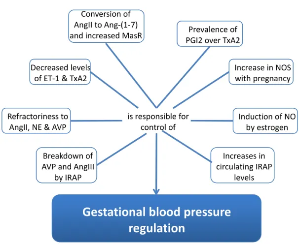
Figure 2 : Regulators of gestational blood pressure control
White boxes depict the mechanisms implicated in blood pressure control during pregnancy. Ang-(1-7) angiotensin-(1-7); AngII, angiotensin II; AngIII, angiotensin III; AVP, arginine vasopressin; ET-1, endothelin-1; IRAP, insulin-regulated aminopeptidase; NE, Norepinephrine; NO, nitric oxide; NOS, nitric oxide synthase; PGI2, prostacyclin; TxA2, thromboxane A2.
Gestational blood pressure
regulation
Decreased levels of ET-1 & TxA2
Breakdown of AVP and AngIII
by IRAP Refractoriness to
AngII, NE & AVP
Conversion of AngII to Ang-(1-7)
and increased MasR Prevalence of PGI2 over TxA2
Increase in NOS with pregnancy Increases in circulating IRAP levels Induction of NO by estrogen is responsible for control of
Chapter 3 - Preeclampsia 3.1 Symptoms
Mild preeclampsia (PE) is diagnosed by an increase in AP above 140/90 mmHg and de novo proteinuria above 300mg/24h after 20 weeks of gestation[58] while severe PE is defined by a diastolic and systolic pressure above 110 and 160mmHg, respectively with the new appearance of proteinuria[59].. As one would expect, the risk of perinatal mortality and morbidity is heightened in cases of severe PE [60]. SPE is characterized by a significant increase in AP (above their already hypertensive values) and de novo proteinuria after 20 weeks of pregnancy[61]. SPE is difficult to diagnose in patients with undiagnosed pre-pregnancy hypertension because, these women can present with normal AP during their first and second trimester as a result of the pregnancy-induced AP decrease. The diagnosis is often made several months after delivery, when AP has still not returned to its normal range.
It is important to understand that PE is a systemic disease that affects virtually every organ, either overtly or subclinically, depending on the cause and severity of the disease. As such, the clinical spectrum of PE varies enormously. This heterogeneity is likely a result of the existence of multiple etiologies[62]. Consequently, several treatment options will likely have to be identified/developed according to these respective subtypes.
PE is normally associated with reversible renal and cardiovascular anatomical and pathophysiological adaptations. The cardiovascular adaptations normally ensue as a result of the increase in afterload[63]. Indeed, high AP is known to cause concentric cardiac hypertrophy in men and non-pregnant women[64]. Similar cardiac findings are observed among preeclamptic patients[65]. Paradoxically, PE is normally associated with a hypovolemic state. Interestingly, preeclamptic women are more susceptible to cardiovascular diseases (CVD) later in life[66, 67]. This may be a result of the development of a pathological cardiac hypertrophy or may be a manifestation of a pre-existing medical condition, which merely progresses with time to CVD, irrespective of whether the patient developed PE, rather than the actual cause of CVD post-PE.
GFR and RBF are normally increased in normal pregnancy as a result of the greater blood volume[28, 30]. However, in PE, both of these components are diminished compared to normal pregnancy[68], resulting in hyperuricemia[69]. Hyperuricemia, if severe, can cause renal failure[70], thereby creating a vicious circle, in addition to being a risk factor for hypertension and CVD[71]. Systemic endothelial dysfunction, commonly observed during PE, can also cause a reduction in both GFR and RBF[72]. Worsening proteinuria or sudden oliguria are signs of severe PE. In very severe cases, kidney failure can arise, necessitating dialysis for the remainder of a woman’s life.
Overt hepatic damage is also usually indicative of severe PE. Generally, 10-15% of PE patients develop HELLP syndrome, which is characterized by hemolysis, elevated liver enzymes and low platelet count[73], and normally requires induction of fetal delivery. A correlation has been reported between low platelet count and perinatal mortality and morbidity[74], as well as between high serum hepatic transaminases levels, a marker of liver damage, and PE severity[75]. HELLP syndrome normally develops in response to over-activation of the coagulation cascade as is observed during PE; thereby promoting the development of microthrombi and giving rise to ischemia and necrosis[35].
Less than 1% of PE cases progress to eclampsia, a disease characterized by convulsions[76]. Those PE patients that do however develop eclampsia will sometimes present with neurological impairment, including headaches, visual symptoms, changes in mental status and lethargy, as a result of cerebral hyperperfusion-induced edema[77]. These patients require close monitoring as an increase in cerebral AP may lead to stroke. Although severe, the prevalence of eclampsia has decreased significantly in the last 20 years because of the use of magnesium sulfate to prevent the progression of PE to eclampsia[78].
Fetal prematurity, resulting from the necessity to induce premature delivery to protect the mother from PE-induced maternal complications is responsible for elevated rates of fetal mortality and morbidity[34]. Fetal symptoms will thus vary
depending on the severity of PE. Since no other treatment exists aside from inducing fetal delivery, fetal consequences are common. These premature neonates may suffer from intra-uterine growth restriction (IUGR). Suboptimal perfusion resulting from PE-induced placental alterations is likely responsible for IUGR [79]. Prematurity and IUGR may have developmental repercussions later in life[34].
Although PE symptoms generally disappear following delivery, these women remain at risk of CVD as previously mentioned earlier in the text. Indeed, several studies have observed an increased risk of CVD in women previously diagnosed with PE[66, 67] and research is now focusing on this issue to better protect these women from prospective diseases. PE will have long-term implications on women’s health and their diagnosis should be used as a marker for underlying diseased state. Similarly, although the apparent fetal consequences of PE are those associated with preterm delivery and IUGR, fetal programming is likely intricately implicated[80-82]. Alterations in placental development, fetal substrate availability and, of importance, the hormonal environment will arise as a result of PE-associated placental hypoxia and oxidative stress. Exposure to high levels of glucocorticoids and nutrient deprivation will alter the expression of transcription factors, receptors, and cell mediators, and in doing so, will increase the fetus’ risk of disorders of adulthood, like CVD, neuroendocrine disorders and psychiatric disorders, among others[83].
3.2 Risk factors of PE
3.2.1 Epidemiology
PE is observed in about 2-7% of healthy nulliparous pregnancies[59, 84]. MacGillivray noted in a well characterized Scottish population that 5.6% of women who experienced a preeclamptic birth were primiparous, while 0.3% of cases occurred during a second pregnancy[85]. It is commonly found in women of a young age, during their first pregnancy[62]. Although an association appears to exist between young age and PE, nulliparity may be the source for this apparent link. It is hypothesized that desensitization or tolerance to paternal antigens is responsible for protecting the mother during future pregnancies with the same mate[86]. Therefore, all factors that modify maternal recognition of paternal antigens tend to have an effect. Indeed, women who have had an abortion[87-89] have a reduced risk of PE while the frequency of PE is superior in women using donor sperm insemination[90] and among those using barrier method contraceptives[91]. Importantly, a pregnancy with a new mate eliminates the protection that was once conferred, lending support to the implication of an immunogenic response as a possible mechanism for PE development[92]. Further supporting this hypothesis is the observation that pregnant women are more likely to develop PE if their partner has previously given rise to a preeclamptic pregnancy[93, 94]. The so-called “dangerous father” therefore demonstrates the existence of paternal factors that contribute to PE risk.
PE prevalence is heightened in women with CVD, including chronic hypertension, metabolic syndrome, obesity, pre-gestational diabetes mellitus, dyslipidemia, and pre-existing thrombophilias[62]. Many of these diseases share common characteristics, like systemic inflammation and endothelial dysfunction. Unfortunately, the rising incidence of CVD will contribute to a further increase in PE prevalence. There is a higher occurrence of PE among women of African descent, which may be due to the higher incidence of hypertension among this group of women[95].
It is estimated that 10-20% of women of childbearing age suffer from hypertension worldwide[83]. This CVD is characterized by an increase in AP, above 140/90mmHg[96]. If left uncontrolled, high AP can lead to stroke and heart attack, among other CVD[97]. Studies have shown a direct link between lowering AP and a decreased risk of CVD and death[98]. Underlying hypertension is an important risk factor for PE, as the prevalence of SPE increases to 10-25% in previously hypertensive women[99, 100]. During pregnancy however, these women also experience a physiological decrease in AP as great as 15-20 mmHg, rendering it difficult to diagnose SPE in previously undiagnosed hypertensive mothers[101]. An increase in AP alone poses a significant risk on fetal outcome[99]. Indeed, perinatal mortality is higher among pregnancies associated with hypertension, with a relative risk of 2.3 compared to normotensive, uncomplicated pregnancies[99]. In an attempt to control AP during pregnancy, care must be taken when choosing a
treatment option, as exposure to anti-hypertensive medications can pose major risks to the fetus. Methyldopa is presently the preferred drug prescribed to hypertensive women during pregnancy, as many others are contraindicated[95, 102]. Inhibitors of the RAS, like AT1 receptor blockers (ARB) or angiotensin-converting enzyme inhibitors (ACEI), have been linked with fetal abnormalities and neonatal death[35]. Interestingly, a recent study by Diav-Citrin et al. fails to observe ARB- and ACEI-mediated teratogenic effects when used during the first trimester[103]. The use of diuretics is also discouraged because of its effect on plasma volume, particularly as PE is associated with a hypovolemic state. Importantly, controlling AP by antihypertensive medication does not treat the disease; it minimizes the risk of developing severe PE. When antihypertensive therapies are not effective at controlling AP, fetal delivery is required, in an attempt to limit negative pregnancy outcomes.
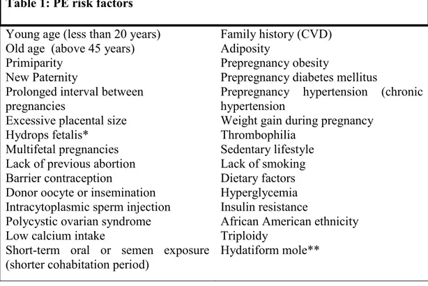
Table 1: PE risk factors Young age (less than 20 years)
Old age (above 45 years) Primiparity
New Paternity Prolonged interval between
pregnancies
Excessive placental size Hydrops fetalis* Multifetal pregnancies
Lack of previous abortion Barrier contraception
Donor oocyte or insemination Intracytoplasmic sperm injection Polycystic ovarian syndrome Low calcium intake
Short-term oral or semen exposure
(shorter cohabitation period) Family history (CVD) Adiposity
Prepregnancy obesity
Prepregnancy diabetes mellitus
Prepregnancy hypertension (chronic hypertension
Weight gain during pregnancy Thrombophilia Sedentary lifestyle Lack of smoking Dietary factors Hyperglycemia Insulin resistance
African American ethnicity Triploidy
Hydatiform mole**
* Hydrops fetalis: Fetal disorder characterized by an abnormal accumulation of fluide in a minimum of two (2) fetal compartments[104].
* Hyaditiform mole: Abnormal pregnancy in which the embryo, amniotic membrane and cord are lacking[105].
3.2.2 Genetic factors
PE is commonly found within members of a same family, providing evidence for the existence of genetics risk factors. For example, a woman born from a preeclamptic birth runs a greater risk (20-40%) of developing PE [106, 107]. Among sisters, the risk can climb to 11-37%[108]. The daughter of a man who was born from a preeclamptic birth is also at a greater risk of suffering from PE[109]. Paternal genes may play a different kind of role in disease development, as depicted by the “dangerous father”, triggering an abnormal immune response against seminal antigens[92, 110]. The link between genetics and PE is far from
simple however. There are various genetic components in play, in addition to their interaction with the environment, which can predispose an individual to this gestational disease[34]. Alone, genetics are unlikely to be responsible for inducing the disease, but they are likely to be implicated in altering an individual’s susceptibility[111].
Association studies have helped elucidate common alleles that are found in women who develop PE. Those extensively studied include methylenetetrahydrofolate reductase (MTHFR), factor V Leiden, prothrombin (factor II), angiotensinogen (AGT), ACE, endothelial NOS (also referred to as NOS3) and human leukocyte antigen (HLA)[112]. MTHFR, factor II and V are involved in inherited thrombophilias, which is a known risk factor for PE. AGT and ACE are critical players in the classical RAS, which has been postulated to be implicated in PE because circulating levels of several RAS components are abnormal in this condition, as mentioned previously. For instance, the AGT T235 polymorphism predisposes women to essential hypertension and, thus, PE, by enhancing plasma and tissue AGT expression, ultimately leading to an increased production of AngII[113, 114]. As previously mentioned, PE is characterized by systemic endothelial dysfunction, rendering the endothelium more responsive to vasoconstrictors and less so to vasodilators. As such, NOS3 is involved in the maintenance of endothelial function. The NOS3 variant Glu298Asp, linked with PE minimize the availability of NO, thereby reducing the potential extent of vasodilation and steering it more towards a vasoconstrictive state[115, 116].
Lastly, HLAs are involved in maternal immune response during pregnancy. The human leukocyte type 2 DR antigens (HLA-DR) are responsible for paternally-derived fetal antigen recognition by the mother. An increase in HLA-DR homozygosity in both parents is associated with the generation of a preeclamptic birth, suggesting that the absence of genetic compatibility between mother and father may play a role in PE initiation[117]. Polymorphisms in other candidate genes have also been identified, including glutathione S-transferase[118].
It is important to understand that maternal and fetal genotypes are not identical. Additionally, the placenta and fetus originate from maternal and paternal genes and thus represents an allograph. An abnormal immune response by the mother could be at the heart of the disease. Interestingly, diseases related to T-cell immunodeficiency, like HIV, have a lower risk of developing PE[119, 120].
3.2.3 Dietary supplementation, stress, smoking and exercise training
To date, the only cure for PE is delivery of the fetus. There are however consequences to this option, as fetal growth and development may be jeopardized in the process if early delivery is chosen. As a result, research has focused on identifying possible pharmaceutical therapies and lifestyle modifications to prevent and control progression of the disease.
Unfortunately, most of the studies investigating the role of dietary supplements in PE have shown no beneficial role. Although oxidative stress has been implicated
in PE development, antioxidant supplementation has been investigated without much success[121]. Interestingly, many of the studies investigating the effects of antioxidants on PE prevention begin supplementation after 14-16 weeks of gestation. At this stage of pregnancy, the processes involved in PE development have already been initiated. Antioxidant supplementation may not be sufficient at this stage to counter the systemic oxidative stress and its damages. Calcium supplementation was shown to be effective only among women with a reduced calcium intake[122]. There is recent evidence indicating that vitamin D supplementation prior to and/or during pregnancy can prevent PE in women[123], although further investigation is required as the exact mechanisms by which vitamin D positively impacts pregnancy remain unclear.
Chronic stress is an environmental factor that can negatively affect CVD, certain cancers, respiratory diseases, etc[124]. Mothers living or working in a stress-ridden environment during pregnancy have been shown to be at a greater risk of delivering prematurely[125]. Additionally, stress at work has been associated with an increased risk of PE[126]. For instance, female resident physicians were more likely to become preeclamptic compared to the wives of their male classmates[127].
Also, although there are many adverse effects of smoking during pregnancy, including fetal growth restriction and placental abruption, it is interesting to note that smokers have a reduced risk of developing PE[128]. The protective effect of smoking is thought to be mediated by carbon monoxide in the placenta[129]. It is
reported that carbon monoxide reduces the production of anti-angiogenic mediators which can cause endothelial dysfunction and thus lead to hypertension and proteinuria[130].
The role of exercise training in modulating PE risk will be discussed in detail in chapter 3.
3.3 Etiologies of PE
PE is a disease of theories; however our understanding of the possible etiologies has increased drastically in the last two decades. There are several mechanisms that have been implicated in the development of PE including abnormal placentation, endothelial dysfunction, oxidative stress, inflammation, immunity and the RAS[62].
3.3.1 Abnormal placentation
The role of the placenta has long been suspected, particularly given that the only treatment for PE is induction of fetal delivery and as such, removal of the placenta. Moreover, the disease may occur during molar and is more frequent during multiple pregnancies, further lending support to a placental role[131, 132]. Placental abnormalities is among the most supported hypotheses for PE development. Indeed, preeclamptic placentas are characterized by the presence of
sclerotic villi, a loss of intermediate villi, a growth in intervillous space and an abundance of syncytial knots[133]. The vasculature is characterized by the presence of foam cells, fibrinoid necrosis and perivascular lymphocyte infiltration, possibly a result of an immunological response[134]. Vascular and thrombotic lesions are detected in vessels on both the fetal and maternal side of the uteroplacental unit and may contribute to the diminished placental perfusion[135] and tissue hypoxia. It is estimated that this reduction in placental blood flow is of the order of 50-70%[136]. Importantly, reduced uterine perfusion pressure has been identified as an animal model of PE[137, 138]. Further aggravating this situation is the abundance of vasoconstrictors, like thromboxane A2 and endothelin-1, in addition to an increased sensitivity to AngII without any changes in that of vasodilators, like prostacyclin (PGI2), NO and Ang-(1-7)[139-141]. The overpowering vasoconstrictive effect, which diminishes placental perfusion, results from a superficial invasion of the spiral arteries by cytotrophoblasts and defective pseudovasculogenesis. Furthermore, the failure of trophoblasts to acquire an endothelial-like adhesion molecule phenotype ensures that the vessels retain their elastic and smooth muscle-like properties, and continue responding to circulating vasoconstrictive substances, further decreasing placental perfusion[135, 142]. Together, adequate placental blood flow is jeopardized, as is fetal growth and development.
The soluble Fms-like tyrosine kinase-1 (sFlt-1) and soluble endoglin (sEng) are anti-angiogenic mediators that have been found to be elevated during PE[143, 144]. Hypoxia causes the placenta to synthesize and release these mediators into the circulation, which is thought to further impact placental development and aggravate hypoxia. Placental sFlt-1 production is also induced by pro-inflammatory cytokines, like TNF-, and its secretion is stimulated by AT1R[145, 146]. In addition to their local effects in the placenta, these mediators enter the circulation and promote systemic endothelial dysfunction, inflammatory responses and oxidative stress.
sFlt1 is a splice variant of the vascular endothelial growth factor receptor (VEGFR or Flt-1), which lacks the transmembrane domain. It therefore circulates in the blood and binds circulating VEGF and placental growth factor (PlGF) which reduces their capacity to attach to their membrane receptors, VEGFR1 (also named Flt-1). As such, the effects on angiogenesis and vascular permeability are blunted[147]. Decreased VEGF availability may also prevent pseudovasculogenesis, since conversion of hematopoietic stem cells into endothelial cells is induced by VEGF[148]. In addition, under hypoxic conditions, a decrease in PlGF gene expression along with an increase in VEGF gene expression in cytotrophoblasts and syncytiotrophoblasts is observed in vitro[149, 150]. Although counterintuitive, the increase in VEGF expression may merely be a compensatory mechanism to counteract the increase in circulating sFlt-1. Finally,
and differentiation[151] by blocking ligand binding. Indeed, the expression of integrin 1, which is normally expressed by endovascular cytotrophoblasts, was significantly reduced in the presence of sFlt-1[151]. Moreover, Zhou et al. also observed an increase in TUNEL staining, thereby demonstrating higher levels of apoptosis[151]. Interestingly however, Flt-1 deletion in an animal model does not affect placentation, suggesting that sFlt-1 during PE may not be implicated in placental alterations. This however cannot be concluded with certainty since sFlt-1 modulates Flt-1, Flk-1 and neuropilin signalling[48, 152]. Moreover, administration of sFlt-1 to pregnant and non-pregnant rats causes hypertension and proteinuria, similarly to the symptoms observed during PE[153]. Similarly, overexpressing sFlt-1 in mice led to the development of PE-like manifestations in pregnant mice, however Lu et al did not observe similar symptoms when transfecting non-pregnant mice[154]. Thus far, sFlt-1 overexpression has been depicted as a deleterious anti-angiogenic state, however lower levels of sFlt-1 during the first trimester have been observed in women who go on to miscarry compared to women who deliver healthy babies[39]. It is important to note that some women with elevated sFlt1 levels do not develop PE, and vice versa, supporting the existence of various etiologies for this disease[143, 155]. Elevated plasma sFlt-1 levels are therefore not specific to preeclampsia.
Endoglin, a coreceptor for TGF 1 and 3, is released by the placenta into the maternal circulation in cases of PE[144]. Antagonization of TGF1 and TGF3 by sEng inhibits TGF signalling pathways, which include eNOS phosphorylation
thereby hindering systemic endothelial function[156-158]. Animal models have shown that sEng alone cannot give rise to all PE features, but does cause an increase in mean arterial pressure (MAP) in pregnant rats[144]. The effects of sEng adrenoviral expression in non-pregnant mice was not however investigated in this study. Animal models in which both sFlt1 and sEng were overexpressed caused severe PE-like features, including a more important increase in MAP compared to sEng alone, proteinuria, HELLP syndrome and fetal growth restriction in pregnant rats and severe vascular injury among their non-pregnant counterparts[144]. These antiangiogenic components thus seem to work together to mediate systemic endothelial dysfunction, inflammation and oxidative stress[159].
Abnormal placentation will negatively impact fetal growth and development, possibly resulting in fetal growth restriction. Fetal growth restriction does not however occur in all women affected by PE, suggesting that the extent of trophoblastic invasion relates to disease severity and that compensatory mechanisms may arise to minimize the effects on the fetus.
3.3.2 Endothelial dysfunction
The endothelium consists of a layer of cells separating vascular smooth muscle cells from circulating blood components. It is responsible for modulating vascular tone and permeability, and plays a critical role in targeting immune cells to the site of injury and controlling the coagulation cascade[160]. Systemic endothelial cell
injury, as is the case in PE, will consequently affect the endothelium’s ability to adequately respond to vasoactive substances, to control vascular permeability and to maintain hemostasis[161]. Systemic endothelial dysfunction is observed biochemically by the synthesis and secretion of markers of endothelial cell injury, like endothelin-1, fibronectin and selectins[162, 163]. Indeed, circulating endothelin-1 levels are greater among PE patients[164]. Additionally, PE patients often present with activation of the coagulation pathway, resulting in microthrombi, and thus contributing to an impaired organ perfusion and endothelial dysfunction[165]. Additionally, the endothelium will acquire new properties, including the ability to produce vasoconstrictors and pro-coagulants like endothelin-1 and thromboxane A2, respectively, further aggravating maternal symptoms[141, 164]. Endothelial dysfunction is present in many vascular beds, and consequently, it has the ability to mediate diffused organ damage, much like what is observed during PE.
Inadequate trophoblastic invasion is believed to be critical for the initiation of PE, and as such, the placental vasculature is likely one of the first tissues affected. Indeed, the endothelial lining of the spiral arteries fails to undergo denudation or remodelling in PE, thus causing the superficial invasion of the maternal vasculature[9, 166-168]. This ultimately leads to placental hypoxia, as the vessels continue to respond to vasorestrictive substances, thereby diminishing placental blood flow. As mentioned previously, anti-angiogenic mediators are released form
the placenta into the circulation and cause systemic endothelial dysfunction by antagonizing VEGF and PlGF. Indeed, decreased levels of VEGF are reported with PE as a result of the increase in circulating sFlt-1[169, 170], although data are inconsistent as some have reported increased levels of VEGF[171, 172]. This discrepancy likely results from the existence of two distinct and publishable VEGF measures[173, 174]. In PE, because of the increase in sFlt-1, a decrease in free circulating VEGF concentrations is often detected. Groups measuring total VEGF concentrations may observe an increase in circulating VEGF, as a result of a compensatory mechanism to counter the higher levels of sFlt-1 normally observed during PE[171, 172]. Elevated levels of circulating VEGF have been linked to transient hypotension while inhibitors of VEGF have been shown to induce proteinuria and hypertension[175]. Similarly, sFlt-1 antagonizes VEGF, and thus reduces the free form of VEGF in the circulation. It is interesting to note the existence of placental derived syncytiotrophoblast microfragments (STBM) in the maternal circulation[176], that appear as a result of necrosis, that may be involved in mediating endothelial cell injury by stimulating an exaggerated maternal inflammatory response.
Circulating components are implicated in promoting endothelial cell activation and thus dysfunction in PE. A whole slew of mediators have been identified, some of which have been mentioned previously, including angiogenic mediators.
Inflammatory mediators and plasma factors, like TNF and AngII, are commonly increased in PE[176-178]. As mentioned previously, there is an absence of
refractoriness to AngII in PE patients compared to normal pregnant controls[179]. Downstream signalling of AngII includes NADPH oxidase activation (which induces oxidative damage), TNF production and sFlt-1 secretion, all of which contribute to endothelial dysfunction[145, 180]. As a results of the increase placental hypoxia and ischemia, concentrations of inflammatory cytokines, like TNF, have been reported to be greater in PE compared to normal pregnancy[181]. Interestingly, in the circulation, TNFimpairs endothelium relaxation[182] via activation of NADPH oxidase[183] and negatively impacts NO signalling and may stimulate the production of endothelin-1[184], further demonstrating the ability of TNF to promote endothelial dysfunction by several means.
Alternately, diminished response to vasodilatory substances, like prostaglandins, NO, acetylcholine and bradykinin has been reported[185-188]. Diminished PGI2 production has also been observed in preeclamptic women[189]. An abnormal vasodilatory response can even be observed prior to the onset of clinical symptoms[36, 187, 190, 191]. Lower flow-mediated dilation and asymmetric dimethylarginine (endogenous inhibitor of eNOS) were observed in women that went on to develop PE, when compared to those with normal pregnancy[36, 191].
For a summary of the circulating factors present in pregnancy and preeclampsia, see table 2.
Table 2 - Circulating factors in pregnancy and preeclampsia
Circulating factor Pregnancy PE
AngII with sensitivity with sensitivity Endothelin-1 with sensitivity with sensitivity
NO , or =
response
PGI2 with response
sFlt-1/sEng Low/absent
STBM Low/absent
Thromboxane A2
TNF
VEGF (free ligand)
VEGF (total ligand)
3.3.3 Oxidative stress
Oxidative stress is implicated in a number of pathologies, including hypertension and obesity. It is the result of an imbalance between reactive oxygen species (ROS) production and the body’s ability to ward them off, via antioxidant defenses such as superoxide dismutase (SOD), catalase and glutathione peroxidase (GPx). ROS are critical for many biological processes, including normal pregnancy. For example, maternal vascular remodeling within the placenta is mediated by ROS, as is fetal growth and development[192]. Conversely, overactivation of NADPH oxidase is observed in PE patients, which contributes to the production of endothelial dysfunction and an exaggerated state of oxidative stress[193]. Indeed, circulating ROS, like superoxide anion, hydrogen peroxide and hydroxyl free
radicals directly and indirectly play a role in the induction of endothelial dysfunction[194]. For instance, they induce lipid peroxidation of the membrane bilayer of endothelial cells and promote oxidation of lipoproteins. Compromising the integrity of the bilayer and the functioning of signaling proteins at the membrane jeopardizes the proper functioning of the cell. Very high levels of lipid peroxidation, originating from the placenta, are observed in the circulation of women who go on to suffer from PE, thereby revealing the placenta as a point of origin for the production of oxidative damage[195, 196]. Indirectly, ROS scavenge NO, thereby minimizing NO stores, and blunting vasodilatory response[197, 198]. In addition, studies have reported that antioxidant defenses are compromised in PE, as seen by a decrease in SOD, GPx and catalase in erythrocytes hemolysates and at the mRNA level in the placenta[45, 199-201]. Moreover, placental ROS levels were found to be elevated in an animal model of spontaneous PE (BPH/5 mice), implicating this system in the production of placental alterations[202]. This increase was associated with a decrease in placental murine SOD expression. In PE, the placental vasculature is also laden with nitrotyrosine, an oxidative bi-product[203]. Oxidative damage observed in PE is proposed to originate as a result of the inadequate placental perfusion and development[204].
3.3.4 Renin-angiotensin system
The RAS is an important mediator in the control of AP and sodium and water handling. Normal pregnancy is associated with cardiovascular and hemodynamic
changes that are mediated by different RAS components. As described above, abnormalities in the RAS are associated with PE (see diagram of the intricate RAS in Figure 2).
Normal pregnancy is associated with an increase in circulating levels of AngII, along with a diminished sensitivity of the AT1R for its ligand[23]. Paradoxically, AngII levels are diminished while maternal sensitivity to this vasoactive substance is increased during PE[205]. A proposed mechanism for this effect is the 4-5 times increase in bradykinin 2 receptors density on platelets and placental omental vessels, compared to healthy pregnancy[206]. Indeed, these can form a heterodimer with the AT1R, which produces an increased sensitivity to AngII[206]. In addition, AT1 auto-antibodies (AT1-AA) have recently been found in the circulation of preeclamptic women[207]. These have a high affinity for the AT1 receptor. As such, AT1-AA and AngII both can stimulate the downstream signalling pathways of the AT1R, which include oxidative stress, endothelial dysfunction[208], sFlt-1 secretion and vasoconstriction[209]. Furthermore, the effects of AngII and AT1-AA appear to be additive[210].
As previously mentioned, ACE2 and Ang-(1-7) are involved in AP regulation during pregnancy. ACE2 converts AngII, a vasoconstrictor, into the Ang-(-1-7), a vasodilator. Modulation of these RAS components may contribute to PE. Indeed, circulating Ang-(1-7) are decreased in preeclamptic women, compared to women with uncomplicated pregnancy[211]. The same group later observed a reduction in
ACE2 mRNA expression and Ang-(1-7) in the uterus of an animal model of PE. Lower levels of Ang-(1-7) likely contribute to the hypertensive state of PE[212]. A diminished level of circulating IRAP has also been observed during PE, compared to normal pregnancy[56]. This is potentially as a result of the necrosis of placental syncytiotrophoblasts, which normally synthesize IRAP during pregnancy[56]. IRAP is critical for glucose uptake and its decrease may be implicated in mediating hyperglycemia and insulin intolerance in PE[213, 214]. In addition, as IRAP is responsible for the degradation of OT, AngIII and arginine vasopressin[54, 55], its decreased circulating levels during PE may thus contribute to the associated vasoconstrictive state and may be involved in inducing pathological cardiac hypertrophy.
3.3.5 Inflammation and immunity
The fetus is analogous to an allograft in the woman’s womb. In order to prevent rejection, the mother’s immune system must adapt. As such, there is a shift away from cell mediated immunity (Th1) towards a humoral one (Th2), in addition to a diminished adaptive immune response[7]. Towards the end of pregnancy, a shift back towards cell-mediated immunity occurs in order to induce the timely delivery of the fetus[7]. Unfortunately, in the case of PE, cell-mediated immunity dominates throughout the pregnancy, resulting in an altered cytokine expression pattern[7], where pro-inflammatory cytokines, such as the tumor necrosis factor alpha (TNFα), interferon gamma (IFN-) and several interleukins (IL) show