RELATIONSHIPS BETWEEN COGNITIVE PERFORMANCE AND
CEREBRAL METABOLISM IN ALZHEIMER’S DISEASE
Fabienne Collette
1, Martial Van der Linden
1, Eric Salmon
21
Neuropsychology Unit, University of Liège, B33 Sart-Tilman, B-4000 Liège
2Cyclotron Research Centre, University of Liège, B30, Sart-Tilman, B-4000 Liège
Address for correspondence: F. Collette, Neuropsychology Unit, University of Liège, B33
Sart Tilman, B-4000 Liège, Belgium
ABSTRACT
The presence of correlations between cerebral metabolism and performance on cognitive tasks (digit span, verbal fluency, Hayling inhibition task and the Mattis dementia rating scale) was assessed in AD patients and elderly controls. Results showed that in both groups the cerebral areas where metabolism was correlated to performance were similar to those found in functional activation studies with young healthy subjects. The comparison of cognitivo-metabolic correlations in AD patients and control subjects showed that the single significant difference was a correlation between posterior cingulate metabolism and performance on the Mattis dementia rating scale in AD patients only. These results are consistent with two neurobiological causes of cognitive impairment in AD : a less efficient functioning of some areas involved in specific cognitive tasks and a deficit of integration of information coming from different areas.
INTRODUCTION
It is now widely acknowledged that an important heterogeneity characterizes Alzheimer’s disease (AD) and that different domains of cognition can be impaired by the disease (see Morris, 1996 for a review), as well as different processes within a single cognitive domain (e.g., Baddeley, Della Sala, & Spinnler, 1991, for the memory domain). However, the neurobiological substrates of cognitive impairment in the disease are not well established at the present time. Consequently, the aim of the present study was to determine by correlation analyses if the cerebral areas involved in the realization of a cognitive task are similar in AD patients and elderly healthy subjects.
As a matter of facts, several studies have been conducted in order to relate the cognitive deficits to the underlying brain dysfunction in AD patients. A first series of studies investigated the relationships in AD patients between cerebral metabolism and the performance in cognitive tasks by correlation analysis using regions-of-interest (ROIs). Haxby et al. (1985, 1990) showed that a lower performance on language tasks was associated to a hypometabolism of left-sided hemispheric regions and impairment of visuospatial function to a right-sided hypometabolism. In another study, Haxby et al. (1988) demonstrated that executive functioning was related to the metabolism of more anterior (frontal) bilateral regions whereas the performances on language and visuospatial tasks were respectively associated to metabolism in left- and right-sided posterior areas. Such relationships between metabolism and neuropsychological performance were also found with very specific cognitive processes. Penniello et al. (1995) showed that, in a writing-spelling task, the presence of phonological errors was associated to hypometabolism in the supramarginal gyrus while lexical errors were to be linked in association with a decreased metabolism in the angular gyrus. With regard to working memory, a study of Perani et al. (1993) showed that the best predictive model for verbal working memory performance in AD included left superior temporal, parietal, frontal associative, and frontal basal areas. The best predictive model for spatial working memory included metabolic values in the right parietal and associative areas. Taken as a whole, these data could indicate that dysfunction
of specific cortical areas (as indicated by lower metabolism) is responsible for precise neuropsychological impairments in AD.
With the development of functional imagery it is now possible to investigate changes in metabolic activity of AD patients during the realization of cognitive tasks. Using such a procedure, Becker et al. (1996) compared single-, three and eight-word recall to a rest condition in AD patients and control subjects. The comparison of three-word recall to rest showed significant activations in brain regions associated with speech and language function (middle and superior temporal gyrus; BA 21/22/42), articulatory rehearsal (Broca’s area) and in a region (post central gyrus; BA 40) close to that associated with phonological storage in activation studies with normal subjects (Paulesu, Frith, & Frackowiak, 1993; Salmon et al., 1996). Contrary to control subjects, the AD patients did not show significant activation in the hippocampal formation, a brain region implicated in long-term memory function. Moreover, the activated cerebral areas are larger in AD patients than in control subjects. When the eight-word was compared to the three-word task, a frontal activation was found in both groups, but not exactly in the same region (AD patients: BA 10; control subjects: BA 9) and AD patients showed a supplementary activation on the left in the supramarginal gyrus (BA 40), in the angular gyrus (BA 39) and in the precuneus (BA 31). In another analysis of these data, Herbster et al. (1996) considered functional connectivity between brain regions activated by the recall of lists of one, three and eight words in AD patients and control subjects. With a principal component analysis, they reported very similar results in both groups, suggesting that AD patients and control subjects could activate a similar neural network during the realisation of verbal memory tasks. Finally, Horwitz et al. (1995) examined functional interactions between multiple brain regions in AD patients and elderly control subjects during a face matching task. They showed that, although AD patients were able to perform the task with the same accuracy as controls, the network of cerebral areas involved in this task differed between both groups: the control subjects used a functional network involving occipital, temporal and frontal areas while the network used by AD patients involved only frontal regions.
However, other recent data have shown that Alzheimer’s disease can also be characterized as a disconnection syndrome. Indeed, there is some evidence for a breakdown of physiological connectivity in AD. This loss of connectivity was demonstrated by Leucher et al. (1992) who
measured the coherence of EEG activity between anterior and posterior brain areas (across the Rolandic fissue) and showed that there exists a loss of coherence in the activity of anterior and posterior brain areas. Moreover, PET studies investigated the pattern of inter-regional correlations of brain activity (Horwitz et al., 1987; Azari et al., 1992). These studies have shown generally lower correlations in AD, but the largest decrease concerned frontoparietal activity, with fewer significant positive correlations between those regions.
Taken as a whole, these data indicates that the neurobiological substrates of cognitive impairment in AD are not really well established at the present time. More specifically, some studies have shown that the cerebral areas involved in the realisation of cognitive tasks are globally similar in AD patients and normal elderly subjects. On the other hand, some data are indicative that the relationship between these areas differs between AD patients and normal elderly subjects. In that context, the aim of the present study was to further investigate the relationships between cognition and metabolism in AD. More precisely, the cerebral areas which are the most related to the performance of cognitive tasks were determined by a correlation analysis between performance on these tasks and cerebral metabolism at rest in AD patients. The cognitivo-metabolic correlations found in AD patients were compared to those observed in healthy subjects. Two tasks considered to explore executive function and requiring controlled processes were administered: verbal fluency and the Hayling inhibition task (Baddeley, 1990; Burgess & Shallice, 1996). The performance of both groups was also assessed on a task requiring more automatic processes (digit span task; Morris, 1994). Moreover, the global cognitive function was measured with the Mattis dementia rating scale (Mattis, 1973). In a previous study, we observed deficits on all these tasks in a group of mildly- and moderately-affected AD patients (Collette, Van der Linden, & Salmon, 1999). We used statistical parametric mapping (SPM; Friston et al., 1995) to look for correlations between cognitive tasks and cerebral glucose metabolism during quiet wakefulness in AD patients and control subjects, avoiding the a priori hypothesis inherent in region-of-interest (ROIs) positioning.
METHOD
Subjects
Two groups of subjects participated in this study: patients with dementia of the Alzheimer type and normal elderly subjects. The selected AD group consisted of sixteen subjects (three males and 13 females) who met the NINCDS-ADRDA criteria for probable Alzheimer’s disease (McKhann et al., 1984). The diagnosis was based on general medical, neurological and neuropsychological examinations. Patients’ age ranged from 65 to 80 years (mean age: 72.19 + 5.56) and their mean MMSE score (Folstein et al., 1975) was 20.82 + 4.04.
Twelve normal elderly subjects served as controls. The normal controls were non-institutionalised, alert and had no history of neurological problems, alcohol abuse or psychiatric disorders. They had normal or corrected vision and normal or corrected hearing. The average age for the control group was 64.75 + 8.11 years, which was significantly different from AD patients [t(26)=2.88, p<0.01]. Given the difference in age between the two groups, ANCOVA analysis was performed in order to exclude a possible effect of age explaining the difference of performance on cognitive tasks.
All patients and control subjects underwent (18F)fluorodeoxyglucose positron emission tomography (18FDG-PET) and neuropsychological examination. AD patients and their relatives, as well as control subjects, gave informed consent to take part in the study, which was approved by the University of Liège Ethics Committee.
Positron emission tomography
Scans were acquired during quiet wakefulness, with eyes closed, on a Siemens 951/31R tomograph (CTI, Knoxville, TN) with collimated septa extended, using the 18FDG autoradiographic technique (Phelps et al., 1979). A transmission scan was acquired for attenuation correction using three rotating sources of 68Ge. Emission scans were reconstructed using a Hanning filter cut-off frequency of 0.5Hz, giving a transaxial resolution of 8.7 mm full width at half maximum (FWHM),
and an axial resolution of 5 mm FWHM for each of the 31 planes, with a total field of view of 10.8 cm in the axial direction.
Neuropsychological tasks
Mattis dementia rating scale (Mattis, 1973)
The Mattis dementia rating scale (DRS) consists of five subscales assessing different cognitive domains: Attention, Initiation/Perseveration, Construction, Conceptualisation and Memory.
Digit span
The forward digit span was tested in auditory modality. Three sequences of each digit length (ranging from 2 to 9) were presented until the patient failed on three sequences within a particular length. The longest sequence correctly recalled on at least two of the three trials represented the digit span of the subject.
Phonemic fluency task
Participants were given 120 sec to generate aloud a list of words beginning with a target letter (letter p) but excluding proper names and variants of a same word. The number of words generated (without errors and repetitions) was recorded. This task requires access to semantic representation but particularly the ability to initiate and sustain word production while maintaining an organised retrieval strategy, as well as inhibitory and shifting attentional mechanisms, and was classically considered to assess executive function (Baddeley, 1990).
Hayling inhibition task (Burgess & Shallice, 1996).
This task assesses the capacity to suppress (inhibit) a habitual response and was initially devised in order to examine both initiation and inhibition processes. The Hayling task consists of sentences in which the final word is omitted, but has a particularly high probability of one specific response. The task is composed of two sections (A and B). In section A (initiation), sentences are read aloud to subjects who have to complete the sentence with the missing word (example: The captain wanted to
stay with the sinking… SHIP). In section B (inhibition), sentences are read aloud to subjects who this time have to complete the sentence not with the expected word but with a word unrelated to the sentence (example: Most cats see very well at… BANANA). Inhibition abilities were measured with the formula section B latencies - section A latencies, which presumably represents the additional thinking time required in having to produce a novel unrelated word rather than a straightforward sentence completion.
Statistical analysis
Performances on cognitive tasks were compared between AD patients and control subjects using ANCOVA with age as covariable.
Images of glucose consumption in both groups were analysed using SPM96 (Friston et al., 1995). Contrary to region-of-interest analysis, SPM enables to consider the entire brain metabolism for correlations between cognitive performance and resting cerebral metabolism (Friston et al., 1995; Friston et al., 1991; Friston et al., 1992). In order to identify region-dependent correlations, a pixel by pixel linear regression analysis partialling out the effect of global metabolism was performed to remove the confounding effect of differences in global metabolism (Friston et al., 1990). In a first analysis, correlations between neuropsychological scores and cerebral metabolism in AD patients only were computed on a pixel by pixel basis by covariance analysis, using age as the confounding covariate and scores at the various cognitive tasks as the variable of interest. Such an analysis showed the cerebral areas which are the most involved in the performance of a given cognitive task in AD patients. In a second analysis, the correlations between metabolism at rest and neuropsychological scores were directly compared between the AD patients and control subjects with a similar procedure1. For correlation analysis, we used a SPM thresholded at p<0.005, with further corrections for multiple comparisons The Table 2 displays the location of pixels with the maximal Z-values comprised in the brain regions found in the correlation analysis. The cerebral areas reported are significant at a cluster level (p<0.05).
RESULTS
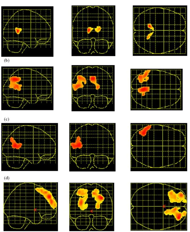
Results of AD patients and control subjects in the different neuropsychological tasks are presented in Table 1. The significant correlations between cerebral metabolism at rest and performance on the cognitive tasks in AD patients are presented in Table 2 and Figure 1.
[Insert Table 1 and 2 and Figure 1 about here]
Mattis dementia rating scale
The mean total score on the Mattis dementia rating scale was significantly lower in the AD patients group than in the control subjects [F(1,25)=21.29, p<0.0005]. A positive correlation was observed in AD patients between the Mattis total score and posterior regions including the cingulate gyrus (BA 23) and the left superior parietal area (BA 7). When the correlation between this measure and metabolism at rest were compared between the AD patients and the elderly control group, results indicated that the posterior cingulate area was correlated to the global cognitive score in AD patients only.
Digit span
The mean digit span level was significantly inferior in the AD patients group in comparison to control subjects [F(1,25)=4.52, p<0.05]. The brain regions which significantly correlated to the digit span performance in the AD patient group comprised the left superior temporal gyrus (BA 22), extending to the left superior parietal region (BA 7) and a region centred on the cingulate gyrus and precuneus (BA 7/23/31). When the correlations between that span performance and metabolism were compared in AD patients and control subjects, no significant difference appeared.
Phonemic fluency
The AD patients gave less exemplars than control subjects [F(1,25)=6.29, p<0.05]. A positive correlation was observed between the performance of AD patients and regional cerebral metabolism in a brain area centred on the left middle temporal gyrus (BA 39), extending to the superior temporal area
(BA 22) and angular gyrus (BA 39). No significant differences between AD patients and control subjects in the cerebral areas involved in the phonemic fluency task were found.
Hayling task
The inhibition ability was lower in the AD patient group relative to control subjects [F(1,25)=12.78, p<0.005]. The brain regions which significantly related to the measure of inhibition in AD patients were the right (BA 8) and left (BA 6/8/10) superior frontal gyrus. Again, no significant differences were found between groups for the correlations between inhibition time and cerebral metabolism.
DISCUSSION
The performance of AD patients is significantly impaired compared to control subjects on digit span, verbal fluency and the Hayling inhibition task as well as on a global deterioration scale (Mattis DRS). In addition, specific patterns of cognitivo-metabolic correlations were found in AD patients as well as between AD patients and control subjects.
First of all, we will consider the comparison of cognitivo-metabolic correlations between both groups. There were no significant differences between both groups for the tasks assessing specific cognitive processes (digit span, verbal fluency and Hayling task). However, the total score of the Mattis DRS was correlated to the metabolism in the posterior cingulate area in AD patients only. This multi-compound score represents the sum of the performance in various domains of cognitive function such as memory and attention. Several studies have shown that the most significant decrease of metabolism in AD occurs in the posterior cingulate cortex (e.g. Minoshima, Foster & Kuhl, 1994; Salmon et al., 1997) and that the posterior cingulate metabolism was correlated to dementia severity, independently of age (Salmon et al., 1998). Moreover, some data suggest that posterior cingulate regions probably play a major role in the interaction between (and integration of) several neural networks sustaining attentional and memory processes (e.g. Fletcher et al., 1996; O’Leary et al., 1997). Taken as a whole, these data could indicate that two different neurobiological substrates are
involved in the cognitive impairment of AD patients. Firstly, the absence of differences between groups on specific cognitive measures could indicate that similar cerebral areas are involved in the performance of AD patients and control subjects, but that these areas are working less efficiently in AD patients. Indeed, in a positive correlation, the cognitive performance of one subject is directly linked to the metabolic level of the cerebral areas involved in the task. Since impairments of specific cognitive processes (such as inhibition) were found in AD patients although cerebral areas similar to control subjects are involved in the tasks, their lower performance appears to be related to a less efficient functioning of the main cerebral areas involved in the task. Secondly, the significant correlation only in AD patients between the metabolic level in the posterior cingulate region and the multi-compound score of the Mattis scale could indicate difficulties of integration of information coming from different cerebral areas in AD. Indeed, several studies have shown that there exists a breakdown of physiological connectivity between anterior and posterior cerebral areas in AD patients (Azari et al., 1992; Leuchter et al., 1992). These difficulties of integration could explain the impairments of AD patients in tasks requiring the intervention of large neural networks comprising the posterior cingulate area, such as episodic memory.
It remains to examine the correlations between metabolism and performance which did not differ between AD patients and control subjects. Digit span performance was correlated to a brain area comprising the left superior temporal gyrus (BA 22) and extending to parietal gyrus (BA 7), as well as to the cingulate gyrus and precuneus (BA 7/23/31). An increase of activity in a region comprising the left superior temporal (BA 22) and posterior parietal (BA 7) regions was already described in activation studies with healthy subjects and was attributed to the processing of verbal information in working memory (e.g. Awh et al., 1996; Paulesu, Frith, & Frackowiak, 1993).
The performance on a verbal phonemic fluency task was correlated in AD patients to the left superior and middle temporal gyrus (BA 22 and BA 39), extending to the angular gyrus (BA 39). Some activation studies with young normal subjects (Frith et al., 1991; Paulesu et al., 1997) already showed that this task does activate the left superior temporal region (BA 22) and the inferior parietal region (BA 39). However, these studies showed that this task also activated prefrontal areas. In the present study, it appears that AD patients (and also control subjects) rely on posterior regions devoted
to the storage of semantic information, without support of frontal areas. Consequently, the lower performance of AD patients would not be due to an executive dysfunction, such as the difficulty to use an efficient search strategy (Troyer et al., 1998), but rather to semantic memory impairments (Randolph et al., 1993). However, the absence of correlations with prefrontal areas could also be due to minor involvement of executive function in the task. Indeed, Philips (1997) showed that the realisation of a keypress random generation task (which requires also executive function) did not interfere with the realisation of the phonemic fluency task.
Significant correlations have been observed between the inhibition time on the Hayling task and the right and left superior frontal area (BA 6/8/10). Inhibition function has been classically related to the prefrontal cortex (Burgess & Shallice, 1996). In a recent PET activation study (Collette et al., 1999), we showed that the inhibition condition of this task was associated to a bilateral activation in the middle and superior frontal gyrus. The existence of correlations in the superior frontal cortex confirms that there exists an involvement of the prefrontal cortex during the suppression of automatic responses. Moreover, the absence of differences in correlations between both groups indicates that the main difficulty of AD patients on the Hayling task relates to the inhibition of a predominant response.
In conclusion, the comparison of cognitivo-metabolic correlations between AD patients and elderly control subjects showed that the lower performance of AD patients in cognitive tasks could be due simultaneously to a less efficient functioning of specific cerebral areas and to a deficit of the integration of information coming from different cerebral areas. Moreover, the pattern of correlation observed in both groups confirms that the performance at the digit span task essentially depends upon temporal and parietal regions. More interestingly, the phonemic fluency task was related to posterior regions involved in semantic processes, suggesting that the prefrontal regions are not essential for the realisation of the task. Finally, the relationship between frontal metabolism and performance on the Hayling task confirms that anterior cortices are essential for inhibitory processes.
FOOTNOTE
1. The correlation between metabolism and cognition will not be directly assessed in control subjects. Indeed, the correlation method is based on the existence of an important variability both in the cerebral metabolic distribution and in the scores in neuropsychological tasks. Since this variability is less important in control subjects, we do not expect significant correlations between metabolism at rest and cognitive performance.
ACKNOWLEDGEMENTS
This work was supported by the Belgian Fund for Scientific Research (FNRS), the “Fondation Medicale Reine Elisabeth”, and the Interuniversity Pole of Attraction P4/22, Belgian State, Prime Minister’s Office, Federal Office for Scientific, Technical and Cultural Affairs. F. Collette is Aspirant at the FNRS.
REFERENCES
Awh, E., Jonides, J., Smith, E.E., Schumacher, E.H., Koeppe, R.A, & Katz, S. (1996). Dissociation of storage and rehearsal in verbal working memory: evidence from positron emission tomography. Psychological Science, 7, 25-31.
Azari, N.P., Rapoport, S.I., Grady, C.L., Schapiro, M.B., Salerno, J.A., Gonzalez-Aviles, A., & Horwitz, B. (1992). Patterns of interregional correlations of cerebral glucose metabolism rates in patients with dementia of the Alzheimer type. Neurodegeneration, 1, 101-111.
Baddeley, A.D. (1990). Human memory: Theory and practice. Hove : Erlbaum.
Baddeley, A.D., Della Sala, S., & Spinnler, H. (1991). The two-component hypothesis of memory deficit in Alzheimer's disease. Journal of Clinical and Experimental Neuropsychology, 13, 372-380.
Becker, J.T., Mintun, M.A., Aleva, K.D., Wiseman, M.B., Nichols, T., & DeKosky, S.T. (1996). Compensatory reallocation of brain resources supporting verbal episodic memory in Alzheimer's disease. Neurology, 46, 692-700.
Burgess, P.W., & Shallice, T. (1996). Response suppression, initiation and strategy use following frontal lobe lesions. Neuropsychologia, 34, 263-273.
Collette, F., Van der Linden, M., & Salmon, E. (1999). Executive dysfunction in Alzheimer's disease. Cortex, 35, 57-72 .
Collette, F., Van der Linden, M., Delfiore G., Degueldre C., & Salmon, E. (1999). The functional anatomy of inhibition processes investigated with the Hayling task. Journal of Neurology, 246 (suppl. 1), I/51.
Fletcher, P.C., Shallice, T., Frith, C.D., Frackowiak, R.S.J., & Dolan, R.J. (1996). Brain activity during memory retrieval. The influence of imagery and semantic cueing. Brain, 119, 1587-1596.
Folstein, M., Folstein, S., & McHugh, P.R. (1975). Mini-mental state: A practical method for grading the cognitive state of patients for the clinician. Journal of Psychiatric Research, 12, 189-198.
Friston, K.J., Frith, C.D., Liddle, P.F., Dolan, R.J., Lammertsma, A.A., & Frackowiak, R.S.J. (1990). The relationship between global and local changes in PET scans. Journal of Cerebral Blood Flow and Metabolism, 10, 458-466.
Friston, K.J., Frith, C.D., Liddle, P.F., & Frackowiak, R.S.J. (1991). Comparing functional (PET) images: The assessment of significant changes. Journal of Cerebral Blood Flow and Metabolism, 11, 690-699.
Friston, K.J., Holmes, A.P., Worsley, K.J., Poline, J.-B., Frith, C.D., & Frackowiak, R.S.J. (1995). Statistical parametric maps in functional imaging: A general linear approach. Human Brain Mapping, 2, 189-210.
Friston, K.J., Liddle, P.F., Frith, C.D., Hirsch, R.S., & Frackowiak, R.S.J. (1992). The left medial temporal region and schizophrenia. Brain, 115, 367-382.
Frith, C.D., Friston, K.J., Liddle, P.F., & Frackowiak, R.S.J. (1991). A PET study of word finding. Neuropsychologia, 29, 1137-1148.
Haxby, J.V., Duara, R., Grady, C.L., Cutler, N.R., Rapoport, S.I. (1985). Relation between neuropsychological and cerebral metabolic asymmetries in early Alzheimer's disease. Journal of Cerebral Blood Flow and Metabolism, 5, 193-200.
Haxby, J.V., Grady, C.L., Koss, E., Horwitz, B., Heston, L., Schapiro, M.B., Friedland, R.P., & Rapoport, S.I. (1990). Longitudinal study of cerebral metabolic asymmetries and associated neuropsychological patterns in early dementia of the Alzheimer type. Archives of Neurology, 47, 753-760.
Haxby, J.V., Grady, C.L., Koss, E., Horwitz, B., Schapiro, M., Friedland, R.P., & Rapoport, S.I. (1988). Heterogeneous anterior-posterior metabolic patterns in dementia of the Alzheimer type. Neurology, 38, 1853-1863.
Herbster, A.N., Nichols, T., Wiseman, M.B., Mintun, M.A., DeKosky, S.T., & Becker, J.T. (1996). Functional connectivity in auditory-verbal short-term memory in Alzheimer's disease. Neuroimage, 4, 67-77.
Horwitz, B., Grady, C.L., Schlageter, N.L., Duara, R., & Rapoport, S.I. (1987). Intercorrelation of regional cerebral glucose metabolic rates in Alzheimer's disease. Brain Research, 407, 294-306.
Horwitz, B., McIntosh, A.R., Haxby, J.V., Furey, M., Salerno, J.A., Schapiro, M.B., Rapoport, S.I., & Grady, C.L. (1995). Network analysis of PET-mapped visual pathways in Alzheimer type dementia. Neuroreport, 6, 2287-2292.
Leuchter, A.F., Newton, T.F., Cook, I.A., Walter, D.O., Rosenberg-Thompson, S., & Lachenbruch, P.A. (1992). Changes in brain functional connectivity in Alzheimer-type dementia and multi-infarct dementia. Brain, 115, 1543-1561.
Mattis S. (1973). Dementia Rating Scale. Windsor : NFER-Nelson.
McKhann, G., Drachman, D., Folstein, M., Katzman, R., Price, D., & Stadlan, E.M. (1984). Clinical diagnosis of Alzheimer's disease: Report of the NINCDS-ADRDA work group under the auspices of departement of health and human services task force on Alzheimer's disease. Neurology, 34, 939-944.
Minoshima, S., Foster, N.L., & Kuhl, D.E. (1994). Posterior cingulate cortex in Alzheimer's disease. The Lancet, 344, 895.
Morris, R.G. (1994). Recent developments in the neuropsychology of dementia. International Review of Psychiatry, 6, 85-107.
Morris, R.G. (1996). Neurobiological correlates of cognitive dysfunction. In R.G. Morris (Ed.), The cognitive neuropsychology of Alzheimer-type dementia. (pp. 223-254). Oxford : Oxford University Press.
O'Leary, D.S., Andreasen, N.C., Hurtig, R.R., Torres, I.J., Flashman, L.A., et al. (1997). Auditory and visual attention assessed with PET. Human Brain Mapping, 5, 422-436.
Paulesu, E., Frith, C.D., & Frackowiak R.S.J. (1993). The neural correlates of the verbal component of working memory. Nature, 362, 342-345.
Paulesu, E., Goldacre, B., Scifo, P., Cappa, S.F., Giraldi, M.C., Castiglioni, I., Perani, D., & Fazio, F. (1997). Functional heterogeneity of left inferior frontal cortex as revealed by fMRI. Neuroreport, 8, 2011-2016.
Penniello, MJ., Lambert, J., Eustache, F., Petit-Taboué, M.C., Barré, L., Viader, F., Morin, P., Lechevallier, B., & Baron, JC. (1995). A PET study of the functional neuroanatomy of writing impairment in Alzheimer's disease : The role of the left supramarginal and left angular gyri. Brain, 118, 697-706.
Perani, D., Bressi, S., Cappa, S.F., Vallar, G., Alberoni, M., Grassi, F., Caltarigone, C., Cipolotti, L., Franceschi, M., Lenzi, G.L., & Fazio, F. (1993). Evidence of multiple memory systems in the human brain. Brain, 116, 903-919.
Philips, L.H. (1997). Do "frontal tests" measure executive function? Issues of assessment and evidence from fluency tests. In P. Rabbit (Ed.), Methodology of frontal and executive function. (pp. 191-213). Hove: Psychology Press.
Phelps, M.E., Huang, S.E., Hoffman, E.J., Selin, S.C., Sokoloff, L., & Khul, D.E., (1979). Tomographic measurement of local cerebral metabolic rate in humans with (F-18) 2-fluoro-2-deoxyglucose: Validation of method. Annals of Neurology, 6, 371-388.
Randolph, C., Braun, A.R., Goldberg, T.E., & Chase, T.N. (1993). Semantic fluency in Alzheimer's, Parkinson's, and Huntington's disease: Dissociation of storage and retrieval failures. Neuropsychology, 7, 82-88.
Salmon E., Collette F., Degueldre C., Lemaire C., Franck G. (1998). Posterior cingulate metabolic impairment is a major characteristic of Alzheimer’s disease, related to both age and dementia severity. Neurology of Aging, 19 (suppl.4), S158.
Salmon, E., & Van der Linden, M. (1997). Anterior cingulate and motor network metabolic impairment in progressive supranuclear palsy. Neuroimage, 5, 173-178.
Salmon, E., Van der Linden, M., Collette, F., Delfiore, G., Maquet, P., Degueldre, C., Luxen, A., & Franck, G. (1996). Regional brain activity during working memory tasks. Brain, 119, 1617-1625.
Talairach, J., & Tournoux, P. (1988). Co-planar stereotaxic atlas of the human brain: 3-dimensional proportional system: An approach to cerebral imaging, Stuttgart : Thieme.
Troyer, A.K., Moscovitch, M., Winocur, G., Leach, L., & Freedman, M. (1998). Clustering and switching on verbal fluency tests in Alzheimer's and Parkinson's disease. Journal of International Neuropsychological Society, 4, 137-143.
TABLES
Table 1
Mean performance (standard deviation) of AD patients and control subjects in neuropsychological tasks
AD patients Control subjects Mattis DRS (total score) 110 (19.33) 138.84 (2.48)
Digit span 4.44 (0.96) 5.42 (1)
Phonemic fluency 15.44 (6.98) 25 (7.69) Hayling (time B-A) 71.06 (39.15) 23.42 (11.80)
Table 2. Correlations between brain metabolism and cognitive performance
Stereotactic coordinates
Region x y z Zscore
Correlations between brain metabolism and Mattis score in AD
Posterior cingulate (BA 23) 26 -54 20 5.47
L superior parietal (BA 7) -40 -44 48 5.03
Cerebral areas correlated to the Mattis score in AD patients but not in control subjects
Posterior cingulate (BA 30) -14 -48 14 4.80
0 -34 16 3.15
Correlations between brain metabolism and digit span in AD
L superior temporal (BA 22) -48 -50 20 3.85
-60 -42 12 3.49
L parietal superior (BA 7) -42 -70 46 3.82
Posterior cingulate (BA 23/31) 18 -58 14 3.84
12 -60 26 3.18
Precuneus (BA 7/31) 2 -58 44 4.19
Correlations between brain metabolism and phonemic fluency in AD
L middle temporal gyrus (BA 39) -42 60 16 3.77
L superior temporal gyrus (BA 22) -54 -50 14 3.60
L angular gyrus (BA 39) -44 -72 38 3.36
Correlations between brain metabolism and Hayling task in AD
R frontal superior (BA 8) 32 50 40 4.08
24 52 44 3.78
8 38 54 3.61
L frontal superior (BA 10/8/6) -32 56 24 3.75
-38 24 58 3.48
-28 36 52 3.32
Note. R = right; L = left; x, y, and z, expressed in mm, are coordinates in the stereotactical space of
Figure 1. SPM for the cerebral areas involved in the different cognitive tasks: Positive correlation (A) with the Mattis total score in AD patients but not control subjects ; (B) with the verbal span task in AD patients ; (C) with the verbal fluency task in AD patients ; (D)with the Hayling task in AD patients.