Structure-biological function study of 17
β-hydroxysteroid dehydrogenase type 1 and reductive
steroid enzymes: inhibitor design targeting
estrogen-dependent diseases
Thèse
Tang Li
Doctorat en médecine moléculaire
Philosophiæ doctor (Ph. D.)
Résumé
La 17β-HSD1 catalyse l’activation de l’œstrogène le plus actif, l’estradiol, ainsi que la désactivation de la dihydrotestosterone, l’androgène le plus puissant. Cette enzyme est considé ré e comme une cible prometteuse pour le traitement des maladies dépendantes des œstrogènes. Malgré des décennies de recherches, aucun inhibiteur ciblant la 17β-HSD1 n’a encore atteint le stade clinique. De plus, le mécanisme de l’inhibition du substrat de la 17β-HSD1, qui peut être utilisé pour faciliter la conception d’inhibiteur, n’est toujours pas bien dé montré de maniè re structurelle. Ici, nous avons Co-cristallisé trois inhibiteurs de différence, à savoir l’EM-139, le 2-MeO-CC-156 et le PBRM, avec la 17β-HSD1 et avons résolu ces structures cristallines. L’inhibiteur ré versible EM-139 s’est révélé moins stable dans le site de liaison aux stéroïdes, avec seulement la fraction du noyau stéroïdien de l’inhibiteur présentant une densité d’électron définissable. La fraction volumineuse de 7α-alkyle de l’inhibiteur, qui limite son activité anti-œstrogénique, n’est pas dé finie dans la densité électronique, peut compromettre l’effet inhibiteur de l’inhibiteur sur l’enzyme. Quant à l’inhibiteur réversible, le 2-MeO-CC-156, il interagit de maniè re similaire que le CC-156 avec l’enzyme. Cependant, avec la présence du groupe 2-MeO, le pouvoir inhibiteur de la 17β-HSD1 est nettement infé rieur à celui du CC-156. L’analyse du complexe ternaire PBRM avec la 17β-HSD1 montre clairement la formation d’une liaison covalente entre l’His221 et la chaîne laté rale bromoethyl de l’inhibiteur, donnant un aperç u des interactions molé culaires
bé né fiques qui favorisent la liaison et l’avè nement de N-alkylation ulté rieur dans le site catalytique de l’enzyme. En outre, le groupe bromoethyl en position C-3 du PBRM justifie son profil non œstrogénique, ralentit son mé tabolisme et assure son action spé cifique de la 17β-HSD1 par la formation d’une liaison covalente avec Nε du ré sidu His221. Nous avons aussi Co-cristallisé la 17β-HSD1 avec l’œstrone ainsi qu’avec
l’analogue de l’œstrone et du cofacteur NADP+, la structure a révélé un mode de liaison inversé de l’œstrone dans l’enzyme, jamais trouvé dans les complexes d’estradiol. L’analyse structurale a démontré que His221 est
le résidu clé responsable de la réorganisation et de la stabilisation de l’œstrone liée de manière inversée, conduisant à la formation d’un complexe sans issue. Ainsi, sur la base du mécanisme d’inhibition du substrat et de l’analyse computationnelle, une novelle entité chimique (SX7) est proposé e qui peut inhiber la 17β-HSD1 et former un complexe sans issue. De plus, avec un grand nombre d’échantillons cliniques, nous avons dé montré la modulation et la corrélation d’expression significative de plusieurs enzymes clés de conversion des sté roïdes, supportant les 17β-HSD1 et 17β-HSD7 ré ductrices comme cibles prometteuses et la nouvelle thé rapie combiné e ciblant les 11β-HSD2 et 17β- HSD7.
Abstract
Human 17β-hydroxysteroid dehydrogenase type 1 (17β-HSD1) catalyzes the activation of the most potent estrogen estradiol as well as the deactivation of the most active androgen dihydrotestosterone, and is considered as a promising target for the treatment of estrogen-dependent diseases such as endometriosis, breast cancer, endometrial cancer and ovarian cancer. Despite decades of research, no inhibitor targeting 17β-HSD1 has yet reached the stage of clinical trials. Moreover, the structure-biological function of the substrate inhibition of 17β-HSD1, which can be used to facilitate the inhibitor design, is still not well demonstrated. Here we co-crystallized three different inhibitors, namely EM-139, 2-MeO-CC-156 and PBRM, with 17β-HSD1 and solved the structures of these complexes. The reversible inhibitor EM-139 showed high mobility in the steroid binding site with only its steroid core moiety could be defined in the electron density. The bulky 7α-alkyl moiety of the inhibitor, which guarantees its anti-estrogenic activity but unable to be defined in the electron density, may compromise the inhibitory effect of the inhibitor on the enzyme. As for the reversible inhibitor MeO-CC-156, it interacts similarly to CC-156 with the enzyme. However, in the presence of the 2-MeO group, it shows much less inhibitory potency to 17β-HSD1 as compared to the CC-156. The analysis of the PBRM ternary complex with 17β-HSD1 clearly shows an unambiguous continuity of electron density from the side chain of His221 to the bound PBRM, demonstrating the formation of a covalent bond between the Nε of
His221 and the C-31 (BrCH2) of the inhibitor. This result provides insight into beneficial molecular interactions
that favor the binding and subsequent N-alkylation event in the enzyme catalytic site. Also, the bromoethyl group at position C-3 of the PBRM warrants its non-estrogenic profile, slows down its metabolism, and secures the specific action of 17β-HSD1 through the formation of a covalent bond with Nε of residue His221. Meanwhile,
we co-crystallized 17β-HSD1 with estrone as well as with estrone and cofactor analog NADP+, revealed a reversely orientated binding mode of estrone in the enzyme, never found in reported estradiol complexes. Structural analysis demonstrated that His221 is the key residue responsible for the reorganization and
stabilization of the reversely bound estrone, leading to the formation of a dead-end complex. Thus, based on the substrate inhibition mechanism and computational analysis, a chemical entity (SX7) is proposed that may inhibit 17β-HSD1 and form a dead-end complex. Furthermore, with large number clinical samples, we demonstrated the significant expression modulation and expression correlation of several key steroid-converting enzymes, supporting the reductive 17β-HSD1 and 17β-HSD7 as promising targets and the new combined therapy targeting 11β-HSD2 and 17β-HSD7.
Table des matières
Ré sumé ... ii
Abstract ... iii
Table des matiè res ... iv
Liste des figures ... vi
Liste des figures, tableaux, illustrations... vii
Liste des abré viations ... viii
Remerciements ... xii
Avant-propos ... xiii
Introduction... 1
Chapitre 1 Combined biophysical chemistry reveals a new covalent inhibitor with a low-reactivity alkyl halide 16 1.1 Ré sumé ... 16
1.2 Abstract ... 16
1.3 Introduction ... 17
1.4 Results and Discussion ... 19
1.5 Conclusion ... 22
1.6 Experimental Procedures ... 22
1.7 Reference ... 25
Chapitre 2 Crystal structures of human 17β-hydroxysteroid dehydrogenase type 1 complexed with the dual-site inhibitor EM-139 ... 38
2.1 Ré sumé ... 38
2.2 Abstract ... 38
2.3 Introduction ... 39
2.4 Materials and Methods ... 39
2.5 Results ... 40
2.6 Discussion ... 41
2.7 Conclusion ... 41
2.8 Reference ... 43
Chapitre 3 Crystal structures of human 17β-hydroxysteroid dehydrogenase type 1 complexed with estrone and cofactor reveal the mechanism of substrate inhibition ... 53
3.4 Materials and Methods ... 55
3.5 Results ... 56
3.6 Discussion ... 59
3.7 Conclusion ... 60
3.8 Reference ... 62
Chapitre 4 Remarkable steroid-converting enzyme and receptor regulations in large number breast tumor samples : molecular correlation and combined therapies ... 78
4.1 Ré sumé ... 78
4.2 Abstract ... 78
4.3 Introduction ... 79
4.4 Materials and Methods ... 80
4.5 Results ... 81 4.6 Discussion ... 84 4.7 Conclusion ... 86 4.8 Reference ... 88 Conclusion...101 Bibliographie ...105
Annexe A Cold-active extracellular lipase: expression in Sf9 insect cells, homogenization, and catalysis ...119
2.1 Ré sumé ...119
2.2 Abstract ...119
2.3 Introduction ...120
2.4 Materials and Methods ...120
2.5 Results and Discussion ...125
2.6 Conclusion ...129
Liste des figures
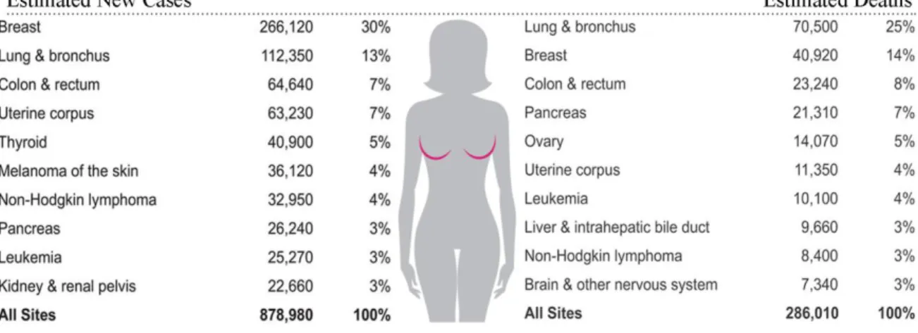
Figure 1. Ten leading cancer types for the estimated new cancer cases and deaths in women in United
States, 2017……….……….1
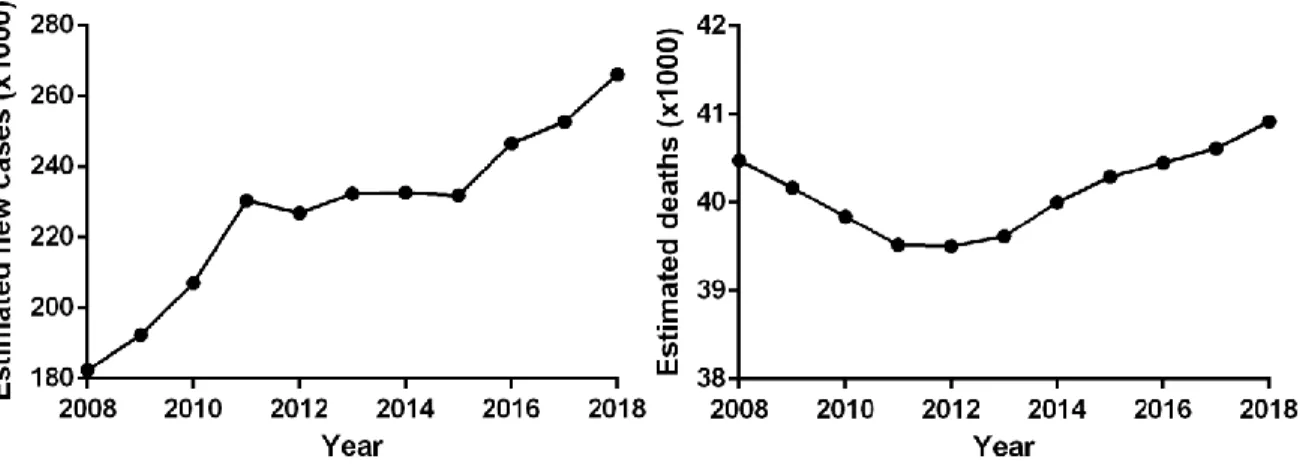
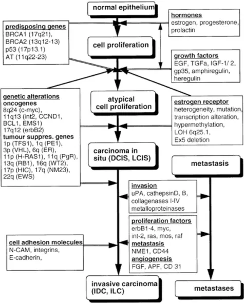
Figure 2. The time course of the estimated new BC cases and deaths in women in the United States……...…2 Figure 3. Model of the multistep carcinogenesis in BC……….………....3 Figure 4. Schematic representations of sex hormones synthesis regulations in pre- (A) and postmenopausal
(B) women……….……….5
Figure 5. Human steroidogenic and steroid-inactivating enzymes in peripheral intracrine tissues………..……..6 Figure 6. Stereo ribbon presentation of human 17β-HSD1 structure………..………8 Figure 7. Two possible stepwise catalytic mechanisms for 17β-HSD1………11 Figure 8. Key inhibitors of 17β-HSD1 from different Series………13
Liste des figures, tableaux, illustrations
Table 1. Previously published 17β-HSD1 structures………...…...……7 Table 2. The ratio of kinetic constants of Human 17β-HSD1 variants vs. that of wild type enzyme……...…...…9
Liste des abréviations
2-MeO-CC-156 2methoxy-16β-(m-carbamoylbenzyl)-E2
3β-diol 5α-androstane-3β,17β-diol
4-dione androstenedione
5-diol 5-androstenediol; androst-5-ene-3β,17β-diol 5-diol-FA 5-diol fatty acid
5-diol-S 5-diol sulfate
ACTH adrenocorticotropic hormone
A-dione 5α-androstane-3,17-dione
ADT androsterone
AIs aromatase inhibitors
AKR aldo-ketoreductase
AR androgen receptor
BC breast cancer
C12E8 octaethylene glycol monododecyl ether
CC-156 16β-m-carbamoylbenzyl-E2
CRH corticotropin releasing hormone
DHEA dehydroepiandrosterone
DHEAS dehydroepiandrosterone sulfate
E1 Estrone
E1S estrogen sulfate
E2 Estradiol
EDD estrogen-dependent disease
EM-139 N-n-Butyl-N-methyl-ll-(16'α-chloro3',17'β-dihydroxyestra-1',3',5'(10')-trien-7'α-yl)undecanamide
epi-ADT epiandrosterone
ER estrogen receptor
EREs estrogen responsive elements
FSH follicle-stimulating hormone
GnRH gonadotropin-releasing hormone
GSC Genome Sequencing Centers
HSD hydroxysteroid dehydrogenase
HTS high throughput sequencing
LH luteinizing hormone
mg microgram
ml microliter
NAD+ nicotinamide adenine dinucleotide
NADP+ nicotinamide adenine dinucleotide phosphate
NIH National Institute of Health
nM nanomolar
OD optical density
PAGE polyacrylamide gel electro phoresis
PBRM 3-(2-bromoethyl)-16β-(m-carbamoylbenzyl)-17β-hydroxy-1,3,5(10)-estratriene
PDB protein data bank
pNPA p-nitro phenyl acetate pNPB p-nitro phenyl butyrate pNPD p-nitro phenyl decanoate pNPL p-nitro phenyl dodecanoate pNPM p-nitro phenyl myristate pNPP p-nitro phenyl palmitate
PPH polyhedron promoter
pro-S prochiral S configuration
RhB rhodamine B
RhB-OOe RhB-olive oil
RNA-seq RNA sequencing
RoDH-1 Ro dehydrogenase 1
SDR short chain dehydrogenase/reductase
SDS sodium dodecyl sulfate
T testosterone
TCGA The Cancer Genome Atlas
Testo testosterone
UGT1A1 uridine glucuronosyl transferase 1A1
UGT2B28 uridine glucuronosyl transferase 2B28
UV ultra-violet
β-DDM n-Dodecyl-β-D-Maltoside
β-ME β-mercaptoethanol
β-OG n-octyl-β-D-glucoside
Remerciements
I would like to convey my immeasurable gratitude to my director of research, professor Sheng-Xiang Lin, for his meticulous guidance and enlightening discussions that helped me overcome all the difficulty and enabled me to present this thesis. I greatly appreciate his trustiness and supports for giving me this inspiring and challenging project. His diligent directions and interactive concept helped me greatly during my study, and I will certainly benefit from that in my future career. I also sincerely appreciate the following person and organisations for supporting my doctoral study to obtain the degree of Doctor of Philosophy (Ph.D.). The Ph.D. study will light up my future career in scientific research and practise.
I would like to thank all the members in Dr. Lin’s Lab. I express my gratitude to Dre. Ming Zhou for her help in protein purification and crystallization; to Mr. Jean-Franç ois Theriault for his help in enzyme kinetics; to Mr. Jian Song for his help in binding study and growing rLcn6 crystals; to Dr. Preyesh Stephen for his help in Crif1-CDK2 project; to Miss. Xiaoye San and Ruixuan Wang for their inspiriting discussion in diagnosis and treatment of breast and ovarian cancers. I would like to thank Dr. Dao-Wei Zhu for his help in familiar with the surroundings and his advices in protein purification and crystallization. As well as Dr. Xiaoqiang Wang, Dre. Dan Xu and Dre. Juliette Adjo Aka for their advices and discussion in cell experiments.
I would like to thank Dr. Donald Poirier. I sincerely appreciate the knowledge from him in the field of medicinal chemistry, especially for the inhibitor design. I would like to thank Dr. Rong Shi for his help in collecting an X-ray diffraction dataset at the CLS synchrotron. I would like to thank Dr. Alexandre Brunet in the Laboratory of Flow-cytometry for his advice in preparing the sample and analysing the results. I also appreciated Dre. Sylvie Bourassa and Dre. Florence Roux-Dalvai for their advice and discussion in mass spectrometry experiment design and sample preparation. I would like to thank Mr. Martin Thibault for his help in analysing image of western blot.
I would like to thank all administration staffs in the Research Center of CHU de Quebec (CHUL). Thank Mme Nicole Almeras for taking care of the registration and financial documents. Thank Mme Marianne Roberge for her help in the order of experiment materials and reagents.
Finally, I am very grateful to my family, especially to my beloved son Guanrui Li and wife Juan Liu as well as my parents. It is your love that encouraging and supporting me during my studies.
Avant-propos
This thesis is submitted to the “Faculté des études supérieures de l'Université Laval” for the requirement of a doctor’s degree in science. The thesis is written in English, except for the summary as well as the abstract of each article, which are in French. Two articles have been published by Journal of Physical Chemistry Letters and Health, respectively. The other three are being submitted for publication or in preparation.
In the introductory section, four major estrogen-dependent diseases were reviewed. The biosynthesis of estrogens, mostly estradiol, and the role of 17β-HSD1 in estrogen activation as well as inactivation of androgen are summarized. The structural and kinetic studies as well as the development of 17β-HSD1 inhibitor design are also discussed. The hypothesis and objectives are described in the end of this chapter. The Chapter I “Tang Li, Rene Maltais, Donald Poirier, Sheng-Xiang Lin. Combined Biophysical Chemistry Reveals a New Covalent Inhibitor with a Low-Reactivity Alkyl Halide. Journal of Physical Chemistry Letters (2017 IF: 8.7). 2018 Aug; 9:5275-5280. doi 10.1021/acs.jpclett.8b02225.” I conducted all the experiments and wrote the manuscript, and I’m the first author of this article. In this chapter, the crystal structures of 17β-HSD1 with two inhibitors (PBRM and 2-MeO-CC-156) were described. This study constructed the first example of N-alkylation between a human enzyme and a low-reactivity alkyl halide derivative, which opens the door to a new design of alkyl halide-based specific covalent inhibitors as potential therapeutic agents.
The Chapter II “Tang Li, Dao-Wei Zhu, Fernand Labrie and Sheng-Xiang Lin. Crystal structures of human 17β-hydroxysteroid dehydrogenase type 1 complexed with the dual-site inhibitor EM-139. Health. 2018 Aug; 10(8):1079-89. doi 10.4236/health.2018.108081.” I processed the crystal diffraction data to solve the complex structure and wrote the manuscript, and I’m the first author of this article. In this chapter, the 17β-HSD1 binary complex with the inhibitor EM-139 was described. The interaction between the steroid moiety of the inhibitor and the enzyme was analyzed. The influence of its bulky 7α-alkyl side chain to its inhibitory effect in 17β-HSD1 was also discussed.
The Chapter III “Tang Li, Preyesh Stephen, Dao-Wei Zhu, Rong Shi, Sheng-Xiang Lin. Crystal structures of human 17β-hydroxysteroid dehydrogenase type 1 complexed with estrone and cofactor reveal the mechanism of substrate inhibition. FEBS Journal. 2019. Doi: 10.1111/febs.14784.” I conducted all the experiments except for the crystallization of the 17β-HSD1-E1 binary complex. I wrote the manuscript, and I’m the first author of this article. In this chapter, the crystal structures of 17β-HSD1 in complex with E1 and with/without cofactor analog NADP+ were described. Based on the E1 binary and ternary complex structures as well as previously published 17β-HSD1 complexes with other ligands, the mechanism of the long observed substrate inhibition of
The Chapter IV “Tang Li, Zhongjun Li, Sheng-Xiang Lin. Remarkable steroid-converting enzyme and receptor regulations in large number breast tumor samples: molecular correlation and combined therapies (Article under submission).” I conducted the data analysis and wrote the manuscript, and I’m the first author of this article. In this chapter, the cDNA sequencing data from the public cohort The Cancer Genome Atlas Breast Invasive Carcinoma (TCGA-BRCA) was extracted and statistically analyzed, and identified several key steroid-converting enzymes which are significantly up-regulated in cancer samples. Close expression correlations of the enzymes were also found, suggesting combined therapy for breast cancer treatment.
In the conclusion, I interactively discussed 17β-HSD1 structure-function study from inhibitor interactions to the mechanism of enzyme regulation. Besides, I also discussed the use of cDNA sequencing data in breast cancer research.
The references of introduction and conclusion are listed after the conclusion section. References of publications are listed after the text of each article.
In the end of the thesis is the appendix “Tang Li, Wenfa Zhang, Jianhua Hao, Mi Sun, Sheng-Xiang Lin. Cold-active extracellular lipase: expression in Sf9 insect cells, homogenization, and catalysis. Biotechnol Rep
(Amst). 2018; 21:e00295. doi:10.1016/j.btre.2018.e00295.” I conducted all the experiments and wrote the
manuscript, and I’m the first author of this article. In this article, I expressed a novel cold-active marine lipase in Sf9 insect cells. After purification, I carefully characterized its enzymatic properties, such as the optimum temperature and pH ranges, substrate specificity, the effects of detergents, organic solvents as well as enzyme inhibitors. These results will facilitate its application in industries.
Introduction
1 Estrogen-dependent disease 1.1 Breast cancer
Breast cancer (BC) is the most commonly diagnosed cancer in women worldwide, and one of the leading cause of cancer death in women1. BC can also occur in man, but it is rare 1. It has estimated that 268,670
patients will be diagnosed BC in 2018 in the United States, among which 99% were women (Figure 1)2. The
estimated number of death from BC in women is 40,920, ranking the second among all estimated deaths from cancers2. The incidence of BC is estimated to increase based on the trend of the past ten years (Figure 2)2-12.
Similar situation was also observed in Canada, about 26,300 female patients will be diagnosed BC, which account for 25% of all cancers in 2017 (Canadian breast cancer statistics 2017, http://www.cancer.ca/en/cancer-information/cancer-type/breast/statistics/). The majority of female patients diagnosed with BC are above 45 years old, and mostly after menopause13. Among incidences of all BCs,
around 60% in premenopausal women and 75% in postmenopausal women are initially estrogen-dependent 14-15. There is a multistep process involved in the occurrence of BC, which starts from normal cells through
hyperplasia, premalignant change, in situ carcinoma, progression of primary BC and to metastasis formation (Figure 3)16. During this progression process, hormones such as estrogen, progesterone and prolactin,
stimulate cell proliferation through their receptors mediated signaling pathways as well as induced genetic damage and mutations16-17.
Figure 1 Ten leading cancer types for the estimated new cancer cases and deaths in women in United States, 2018 (Siegel et al., 2018).
Figure 2 The time course of the estimated new BC cases and deaths in women in the United States (Jemal et al., 2008-2010; Siegel et al., 2011-2018).
Estradiol (E2) is the most biologically potent natural estrogen. In estrogen dependent human breast cancers, E2 plays a critical role in the proliferation and development of carcinoma cells and it is actually essential for some of these carcinomas to continue growth18. The primary biological effects of estrogen are mediated by two
distinct nuclear receptors, estrogen receptor (ER)α19 and ERβ20, which encoded by unique genes and function
in the nucleus as ligand-dependent transcription factors. ERα is mainly responsible for the effects of estrogens on normal and malignant breast tissues. Its role in promoting proliferation of BC cells is well characterized, through either membrane and cytoplasmic signaling cascades21 or transcriptional regulation22. In contrast, the
role of ERβ in BC is not clearly understood but seems to act as an antagonist of ERα activity, attenuating the proliferation stimulation effect of estrogen23-25.
Figure 3 Model of the multistep carcinogenesis in BC (Beckmann et al., 1997). 1.2 Endometrial cancer
Endometrial cancer is one of the most common gynecologic malignancies. It ranks to be the fourth most diagnosed cancers in women after breast, lung, and colorectal cancers, and was expected to have more than 63,000 new cases in US in 2017 (Figure 1)12. The death rate for endometrial cancer almost doubled during the
past two decades26. Endometrial cancer is commonly classified into two types based on the dualistic model of
endometrial cancer tumorigenesis described by Bokhman27. Type I commonly develops in women before
menopause in an estrogen-dependent manner. In contrast, type II endometrial cancer majorly develops in postmenopausal women in an estrogen-independent manner28. The pathogenesis of type I endometrial cancer
is through atypical endometrial hyperplasia, whereas type II endometrial cancer is proposed to be generated directly from normal endometrium28. Most patients diagnosed with endometrial adenocarcinoma are between
the ages of 50 and 60 years, and 90% of women diagnosed with endometrial cancer are after age of 50, mostly after menopause26, 29. About 80% of endometrial cancers are estrogen-dependent30 and the most
potent estrogen, estradiol (E2), is suggested to play an important role in the pathogenesis of the disease by increasing the mitotic activity of endometrial cells31.
1.3 Endometriosis
Endometriosis is an estrogen-activated gynecological disease characterized by the presence of endometrial-like tissue growing outside the uterine cavity, typically on the pelvic peritoneum, ovaries, and uterosacral ligaments, and in the rectovaginal septum and vesico-uterine fold32. Severe disease may lead to deformation
of pelvic anatomy and extensive pelvic adhesions, often associated with pelvic pain and infertility32.
Endometriosis is initially considered largely as a benign condition, while the wide opinion nowadays is that endometriosis is a neoplastic condition which can develop into specific type of invasive ovarian cancer33-34. It is
estimated that 6 to 10% of diagnosed endometriosis are in premenopausal women, whereas the frequency rises up to 50% of women with infertility32. Endometriosis is a multifactorial disease. Its pathogenesis involves
estrogen overexposure, angiogenesis, inflammation, genetic predisposition, and environmental exposure to pollutants35-40. It has been demonstrated that estrogen plays a central role in the development and
maintenance of endometriosis by promoting the growth of ectopic tissue41. In premenopausal patient, the
depression of E2 levels through gonadotropin-releasing hormone analogues (GnRH-a) leads to the relieving of pains and regression of endometriotic lesions, which relapsed with the recovery of E2 when the therapy discontinues42. While in postmenopausal women, the administration of hormone replacement therapy may
lead to the relapse of endometriosis43.
1.4 Ovarian cancer
Ovarian cancer is the fifth most lethal of all gynecological malignancies in western country with more than 14,000 estimated death in 2017 in The United States (Figure 1)12. As more than 80% of all diagnosed ovarian
cancers are in women above age 50, it is mainly considered to be a disease of postmenopausal women44.
About 90% of malignant ovarian tumors are epithelial ovarian cancer45. Epidemiological data show that
estrogen exposure and metabolism are involved in the stimulation and pathogenesis of ovarian cancer, and patients taking estrogen-only hormone replacement therapy have a higher risk of ovarian cancer44, 46-48. Cell
studies confirmed that ovarian cancer cells share several estrogen regulation pathways with other estrogen-associated cancers such as endometrial cancer and breast cancer, and anti-estrogen intervention suppresses the proliferation of ovarian cancer cells in vitro and in vivo49-51. Moreover, estrogen was demonstrated to
2 Origins of estradiol
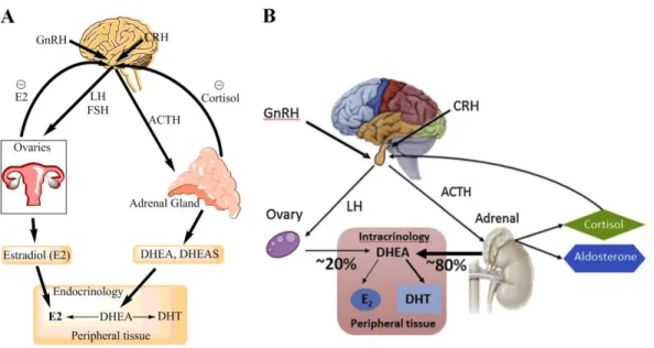
The origins of E2 in women can be divided into two sources, one is secreted from the ovary, and another is locally biosynthesized from the adrenal precursor dehydroepiandrosterone (DHEA), dehydroepiandrosterone-sulfate (DHEAS) and androstenedione in the peripheral tissues51. In premenopausal women, circulating E2 is
produced primarily by the ovaries53, and DHEAS is produced primarily by the adrenal glands54. As for the
DHEA, half of it is produced by adrenal glands, 20% originates from the ovaries and the other 30% is converted from DHEAS in peripheral tissues by sulfatase55. The production of androstenedione is equally
contributed by the adrenals and the ovaries56 (Figure 4A). After menopause, when the ovaries become
atrophied and cease to act, E2 no longer functions as a circulating hormone. Thus, E2 in postmenopausal women is produced only from precursor steroids of the adrenal glands in an intracrine manner to peripheral sites, which include breast, bone, vascular smooth muscle, and various sites in the brain (Figure 4B)51, 57.
Moreover, it is increasingly being recognised in EDDs that these tumor tissues are not just passively dependent on circulating levels of E2 but rather generate it locally from precursors in an active fashion58-59.
Figure 4 Schematic representations of sex hormones synthesis regulations in pre- (A) and postmenopausal (B) women. GnRH, gonadotropin releasing hormone; LH, luteinizing hormone; FSH,
follicle-stimulating hormone; CRH, corticotropin releasing hormone; ACTH, adrenocorticotropic hormone; T, testosterone; DHT, dihydrotestosterone; DHEA, dehydroepiandrosterone; E2, estradiol (Labrie 2015).
3 The role of 17β-HSD1
17β-HSD1 belongs to the short-chain dehydrogenase/reductase (SDR) family60. The major function of this
enzyme is the activation of estrone (E1) to the most potent estrogen E2 (Figure 5) 61, which is known to play a
pivotal role in the occurrence and development of estrogen-dependent diseases (EDDs). It can also catalyze the conversion of DHEA into 5-androstene-3β,17β-diol (5-diol), which has been suggested to be the main estrogen after menopause62. Beside the ability of activating estrogen, 17β-HSD1 can also inactivate
androgens. It has been demonstrated that 17β-HSD1 can transform the most potent androgen dihydrotestosterone (DHT) into a weak estrogen 5ɑ-Androstane-3β,17β-diol (3β-diol), a reaction which has been proposed to become more important after menopause and may be involved in aromatase inhibitor (AI) resistance63-65. 17β-HSD1 is the most active enzyme in terms of the production of E266-67. The over-expression
of 17β-HSD1 as well as the increased estrogen/androgen ratio indicates the pivotal role of the enzyme in breast cancer68-69, endometrial cancer30, 70, endometriosis 71, and ovarian cancer72. Thus, inhibition of
4 Structural studies of 17β-HSD1
17β-HSD1 is the first human steroid-converting enzyme whose three-dimensional structure has been solved. 17β-HSD1 consists of 328 amino acids with a molecular weight of 34.5kDa. This membrane-associated enzyme is acting as a homodimer and possesses a conserved Tyr-X-X-X-Lys sequence as a SDR family member and a Ser residue at the active site66, 73. The first crystallization of human estrogenic 17β-HSD1 was
reported by Zhu and co-workers in 199374. The three-dimensional structure of the enzyme was published in
199575. Since then, there are 22 17β-HSD1 structures deposited into the protein data bank (PDB), some in
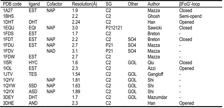
complex with substrate or inhibitor, some in complex with cofactor, and some in combination with cofactor and substrate/inhibitor (Table 1). This has led to the atomic level description of the substrate and cofactor binding cavities of the enzyme and a detailed understanding of its mechanism of action, as well as the molecular basis for the estrogen-specificity of the enzyme76-78.
The core of 17β-HSD1 structure is consisting of seven-stranded parallel β-sheet (βA to βG) surrounded by six parallel α-helices (αB to αG), evenly distributed by the two sides of the β-sheet (Figure 6). The structure of the protein generally forms into two segments the first segment, βA to βF, is a classic Rossmann fold, responsible for cofactor binding; the second segment, βD to βG, is partly in the Rossmann fold, governs steroid substrate binding75. The C-terminus of 17β-HSD1 (285-327) cannot be defined in all published structures and residues
190-199 have very poor density or even no density in many structures (1FDS, 1FDU, 1FDV, 1FDW, 1JTV, 1QYV, 1QYW, 1QYX, 3DEY, 3KLM, 3KLP, 3KM0).
Table 1 Previously published 17β-HSD1 structures
PDB code ligand Cofactor Resolution(Å ) SG Other Author βFαG’-loop
1A27 EST NAP 1.9 C2 Mazza Closed
1BHS 2.2 C2 Ghosh Semi-opend
1DHT DHT 2.24 C2 Han Opened
1EQU EQI NAP 3.0 P212121 Sawicki Closed
1FDS EST 1.7 C2 Breton -
1FDT EST NAP 2.2 C2 SO4 Breton Closed
1FDU EST NAP 2.7 P21 SO4 Mazza -
1FDV NAD 3.1 P21 SO4 Mazza -
1FDW EST 2.7 C2 Mazza -
1I5R HYC 1.6 C2 GOL Qiu Closed
1IOL EST 2.3 C2 Azzi Opened
1JTV TES 1.54 C2 GOL Gangloff -
1QYV NAP 1.81 C2 GOL Shi -
1QYW 5SD NAP 1.63 C2 GOL Shi -
1QYX ASD NAP 1.89 C2 GOL Shi -
3DEY DHT 1.7 C2 GOL Mazumdar -
3HB4 E2B 2.21 C2 Mazumdar Closed
3HB5 E2B NAP 2.0 C2 Mazumdar Closed
3KLM DHT 1.7 C2 GOL Aka -
3KLP B81 2.5 C2 Mazumdar -
3KM0 AOM NAP 2.3 C2 Mazumdar -
EST, estradiol; NAP, NADP; B81, 5-Androstenediol; AOM, 3β,17β-diol (3β-diol); 5SD, 5α-Androstane-3,17-dione (5α-Adione); ASD, 4-Androstene-5α-Androstane-3,17-dione (4-dione); TES, testosterone (T); AND,
Dehydroepiandrosterone (DHEA); EQI, Equilin; HYC, EM-1745. SG, space group.
Figure 6 Stereo ribbon presentation of human 17β-HSD1 structure. The α-helices are represented as
magenta coils and designated as αB to αH, β-strands are blue arrows and marked as βA to βF, and loops and turns are drawn as gray ropes. The N-terminus and the C-terminus of the protein molecule are indicated (Ghosh et al., 1995).
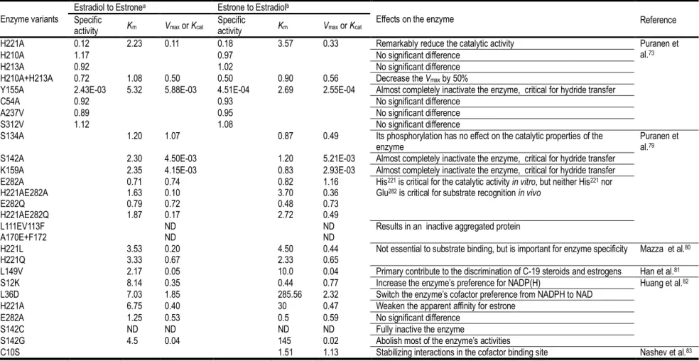
Table 2 The ratio of kinetic constants of 17β-HSD1 variants vs. that of wild type enzyme
a, NAD+ was used as cofactor in the kinetic tests. b, NADPH was used as cofactor in the kinetic tests. ND, undetectable. Some of the 17β-HSD1 kinetic data were
reported by Jin et al.67
Enzyme variants
Estradiol to Estronea Estrone to Estradiolb
Effects on the enzyme Specific
activity Km Vmax or Kcat Specific activity Km Vmax or Kcat Reference
H221A 0.12 2.23 0.11 0.18 3.57 0.33 Remarkably reduce the catalytic activity Puranen et al.73
H210A 1.17 0.97 No significant difference H213A 0.92 1.02 No significant difference H210A+H213A 0.72 1.08 0.50 0.50 0.90 0.56 Decrease the Vmax by 50%
Y155A 2.43E-03 5.32 5.88E-03 4.51E-04 2.69 2.55E-04 Almost completely inactivate the enzyme, critical for hydride transfer C54A 0.92 0.93 No significant difference
A237V 0.89 0.95 No significant difference S312V 1.12 1.08 No significant difference
S134A 1.20 1.07 0.87 0.49 Its phosphorylation has no effect on the catalytic properties of the
enzyme Puranen et al.79
S142A 2.30 4.50E-03 1.20 5.21E-03 Almost completely inactivate the enzyme, critical for hydride transfer K159A 2.35 4.15E-03 0.83 2.93E-03 Almost completely inactivate the enzyme, critical for hydride transfer E282A 0.71 0.74 0.82 1.16 His221 is critical for the catalytic activity in vitro, but neither His221 nor
Glu282 is critical for substrate recognition in vivo
H221AE282A 1.63 0.10 3.70 0.36 E282Q 0.79 0.72 0.48 0.73 H221AE282Q 1.87 0.17 2.72 0.49
L111EV113F ND ND Results in an inactive aggregated protein
A170E+F172 ND ND
H221L 3.53 0.20 4.50 0.44 Not essential to substrate binding, but is important for enzyme specificity Mazza et al.80
H221Q 3.33 0.67 2.33 0.65
L149V 2.17 0.05 10.0 0.04 Primary contribute to the discrimination of C-19 steroids and estrogens Han et al.81
S12K 8.14 0.35 0.44 0.77 Increase the enzyme’s preference for NADP(H) Huang et al.82
L36D 7.03 1.85 285.56 2.32 Switch the enzyme’s cofactor preference from NADPH to NAD H221A 6.75 0.40 30 0.47 Weaken the apparent affinity for estrone
E282A 1.25 0.53 0.5 0.59 No significant difference S142C ND ND ND ND Fully inactive the enzyme
S142G 4.5 0.04 145 0.02 Abolish most of the enzyme’s activities
4.1 Substrate recognition
The substrate recognition domain of 17β-HSD1 structure is buried under the flexible loop located between βF and αG’, and delimited by the C-terminal region. The tunnel-like substrate binding cavity is composed majorly by hydrophobic residues, such as Leu96, Val143, Met147, Leu149, Pro150, Pro187, Val225, Phe226, Phe259, Leu262,
Leu263 and Met279, as well as polar residues Asn152 and Tyr218. The βFαG’-loop acts as a lid covering the entry
of the cavity. This segment is highly flexible and unable to be defined in twelve 17β-HSD1 structures. While in the rest ten structures, it shows three possible conformations, including the closed, semi-opened and opened conformation (Table 1). Interestingly, all structures with the presence of cofactor analog NADP+ adopt a closed conformation, whereas structures only with natural steroid ligands exhibit an opened conformation, which suggests the modulation role of cofactor on the conformation of the loop. Moreover, the loop region in structures complexed with inhibitor CC-156 (E2B) and EM-1745 also has a close conformation even without cofactor. Only the apoenzyme has a semi-opened conformation at this flexible loop region. In the close conformation, residue Phe192 from the loop region forms a T-stacking conformation with residue Tyr155,
providing extra contacts for stabilizing the bound ligand84. The roles of residues from the active site of
17β-HSD1 have been investigated by mutagenesis and kinetic experiments which are summarized in Table 2. Residue His221 as well as Tyr155/Ser142 are critical for steroid substrate recognition through their hydrogen
bonds with the O3 and O17 of the ligand, respectively. Residue Glu282 is supposed to play the same important
role as the His221 does since it might also form a hydrogen bond with the O3 of the bound steroid79, as showed
in the E2 complex structure76. However, the variant E282A in Huang et al.’s experiment did not show any
significant modification in kinetics82. In contrast, residue Leu149 plays an important role for the discrimination of
C-19 steroids and estrogens. Steroid ligand is stabilized by hydrogen bonds between O3 and His221/Glu282 at
the recognition end, as well as between O17 and Tyr155/Ser142 at the catalytic end of the cavity.
4.2 Catalytic mechanism of 17β-HSD1
The kinetics of 17β-HSD1 follows the common chemical mechanism: a reversible hydride transfer from NADPH to a ketosteroid or a hydride transfer from a hydroxysteroid to NADP+, which is achieved by a proton shift for charge equalization. Based on mutational and structural studies, three conserved amino acids, Tyr155,
Lys159 and Ser142 (catalytic triad), and a water molecule have been identified to be essential for the catalytic
process73, 75-76. Previous kinetic studies, which was measuring the rate of isotopic exchange between
substrate-product pairs while varying concentrations of unlabeled reactants, demonstrated that the binding of substrate and cofactor is random during the reaction85. Therefore, three hypotheses of the catalytic
oxyanion (Figure 7)86. The proton relay is mediated by the phenyl ring of Tyr155, an electrostatic interaction
between the protonated side chain of Lys159 and a hydrogen-bond network involving Lys159, Asn114 and two
water molecules87. Phe192 may also involve in this step by forming a T-shape conformation with Tyr155 to
increase the acidity of the phenol group of Tyr155 (84).
Figure 7 Two possible stepwise catalytic mechanisms for 17β-HSD1. (A) In the first step the prochiral S
configuration (pro-S) hydride of NADPH is transferred to the α-face of E1 at the planar C17 carbon (A1), resulting in an energetically favorable aromatic system; subsequently the resultant oxyanion is protonated by the acidic OH group of Tyr155 (A2). (B) In the first step the keto oxygen of E1 is protonated by the acidic OH of
Tyr155 (B1); then the resultant carbocation accepts the pro-S hydride of NADPH at the α-face (B2). Hydrogen
bonds are represented in dashed lines (Marchais-Oberwinkler et al., 2011)..
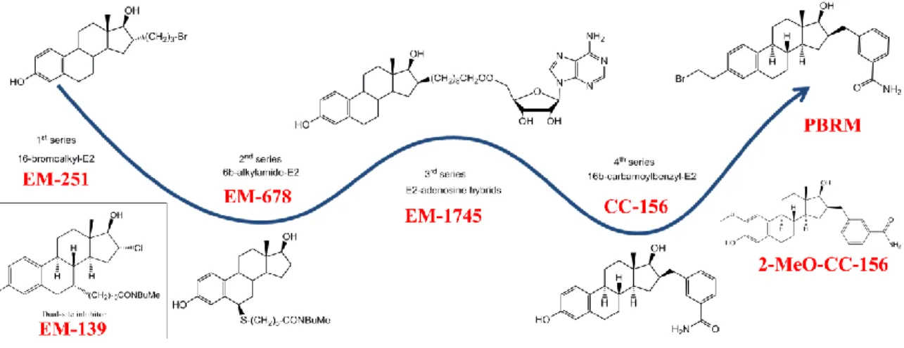
4.3 Inhibitors of 17β-HSD1
The development of inhibitors of 17β-HSD1 began in the 1970s and gradually gained momentum thereafter before culminating in the first decade of the 2000s88. Despite the number of years of research, no inhibitor has
yet reached the stage of clinical trials. The general properties of a good inhibitor should be highly potent and non-estrogenic. Also, it should be selective to HSD1 over the other HSD isozymes, especially 17β-HSD2, which catalyzes the reverse reaction (eg. oxidation of estrogens)89. The development of 17β-HSD1
were E2 derivatives bearing a bromoalkyl side chain at the 16α-position represented by the compound EM-25190. This irreversible competitive inhibitor EM-251 on 17β-HSD1 has an IC50 of about 320 nM, but was
proven to have estrogenic activity on the estrogen sensitive human breast cancer cell line ZR-75-191. A
modification at the C6 position of E2 has led to the development of a second series of reversible inhibitors. These inhibitors have a thiaheptamamide side chain at the 6β-position of E2, and were represented by the compound EM-678 (IC50=0.17μM) which was found to be more potent than the substrate E1 itself92. Similar as
the first series inhibitors, it also has an estrogen effect92-93. Based on the binding energies of both the cofactor
and substrate sites94, as well as the three dimensional-structure of 17β-HSD175-76, a third series of inhibitors
from E2-adenosine hybrids were developed. These molecules are represented by compound EM-1745. This compound has an E2 moiety to interact with the substrate-binding site and an adenosine moiety to interact with the cofactor binding site, which is connected by an eight methylene groups side chain95. Though it has a
high inhibitory activity on purified 17β-HSD1 (IC50=52nM), there are some major drawbacks such as difficulty to
penetrate the cell membrane and weak competition ability against NADPH in intact cells 96. Further studies
focused on a benzyl group at the 16β-position of E2, which is proven to be efficient in improving the inhibitory activity, yielded the 16β-m-carbamoylbenzyl-E2 (CC-156), which is the most potent 17β-HSD1 inhibitor by far
with an IC50 value of 44nM for the conversion of E1 into E297. However, this fourth series of compounds was
demonstrated to have estrogenic activity. It stimulated the proliferation of estrogen receptor positive cell line MCF-7 and T-47D cells97. To reduce the unwanted estrogenic activity of CC-156, a series of modification at
position 2, 3 and 7 have been made and assessed, yielding the compound 18 (2-MeO-CC-156)97 which is less
potent (IC50 of about 230nM) than CC-156 but bearing no estrogenic activity, and a new potent nonestrogenic
compound named as 3-(2-bromoethyl)-16β-(m-carbamoylbenzyl)-17β-hydroxy-1,3,5(10)-estratriene (PBRM) 98-99. The latter did neither inhibit other 17β-HSDs nor CYP3A4100, and demonstrated to form a covalent bond
with 17β-HSD1. A long delay period (i.e. 3-5 days) was required to restore the 17β-HSD1 activity in cells after they had been treated with PBRM101. Moreover, further investigation demonstrated its efficiency in both breast
Figure 8 Key inhibitors of 17β-HSD1 from different Series (Poirier 2011).
Other than the inhibitors with a steroidal scaffold, several classes of non-steroidal 17β-HSD1 inhibitors have also been reported, such as the phytoestrogens102-103, gossypols104-105, thiophenepyrimidinones106,
(hydroxyphenyl)naphthols107-109, and bis(hydroxyphenyl)heterocycles110-112. Among these non-sterodial
inhibitors, the bicyclic substituted hydroxyphenylmethanones (BSHs) exhibited high inhibitory activity toward the 17β-HSD1 enzyme113-114. The following structural optimized
(5-(3,5-dichloro-4-methoxyphenyl)thiophen-2-yl)(2,6-difluoro-3-hydroxyphenyl)methanone displayed a subnanomolar IC50 towards the enzyme as well as
high selectivity over other enzymes, especially the 17β-HSD289, and estrogen receptors115, making it a
promising candidate for following development as a therapeutic agent.
Beside the traditional 17β-HSD1 inhibitors, a series of E2 derived pure antiestrogens bearing a 7α-alkylamide side chain and a D-ring modification (a halogen atom or a double bond) were reported to exert potent inhibitory effects on 17β-HSD1 activities116. These compounds were defined as dual-site inhibitor which represented by
compound EM-139116. Although the inhibition on 17β-HSD1 activities was obtained with this series of
inhibitors, the lack of selectivity for other enzymes compromised their potential in clinical utilities117.
5 The role of 17β-HSD7
17β-HSD7 is another important multi function enzyme in the reductive 17β-HSDs. Like 17β-HSD1, it catalyzes the formation of E2 from E1 and performs a more significant role in the inactivation of DHT into 3β-diol118-119.
17β-HSD7 was reported to be primarily involved in cholesterol synthesis120-121, and was suggested to be
predominantly involved in cholesterol metabolism rather than in sex steroid synthesis 122-123. However,
experiment conducted by Mr. Thé riault in Prof. Lin’ lab demonstrated that inhibiting E1 to E2 activity of the enzyme by inhibitor is not blocking its zymosterol to zymosterone activity (unpublished data). Moreover, unlike aromatase, which converts testosterone (T) to E2 and is mostly expressed in stromal cells, 17β-HSD7 is
principally expressed and modulated in epithelial cancer cells such as MCF-7 and T47D124. Furthermore,
recent in vitro and in vivo experiments demonstrated that inhibition of 17β-HSD7 can induce cell cycle arrest and trigger cell apoptosis in BC cells, and auto-downregulation feedback of the enzyme, leading to significant shrinkage of xenograft tumors118, 124. Furthermore, recent kinetic study showed that 17β-HSD7 has a Km value
of 5.2±0.4 μM which is much higher than the value of 17β-HSD1 (0.03± 0.01 μM); while the kcat value of
17β-HSD7 (2.9± 0.4 s-1) is much lower that the value of 17β-HSD1 (0.0063± 0.0003 s-1)67, 125. As a result, the Kcat/Km
value of 17β-HSD7 is 80,000 times lower than the value of 17β-HSD1, indicating that these two reductive steroid enzymes may responsible of the E1 to E2 conversion at different substrate (E1) levels.
6 Statistical Analysis of RNA sequencing Data in Cancer Research
DNA sequencing technologies have been advanced during recent years due to the development of high throughput sequencing (HTS) technologies which can sequence multiple DNA molecules in parallel126. They
enable simultaneous sequencing of millions of DNA molecules and are widely applied on genomics, epigenomics and transcriptomics127. RNA sequencing (RNA-seq) provides a profound advantage over other
methods on cancer diagnosis and classification, prediction of response to therapy and prognosis, as well as unveiling the molecular bases of tumorigenesis128. Moreover, transcriptomic profiling through RNA-seq will
facilitate the development of personalized treatment for cancer patients through the molecular classification of subtypes128. The Cancer Genome Atlas (TCGA) is a community resource project launched in 2005 by the
National Institute of Health (NIH) as a pilot project aiming to discover and catalogue major cancer-causing genome alterations in large cohorts through large-scale genome sequencing and integrated multi-dimensional analyses. The Genome Sequencing Centers (GSCs) of TCGA performed large-scale DNA sequencing on two complementary DNA (cDNA) samples from every TCGA cancer case: one from the tumor specimen and the second from non-malignant tissue to serve as a control. The TCGA database is currently the largest database of cancer genetic information of over 30 kinds of human tumours129. TCGA database provides the most
complete clinical information of each patient, and is widely used in many studies130-131.
7 Working Hypothesis and Research Objectives 7.1 Hypothesis
7.1.1 PBRM inhibiting 17β-HSD1 activity would be through the formation of a covalent bond with the enzyme. The interactions of the three inhibitors (PBRM, 2-MeO-CC-156 and EM-139) with 17β-HSD1 would have significant difference.
7.1.2 The substrate inhibition of 17β-HSD1 would be due to the formation of a dead-end complex which is involving the binding of a reversely oriented E1 and the enzyme.
7.1.3 The analysis of RNA sequencing data would unveil potential new target and combined therapy for breast cancer treatment.
7.2 Objectives
Objective one: To elucidate the structural detail of representative inhibitors interacting with 17β-HSD1, such
as EM-139, 2-Meo-CC-156 and PBRM. To achieve this, we have expressed and purified the recombinant 17β-HSD1 protein with Sf9 cells, which then was used in co-crystallization with these inhibitors in the presence or absence of cofactor analog NADP+. The crystal structures of the three complexes were determined and analyzed.
Objective two: To identify the mechanism of the substrate inhibition of 17β-HSD1 and in silico design of
inhibitors based on this information. To reach this goal, we have co-crystallized the purified 17β-HSD1 with E1, in the presence or absence of cofactor analog NADP+. After determination of the binary and ternary complex structures, a comparative analysis with previously reported E2/testosterone complexes will be performed to elucidate the substrate inhibition mechanism, followed by computer assisted inhibitor design.
Objective three: To use RNA-seq data from large number clinical samples from TCGA-BRCA cohort to
Chapitre 1 Combined biophysical chemistry
reveals a new covalent inhibitor with a
low-reactivity alkyl halide
1.1 Résumé
La 17β-HSD1 joue un rôle central dans la progression des maladies liées aux œstrogènes en raison de son implication dans la biosynthèse des œstrogènes, en particulier de l’estradiol, constituant une cible thé rapeutique importante pour le traitement endocrinien. Auparavant, le composé principal 16β-(m-Carbamoylbenzyl)-E2 (CC-156) é tait dé crit comme un puissant inhibiteur de 17β-HSD1 dans la transformation de l’œstrone en estradiol. Cependant, l’activité œstrogénique de l’inhibiteur a compromis son potentiel de dé veloppement ulté rieur. Une modification à la position C-2 du CC-156 a produit un inhibiteur non œstrogénique, le 2-MeO-CC-156, avec beaucoup moins de puissance d’inhibition que celle d’origine. Des recherches plus poussé es à la position C-3 du CC-156 donnent un nouvel inhibiteur irré versible, non estrogé nique, puissant et sté roïdien, le 3-(2-bromoethyl)-16β-(m-carbamoylbenzyl)-hydroxy-1,3,5(10)-estratriene (PBRM). Dans cette publication, nous rapportons les structures des complexes ternaires de la 17β-HSD1 avec le NADP+ et l’inhibiteur 2-MeO-CC-156 ou le PBRM.
1.2 Abstract
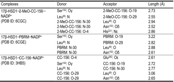
17β-Hydroxysteroid dehydrogenase type 1 (17β-HSD1) plays a pivotal role in the progression of estrogen-related diseases for its involvement in the biosynthesis of estrogens, especially estradiol, constituting a valuable therapeutic target for endocrine treatment. Previously, the lead compound 16β-(m-Carbamoylbenzyl)-E2 (CC-156) was described as a potent 17β-HSD1 inhibitor of the transformation of estrone into estradiol. However, the estrogenic activity of the inhibitor compromised its potential for further development. A modification at the position C-2 of CC-156 produced a non-estrogenic inhibitor 2-MeO-CC-156, with a much less potency as compared with the original one. Further investigation at the position C-3 of CC-156 yield a new potent and steroidal non-estrogenic irreversible inhibitor 3-(2-bromoethyl)-16β-(m-carbamoylbenzyl)-17β-hydroxy-1,3,5(10)-estratriene (PBRM). In the present paper, we report structures of the ternary complexes of 17β-HSD1 with NADP+ and inhibitor 2-MeO-CC-156 and PBRM. In the 17β-HSD1-2-MeO-CC-156-NADP+ complex, the presence of a methoxy group at C-2 of the inhibitor significantly reduces its estrogenic effect in estrogen-depended cancer cells, however it also impedes the essential hydrogen bond at the recognition end of the ligand binding pocket, significantly decreasing its inhibitory activity to the enzyme. For the 17β-HSD1-PBRM-NADP+ complex, the hydrogen bond between O-19 of the inhibitor and Oγ of Ser142 is much weaker as
selectivity of 17β-HSD1 through the formation of a covalent bond with Nε of residue His221, suggesting its
potential as a therapeutic agent for EDDs.
1.3 Introduction
Covalent inhibitors (CIs) are more beneficial than noncovalent ones because of the reduced risk of drug resistance, extended inhibition effect, increased efficiency with lower doses, and fewer side effects1. However,
despite these advantages, toxicity issues encountered with the first generation of CIs related to their high reactivity, low specificity of action, and some immunogenicity response resulted in resistance from the pharmaceutical industry2. Nevertheless, the approval of more specific and safe targeted CIs in the past decade
led to a resurgence of interest in the pharmaceutical research field3-4. However, the design of such inhibitors
remains a challenge, considering that a high binding affinity for the targeted protein, as well as an inherent reactivity, are two essential elements that must be combined in a single molecular entity to obtain a valuable drug candidate. Even if some covalent drugs have been documented bearing a low-reactivity group that could lead to alkylation in a particular molecular context5, the electrophilic group incorporated into CI is generally
highly reactive (α,β-unsaturated ketone, α-haloketone, cyanamide, fluorophosphate, and epoxide), with the inconvenience of increasing the risk of off-target and nonspecific tagging6. The use of less reactive electrophile
groups is thus suitable for increasing the level of CI specificity2, 7-9.
Most CI drugs are based on the reactivity of cysteine10, the strongest nucleophile among natural amino acids
(AAs), allowing the alkylation of a large diversity of electrophiles11. However, because of its low abundance or
an inaccessible position in the enzyme catalytic site, other nucleophilic residues have been exploited for covalent inhibition, such as lysine, serine, tyrosine, threonine, aspartate, and glutamate12-13. One uncommon
case is the histidine (His) residue, which, despite its good nucleophilicity and its presence at the catalytic site of many enzymes14, has been very rarely exploited in CI design15.
17β-hydroxysteroid dehydrogenase type 1 (17β-HSD1) catalyzes the final step of the transformation of estrone (E1) to estradiol (E2), the most potent estrogen, and is considered a promising therapeutic target for endocrine treatment16-21. This enzyme also catalyzes the reduction of dehydroepiandrosterone (DHEA) into
5-androstene-3β,17β-diol (5-diol) and dihydrotestosterone (DHT) into 5α-androstane-3β,17β-diol (3β-diol), which has been suggested to become more important after menopause, and may be involved in aromatase inhibitor resistance16, 21-23. It is well-known that E2 stimulates breast cancer and also plays a crucial role in other
estrogen-related diseases such as ovarian cancer, endometriosis, and endometrial cancer24-25. Thus, the
blockade of the biosynthesis of E2 is considered to be a valuable therapeutic approach for treating estrogen-dependent diseases24-26.
Previous reports have described 16β-(m-carbamoylbenzyl)-E2 (CC-156) (Figure 1.1) as a potent competitive and reversible inhibitor of 17β-HSD1 with an IC50 value of 44 nM27. Unfortunately, this compound has an
estrogenic activity observed by the proliferation of the stimulation of estrogen receptor (ER) positive cell lines MCF-7 and T-47D27. To reduce this unwanted estrogenic activity, further development was then engaged to
modify the E2 scaffold of CC-156. The addition of a methoxy (MeO) group at position C-2 of CC-156, which produced 2-MeO-CC-156 (Figure 1.1), was efficient in attenuating estrogenic activity but was unfavorable for enzyme inhibition27. A more promising strategy next focused on the chemical modification of the C-3 phenolic
group, which is known to be important for the binding of the E2 scaffold to ER28, and resulted in the discovery
of PBRM (Figure 1.1), the first nonestrogenic irreversible inhibitor of 17β-HSD129. Further investigations
demonstrated the PBRM efficiency in both breast cancer cells and human breast tumor xenografts in nude mice30-31, as well as interspecies differences of 17β-HSD1 inhibition32. Kinetic studies classified PBRM as a
competitive and irreversible inhibitor of 17β-HSD1, and a covalent binding of PBRM with 17β-HSD1 was then demonstrated by using a 17α-tritiated derivative of PBRM32. Furthermore, a molecular modeling study
investigating interspecies inhibitory activity of PBRM noted His221 as a potential key AA involved in the
formation of a covalent bond with the bromoethyl side chain. Interestingly, as an indication of the applicability of the bromoethyl group for developing a specific CI drug, PBRM possesses the expected properties of a CI, such as an extended inhibition action and a very low promiscuity rate33. The bromoethyl side chain also
provides a reduced in vitro CYP metabolism in comparison to its phenolic analog (CC-156), which is translated by a higher in vivo bioavailability for PBRM29.
Despite the indirect evidence of an alkylation between PBRM and 17β-HSD1, the existence and the exact configuration of expected covalent bonds remain to be proven. This was especially significant, considering the predicted low reactivity between a His residue and a primary alkyl halide, even more in a physiological environment32. Obviously, the demonstration of the capacity of a common and accessible functional group like
a primary alkyl halide to act as a reagent for the N-alkylation of an enzyme could demonstrate the viability of such a weak electrophile group for the development of a new type of selective CI. In fact, very few documented examples of an enzyme alkylation by a primary alkyl halide derivative have been reported to date, including a case of O-alkylation from a carboxylate group of Asp106 residue for haloalkane dehalogenase
tagging34 and a suspected S-alkylation from Met193 of 16α-bromopropyl-E2 leading to an irreversible inhibition
of 17β-HSD135-36. Importantly, the primary alkyl halide electrophile group must not be confused with activated
alkyl halide units, like the highly reactive N-ethylhalide of “nitrogen mustard” agents, which form a covalent bond via the formation of an intermediate aziridinium very reactive species that reacts with the DNA nitrogenous base37, or with benzyl halide38-39 and α-halo ketone40 groups, which are not specific, albeit useful
1.4 Results and Discussion
Analysis of PBRM molecular interactions before the His221 N-alkylation
The inhibitor PBRM bears a bromoethyl side chain at position C-3, making the inhibitor a little longer compared to CC-156’s. However, since the core structure of PBRM, especially the C-16 benzylamide moiety, is the same as that of CC-156, we expected the major conformational modifications during the binding of PBRM had happened at the recognition end (His221, Glu282) of the steroid binding site43-44. We were therefore interested in
investigating the interaction of the bromoethyl chain with His221 before the N-alkylation event. Besides, the
Met279 could possibly act as a nucleophile over the bromoethyl chain, considering that the distance between
the C-3 of phenolic OH at C-3 in CC-156 and Met279 (3.97 Å ) is similar to with His221 (3.45 Å ) (data from
CC-156 ternary complex45).
The pseudo PBRM complex structures were visually built from the CC-156 ternary complex structure using
SeeSAR software. In the CC-156 complex, the Glu282 side chain faces the binding site to make a hydrogen
bond with the inhibitor, leaving no space to build the bromoethyl side chain on CC-156. Since Glu282 is a
solvent-exposed flexible residue with a high average B-factor value of 40.6 Å 2, we modeled its conformation to
have the side chain exposed to the solvent (as described in the Experimental Section). Moreover, with the existence of side chains from His221 and Met279, the bromide from generated poses of PBRM maintained at
least a van der Waals distance from them, which is too long to overcome the force field limitations that do not allow for covalent reactions between the bromoethyl moiety and the side chains. To explore the possible positions of the bromoethyl side chain before the subsequent N-alkylation reaction, residues His221 and Met279
were mutated into Ala, which has a smaller side chain. The best poses with the highest estimated affinity using this binding site conformation are presented in Figure 1.2 A,B. The distance from the CH2 of the bromoethyl
side chain to the NH of the His221 side chain is about 2.5 Å , whereas that distance to the S of Met279 is 2.0 Å .
This result urges us to engage co-crystallization experiment for 17β-HSD1-PBRM to clarify the mechanism. Because no example of N-alkylation between an enzyme and a primary alkyl halide has been reported to date and also to rule out the possibility of the Met279 of 17β-HSD1 to act as nucleophile over the primary alkyl halide
(Figure 1.2), we thus seized this opportunity and engaged cocrystallization experiments of PBRM with 17β-HSD1 to prove the capacity of such a weak electrophile to form a covalent bond with the suspected His221 residue, an AA rarely exploited in design of CI drugs15.
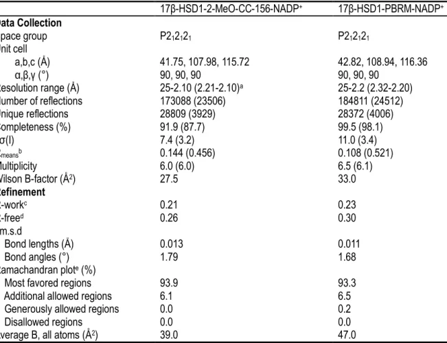
Structure determination of enzyme-inhibitor complex crystals
HSD1–PBRM–NADP+, were refined to 2.1 and 2.2 Å , respectively. The two models show good
stereochemistry48, and the quality of the final refined models can be accessed from the statistics in Table 1.1.
The models of 17β-HSD1 with PBRM (F0D) and 2-MeO-CC-156 (F0A) ternary complexes show very clear electron density for almost all residues, except for the C-terminal end of the protein (residues 286–327) as well as the flexible loop region from Ala191 to Gly198, as observed in other 17β-HSD1 complexes21, 43, 49-50. The
active-site structure of both inhibitor complexes for the A subunit is shown in Figure 1.3.
Comparison of 2-MeO-CC-156 and CC-156 ternary complexes
The presence of a methoxy group at position C-2 in 2-MeO-CC-156 introduces a strong hydrophobic interaction with residue Leu262 with the distance of 3.15 Å between C-32 (CH3 of MeO) of inhibitor and Cδ of
the AA residue. This interaction causes the inhibitor to shift 1.04 Å at the O-4 end and to rotate by approximately 4.8° at the steroid core and 3.5° at the benzylamide ring, as compared to the position of the CC-156 complex when superimposing the 2-MeO-CC-156 complex with the previously reported CC-156 ternary complex (PDB ID 3HB5) by Cα atoms (Figure 1.4A)45.
The side chain of Glu282, used to make a hydrogen bond with the inhibitor in the CC-156 ternary complex,
adopts a conformation facing the outside of the protein. Thus, no hydrogen bond can form between the AA residue and 2-MeO-CC-156. Besides, the movement of the O-4 at the end of 2-MeO-CC-156 forces the imidazole side chain of His221 to shift away by 1.49 Å for the Nε as compared with the position of the Cε of
His221 in CC-156 complex. The hydrogen bond between the inhibitor and His221, which is important for ligand
recognition and orientation51, is established in the 2-MeO-CC-156 complex with a distance of 2.86 Å (Table
1.2). However, the movement of the side chain of His221 toward the solvent leads to the decrease of its stability
(average B-factor of 49.0 Å 2 of the AA residue as compared with 39.0 Å 2 of the subunit) compared with its
counterpart (average B-factor of 30.7 Å2 of the AA residue as compared with 29.8 Å 2 of the subunit) in the
CC-156 complex.45 Indeed, when 0.1 μM inhibitor concentration was used, 2-MeO-CC-156 inhibited 37% of the
transformation of E1 into E2, whereas CC-156 inhibited 77% of the same reaction27. This is in agreement with
the relatively high flexibility of the bound 2-MeO-CC-156 (average B-factor, 54.9 Å ) as compared to CC-156 (average B-factor, 35.6 Å ). The hydrogen bonding with Ser142 is conserved in the 2-MeO-CC-156 complex, as
in the CC-156 ternary complex.
For the benzylamide ring, the π–π interaction between Tyr155 and the ring is conserved (Figure 1.4A). The
distance between Cε2 of Tyr155 and C-23 of 2-MeO-CC-156 is 3.50 Å , and the distance between the centroid of
O-29 of the carboxamide (CON) group of the inhibitor acts as an acceptor forming a hydrogen bond with N of Leu95, whereas the N-30 (CON) acts as a donor forming two hydrogen bonds with O of Leu95 and Oδ of
Asn152 (Table 1.2). Thus, the CON group in 2-MeO-CC-156 adopts a conformation of 180° flip, as compared
with that in the CC-156 ternary complex (Figure 1.4A).
Enzyme interaction with NADP+ in ternary complexes
Similar to the previously described model50, only the adenine ring, the ribose and phosphate groups of NADP+
molecule can be unambiguously identified in the electron densities of the 17β-HSD1−2-MeO-CC-156−NADP+
ternary structure (Figure 1.3A). The NMN moiety of the NADP+ molecules missing from the densities was
omitted from the final models. It indicates that the major interaction between the NADP+ and enzyme happens
at the ADP part, in agreement with previous the structure-function study44. As compared with the NADP+
molecule in CC-156 ternary complex, the 2’-phosphate group attached to the adenine ribose in 2-MeO-CC-156 ternary complexes has moved 3.7 Å toward the position of Nη of Arg37 in CC-156 complex, and is stabilized by
the water bridged hydrogen bond with the N of Arg37 and Asp38 as well as the Oγ of Thr41. As a result, the side
chain of Arg37 has moved to the protein surface and stabilized by forming a hydrogen bond with Oδ of Asp38.
Two important hydrogen bonds between the adenine ring and residues Asp65 and Val66 are conserved in
2-MeO-CC-156 ternary complex, as well as the hydrogen bond between the O-3 attached to the adenine ribose and Oγ of Ser12. No obvious different interaction was observed at the NADP+ binding site in the PBRM
complex, as compared with that of the 2-MeO-CC-156 complex. Similarly, the electron density map of the nicotinamide and the attached ribose of the NADP+ molecule are unable to define (Figure 1.3B). The
hydrogen bonds with surrounding residues Ser11, Ser12, Asp65 and Val66, as well as the water bridged hydrogen
bond with residues Asp38 and Thr41 stabilized the ADP moiety of the NADP+ molecule. No direct interaction
was observed between the bound inhibitor and cofactor molecule in the ternary complex.
Comparison of PBRM and CC-156 ternary complexes
In the 17β-HSD1–PBRM–NADP+ ternary structure, an unambiguous continuity of electron density from the
side chain of His221 to the bound PBRM is observed in both subunits, indicating the formation of a covalent
bond between the Nε of His221 and the C-31 (BrCH2) of PBRM (Figure 1.3B). The structure overlay of the
complex with CC-156 complex shows a slight shifting at the C-3 end of PBRM (0.66 Å ) as well as the imidazole side chain of His221 (0.89 Å ) as compared with the positions of their counterparts in the CC-156
complex, indicating the dynamic process favoring the formation of the covalent bond between them. The slight movement of the steroid core of PBRM and side chain of His221 is caused by the formation of their covalent