Induction by estrogens of methotrexate resistance in MCF-7 breast
cancer cells
Paul A.Thibodeau, Nathalie Bissonnette, Suzanne Kocsis Be´dard, Darel Hunting and Benoit Paquette1
Department of Radiobiology, Faculty of Medicine, Universite´ de Sherbrooke, 3001 12th Avenue North, Sherbrooke, Que´bec J1H 5N4, Canada
1To whom correspondence should be addressed Email: [email protected]
Development of drug resistance is a major factor that limits the effectiveness of chemotherapy treatments. In this study, we determined whether estradiol or its metabolites 2-, 4- and 16α-hydroxyestrone could enhance the develop-ment of methotrexate resistance in the breast carcinoma cell line, MCF-7. Cells were incubated with the estrogens at a concentration of 10–8 M for 12 cell doublings and enhancement of methotrexate resistance was measured with the Luria–Delbru¨ck assay. The most efficient estrogens were the 4-hydroxyestrone and 16α-hydroxyestrone, which both stimulated methotrexate resistance by 88-fold as com-pared with the control without estrogen. 2-Hydroxyestrone had an enhancement factor of 33-fold, whereas estradiol showed a slight effect with an enhancement factor of 3.2-fold. To determine whether the estrogen receptor was involved in the development of resistance, expression of the pS2 gene, which contains an estrogen-responsive ele-ment, was measured. Both estradiol and 16 α-hydroxyes-trone stimulated expression of the pS2 gene. In contrast, 2- and 4-hydroxyestrone did not increase the level of pS2 mRNA. This suggests that tumors classified as estrogen receptor negative could also develop methotrexate resist-ance as the result of exposure to estrogens. The status of the tumor suppressor gene p53 was analyzed in methotrexate sensitive and resistant clones. In all the methotrexate resistant clones analyzed, the western blots indicated that the p53 protein was still present and transcriptionally competent, as measured by its capacity to stimulate tran-scription of the p21waf1/cip1gene following UVB irradiation. However, the basal level of p53 was higher in resistant clones and addition of 2- or 4-hydroxyestrone increased
p53 to levels equivalent to those observed following UVB
irradiation. However, this induction of p53 accumula-tion by estrogens failed to stimulate the transcripaccumula-tion of
p21waf1/cip1, which indicates that a transcriptionally inactive form of p53 accumulated in methotrexate resistant cells.
Introduction
Development of drug resistance is a major factor limiting the effectiveness of chemotherapy treatments. Methotrexate (MTX), a folic acid analog and a potent inhibitor of the
Abbreviations: DCC, dextran-coated charcoal; LD50, drug concentration lethal for 50% of the cells; MEM, minimal essential medium; MTX, methotrex-ate; SDS, sodium dodecylsulfmethotrex-ate; SSC, standard saline citrate.
enzyme DHFR, is commonly used for treatment of breast cancer. Several mechanisms associated with increased resist-ance to MTX have been identified, such as diminished drug uptake (1), expression of an altered DHFR enzyme with a reduced affinity for MTX (2), and increased levels of the target enzyme, DHFR (3).
The genotoxic and carcinogenic potential of some estrogens has been reported (4–8) and the most potent estradiol metabol-ites seem to be 2-, 4- and 16α-hydroxyestrone. However, the possible role of these hormones in the induction of drug resistance is still largely unknown. Among the estrogens potentially involved in breast cancer, only estradiol has been the subject of studies on MTX resistance. In wild-type MCF-7 cells, this hormone can increase resistance to MTX by decreasing cytoplasmic membrane fluidity, which contributes to the decreased transport of MTX and results in a lower steady-state concentration of MTX in cancer cells. However, this effect was observed only at estradiol concentrations 10-to 100-fold higher than physiological levels (i.e. 10–7 and
10–6M) (9,10). Nevertheless, an increased resistance to MTX
was also observed at physiological concentrations (10 nM) of estradiol in MCF-7 cells already resistant (11). These MTX-resistant cells, obtained after a step-wise selection in MTX medium, showed a 50-fold increase in the level of DHFR enzyme and a 1000-fold increased resistance to MTX. Incuba-tion with estradiol resulted in an addiIncuba-tional 1.5- to 3.0-fold increase in their already elevated level of DHFR. Although results were not shown, these authors also mention that a smaller (30%) induction in DHFR activity by estradiol was found in the wild-type MCF-7 cells (11,12). Thus, although the presence of estradiol has been shown to increase resistance to MTX in cells that are already resistant, the involvement of this hormone and its major metabolites in the development of MTX resistant cells prior to exposure to MTX needs to be investigated.
Increased resistance to MTX can result from genomic modifications, the most common being amplification of the
dhfr gene. Accumulation of DNA modifications is favored in
cancer cells since their genome is unstable (13). This genomic instability is characterized by the emergence of DNA modifica-tions by few or many cell doublings after exposure to carcino-gens. The DNA modifications that result from genomic instability do not appear randomly in the genome (14,15). This suggests that genomic instability may induce a persistent ‘competent state’ for mutations at specific loci in mammalian cells (15,16).
Cancer cells appear to have the capacity to modulate their level of genomic instability in response to external agents or stress. Indeed, an in vivo enhancement of genomic rearrange-ments (a type of genomic instability) was measured in mouse 10T12 cells transformed in vitro by X-rays (15). A high
frequency of genomic rearrangements (50–100%) was found in cells derived from tumors that grew in vivo, whereas the frequency was very low (,10%) in the same transformed cells
cultured only in vitro. This suggests that agents present in the C3H mouse may enhance genomic instability. A similar enhancement of genomic instability has been reported with Fisher rat embryo fibroblasts subjected to temporary anoxia (17), and seems to involve a specific endonuclease different from those involved in apoptosis (18).
Identification of agents that can modify the level of genomic instability could be useful to block the development of drug resistance. Recently, we have demonstrated that estradiol can enhance the phenotype of genomic instability, as detected by the appearance of new genomic rearrangements in minisatellite sequences (19).
We have determined the potential of estradiol and the metabolites 2-, 4- and 16α-hydroxyestrone to enhance the development of MTX resistance. The carcinoma cell line MCF-7 was incubated for 12 cell doublings in the presence of each estrogen. The hormone was then removed and the MTX resistant cells were isolated in the selection medium. The most efficient estrogens were the 4- and 16α-hydroxyestrones, which stimulated the development of methotrexate resistance in MCF-7 cells by 88-fold as compared with the control. The 2-hydroxyestrone was also potent with an enhancement factor of 33-fold whereas estradiol showed a slight effect with an enhancement factor of 3.2-fold.
Materials and methods Cells and culture conditions
The breast carcinoma MCF-7 cell line was obtained from the American Type Culture Collection (HTB-22; Rockville, MD) and was grown as recommended by the supplier. The culture medium consisted of minimum essential medium with phenol red (A-6770; Sigma, St Louis, MO) supplemented with sodium pyruvate (1 mM), bovine insulin (10µg/ml), 10% fetal bovine serum (1020– 90, lot no. MB98212; Intergen, New York, NY), penicillin (50 U/ml) and streptomycin (50 µg/ml). For the Luria–Delbru¨ck assay, endogenous estrogens were eliminated from fetal bovine serum using dextran-coated charcoal (DCC), which consists of 0.25% charcoal (Norit A), 0.025% dextran (mol. wt 80 700 Da) and 0.01 M Tris, pH 8.0. An aliquot of 40 ml of DCC solution was mixed overnight with 200 ml of fetal bovine serum, centrifuged (2500 g for 15 min), and then the serum was sterilized by filtration (bottle filter, 0.22 µm; Costar). This method eliminates 97% of the hormone as measured using radioactive estradiol at a concentration of 10–8M as a marker. During the selection with MTX, the DCC-treated FBS was also dialyzed. LD50determination
The LD50 (LD50: MTX concentration lethal for 50% of the cells) was determined by plating, in triplicate, 1000 MCF-7 cells in p100 Petri dishes, which were incubated 8 h later with MTX at concentrations that ranged from 1 to 20 nM. The medium with MTX was changed every 3 days, and 12 day colonies were stained and counted. The results from two separate experiments gave a LD50for MCF-7 cells of 7.5 nM.
Determination of the rate of development of methotrexate resistance induced by estrogens with the Luria–Delbru¨ck assay
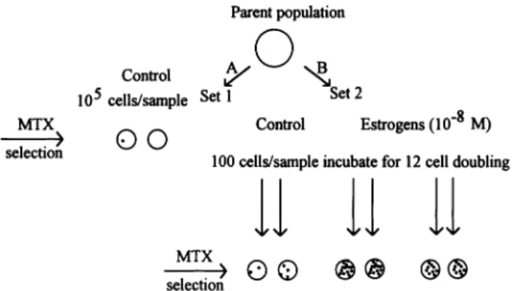
The analysis is based on the variation between the number of MTX resistant colonies arising from parallel cultures as described in Figure 1 (20). This figure shows a schematic diagram of a fluctuation experiment and the hypothetical results one could obtain. The first set of 10 Petri dishes (Figure 1A) contained replicate samples in which 105cells from the parent population were plated directly into selection medium (93LD50of MTX) and analyzed for the number of resistant colonies that emerged. The colonies on these plates represented rare pre-existing MTX resistant cells. Among these plates, the variation in the number of colonies per plate should have exhibited a Poisson distribution, and the mean number of colonies per plate reflected the prevalence of resistant variants in the parental population. Replicate plating from the same parent population should have only shown variation because of random sampling.
For the second set (Figure 1B), 100 viable cells (a number small enough to virtually eliminate pre-existing resistant variants) were plated in 20 Petri dishes and allowed to propagate for 12 cell doubling with one of the estrogens (i.e. estradiol or its metabolites, 2-, 4- or 16α-hydroxyestrone) at a concentration of 10–8M. As a control, the assay was performed with culture
Fig. 1. Schematic diagram of the Luria–Delbru¨ck assay. The first set of 10
Petri dishes (A) contains replicate samples in which 105cells from the parent population were plated directly into selection medium (93LD50of MTX) and analyzed for the number of resistant colonies that emerged. The colonies on these plates represented rare, resistant, pre-existing MTX resistant cells. For the second set (B), 100 viable cells (a number small enough to eliminate pre-existing variants) were plated in 20 Petri dishes and allowed to propagate for 12 cell doublings with one of the estrogens (i.e. estradiol or its metabolites 2-, 4- or 16α-hydroxyestrone) at a concentration of 10–8M. As a control, the assay was performed with culture media containing 0.01% ethanol. After exposure to the estrogen, individual populations (105cells) were then transferred to fresh plates and placed in selection medium (93LD50of MTX). Statistical analysis of the variation of MTX resistant colonies in parallel cultures allowed calculation of the rate of appearance of spontaneous and estrogen-induced mutants. The mutation rate was calculated according to the formula adapted by Capizzi and Jameson as described in Materials and methods.
medium that contained 0.01% ethanol, the solvent used to solubilize the estrogens. Individual populations (105 cells) were then transferred to fresh plates and placed in selection medium (93LD50of MTX). Statistical analysis of the variation of MTX resistant colonies in parallel cultures allowed calculation of the rate of appearance of spontaneous and estrogen-induced mutants. The mutation rate was calculated according to the following formula adapted by Capizzi and Jameson (21):
Cr5 (CµNt)ln(CµNt)
whereµrepresents the mutation rate per cell per generation, C is the number of Petri dishes in a series, r is the average number of mutants per series and Nt is the total number of cells. These latter two parameters were adjusted for the corresponding cloning efficiency. Once r, Nt, C and Cr are known, the mutation rate,µ, is obtained with the help of a table provided by Capizzi and Jameson (21). The Luria–Delbru¨ck assay was performed twice. The cloning efficiency for the wild-type MCF-7 cells was 65%, and for the MTX resistance cells it ranged from 29 to 60%.
pS2 mRNA induction
MCF-7 cells were incubated with the estrogens (10–8M) for 5 days, and then the RNA was isolated using the acid guanidium–thiocyanate extraction method (22). Aliquots of 15 µg of RNA was separated on a 1% agarose–2% formaldehyde denaturing gel and transferred to a nylon membrane (Hybond-N1; Amersham, Arlington Heights, IL) in 0.04 N NaOH with a vacuum transfer system (Tyler Research Instruments, Edmonton, Canada) for.5 h. The membrane was neutralized in 53 SSC and prehybridized for 16 h at 65°C in 120 mM Tris, pH 7.4, 600 mM NaCl, 8 mM EDTA, 0.1% sodium pyrophosphate, 0.2% SDS and 625 µg/ml of heparin. Hybridization was performed at 65°C overnight in the same buffer containing 53105 c.p.m./ml of32P-labeled probe. Probes were generated using random oligonucleotide primers and purified pS2 (0.6 kb) or GAPDH (1.0 kb) DNA. Following hybridization, the blots were washed for 20 min at room temperature in 23 SSC, and then for 60 min and 30 min at 65°C in 0.13 SSC and 0.1% SDS. The washed blots were exposed to a Kodak XAR-5 film for 2–16 h. The films were scanned with an UMAX Power Look II scanner and the relative intensity of the pS2 and GAPDH bands were determined with the NIH image 1.58 f program.
Extraction of genomic DNA
DNA was extracted according to a salting-out procedure described by Miller et al. (23). Briefly, ~53107 cells were washed in PBS, scraped with a policeman and centrifuged. The cell pellet was resuspended in 3 ml of a lysing buffer containing 10 mM Tris–HCl, 400 mM sodium chloride and
2 mM EDTA. To this cell suspension, 0.1 ml of sodium dodecylsulfate (20%) and 0.5 ml proteinase K (10 mg/ml, DNase free) were added and incubated overnight at 37°C or at 50°C for 3 h. The DNA was precipitated by the addition of 1.2 ml of 5 M sodium chloride. The tube was agitated for 1 min, centrifuged at 2500 g for 15 min and then the supernatant was transferred to another tube. The DNA was precipitated with 2.5 vol of 95% ethanol, the tube was gently inverted for 30 s, and the DNA was spooled out and air-dried briefly. The DNA was dissolved in TE buffer (10 mM Tris–HCl, 1 mM EDTA, pH 8.0) and RNase A (0.05 ml of a 10 mg/ml solution) was added and incubated for 1 h at 37°C. The DNA was precipitated a second time with ethanol as described above and redissolved in TE buffer.
Southern blot analysis
Aliquots of 10µg of each DNA sample were digested by EcoRI according to the manufacturer’s recommendations (New England Biolabs, Beverly, MA). Digested DNA was separated in 0.8% agarose gels at 40 V for 4 h, and then transferred onto a nylon membrane (Hybond-N1; Amersham) with a vacuum transfer (Tyler Research Instruments) in 0.4 M NaOH. The membranes were briefly neutralized in 53 SSC, prehybridized for 36 h at 67°C in 63 SSC, 53 Denhardt’s, 1% SDS and 250µg/ml ssDNA, and hybridized for 18 h at 67°C in the same solution containing 53105c.p.m./ml of32P-labeled probe (DHFR or GAPDH). The membranes were washed twice for 15 min at 65°C in 23 SSC/0.1% SDS (once for 30 min in 13 SSC/0.1% SDS and once for 45 min in 0.23 SSC/0.1% SDS) and briefly in 0.23 SSC. The washed filters were exposed to X-ray film (BioMax; Kodak, NY) at –80°C for 2–7 days. The films were scanned with an UMAX Power Look II scanner and the relative intensity of the dhfr and gapdh bands were determined with the NIH image 1.58 f program.
Irradiation and cellular lysate preparation
Cells were irradiated with a60Co source at 15 cGy/s (gamma radiation) or with UVB (302 nm) at a dose rate of 20 W/s. Cells were washed and scraped in buffered saline. Total cellular protein was extracted with lysing buffer (25 mM HEPES, pH 7.8, 2 mM EDTA, 1% NP40, 15% glycerol, 1 mg/ ml phenylmethanesulfonyl fluoride, 10 µg/ml of peptide inhibitor cocktail [leupeptin, chymostatin and pepstatin A] and 2 mM dithiothreitol). Electrophoresis and immunoblotting
SDS–PAGE was performed using a 5% stacking gel and a 12% separating gel. Equal amounts of protein from different samples were placed in boiling water for 4 min in the presence of SDS gel sample buffer (125 mM Tris– HCl, pH 6.8, 4% SDS, 0.01% bromophenol blue, 10% 2-mercaptoethanol and 15% glycerol) and electrophoresed for 90 min at 160 V. After transfer onto nitrocellulose (Hybond ECL; Amersham), the membrane was first blocked with 8% powdered skimmed milk in TBS (10 mM Tris–HCl, pH 8.0, and 150 mM NaCl) for 1–2 h and incubated with the appropriate first antibody overnight. Visualization of the second antibody was performed with a chemiluminescence detection procedure (PIERCE) according to the manufac-turer’s protocol (Chromatographic Specialties, Brockville, Canada). Antibodies
The antibodies were DO-1 [p53 (24)] and anti-p21waf1/cip1(Oncogene Science, Cambridge, MA).
Results
Induction of MTX resistance by estrogens
To quantify the induction of MTX resistance by the estrogens, we measured the corresponding mutation rate with a Luria– Delbru¨ck assay (20). The experimental set-up is illustrated in Figure 1. Briefly, 100 viable MCF-7 breast carcinoma cells were plated and allowed to propagate for 12 cell doublings in a total of 40 Petri dishes that contained one of the estrogens (i.e. estradiol or its metabolites 2-, 4- or 16α-hydroxyestrone) at a physiological concentration of 10–8 M. As a control, the
assay was performed with culture medium containing 0.01% ethanol, the solvent used to dissolve the estrogens. After 12 cell doublings, the hormones were removed and individual populations (105 cells) in each Petri dish were transferred to
fresh plates in medium that contained 93LD50 of MTX. To verify that the cells had grown for an equivalent number of cell doubling in each series of Petri dishes, cell numbers were determined prior to beginning the selection. The maximum variation between the different series of Petri dishes was 0.27
cell divisions. To determine the mutation rate with a Luria– Delbru¨ck assay, it was also necessary to know the number of reproductively competent cells plated in the selection medium. Therefore, at the beginning of the selection with MTX, control Petri dishes were plated in medium without MTX to determine the number of reproductively competent cells. These controls confirmed that none of the estrogens significantly altered the reproductive capacity of MCF-7 cells (data not shown).
The events that led to the development of MTX resistance were either spontaneous (control without estrogen) or estrogen induced. In a given series of Petri dishes, each expanding parallel culture had the same probability of generating resistant mutants with each cell division. In some cultures, the MTX resistance occurred early and many of the progeny of the resistant cell were present to form MTX resistant colonies. In others, the event occurred during one of the last cell divisions, thus generating few MTX resistant progeny. Statistical analysis of this variation allowed calculation of the rate of appearance of spontaneous and estrogen-induced mutants. The mutation rate was calculated according to the formula adapted by Capizzi and Jameson (21) and described in Materials and methods.
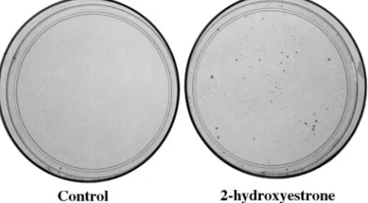
Figure 2 shows typical MTX resistant colonies that have grown in the selection medium from cells previously exposed to 2-hydroxyestrone. The rate of MTX resistance per cell per cell doubling, induced by either estradiol or 2-, 4- or 16α -hydroxyestrone, can be found in Table I. Our results clearly demonstrate that the estradiol metabolites enhanced the devel-opment of MTX resistance in the breast cancer cell line MCF-7. The most efficient estrogens were the 4-hydroxyestrone and 16α-hydroxyestrone, which stimulated the appearance of MTX-resistant MCF-7 cells by 88-fold. The 2-hydroxyestrone was also efficient with an enhancement factor of 33-fold, whereas estradiol showed a slight effect with an enhancement factor of 3.2-fold. These data cannot be explained by an increased rate of cell proliferation that occurs during exposure to the estrogens, because the maximum variation in cell number was equivalent to only 0.27 cell divisions between the different series of Petri dishes at the end of the incubation with the hormones.
Absence of pre-existing MTX resistant cells
To test whether the MTX-resistant cells actually appeared as a result of incubation with estrogens and not simply as a result of selection, the number of pre-existing MTX resistant cells in the parent population of MCF-7 cells was determined. The MCF-7 cells (13105) from the parent population were directly
incubated in the selection medium (i.e. 93LD50 of MTX) without exposure to the estrogens, and the number of MTX-resistant colonies were counted. After selection, an average of only 1.8 MTX resistant colonies per 13105MCF-7 cells plated
was measured. During the determination of MTX resistance induced by estrogen, 100 viable cells were plated per Petri dish; therefore, the corresponding number of pre-existing MTX resistant cells plated during our mutation assay was only 0.0018 per 100 cells. Consequently, we can conclude that virtually no pre-existing MTX resistant cells were present in the 100 cells plated per Petri dish at the beginning of the Luria–Delbru¨ck assay.
The absence of pre-existing MTX resistant cells was also verified by analyzing the maximum number of MTX resistant colonies that emerged at the end of the mutation assay. If one of the 100 cells plated at the beginning of the Luria–Delbru¨ck
assay was already resistant, and grew at the same rate as the other cells, the ratio of the pre-existing MTX resistant cell progeny to the other cells would not change. Consequently, at the end of the incubation with the estrogens, 103 of the 105
cells (i.e. 1/100 5 103/105) transferred into the selection medium would represent pre-existing MTX resistant cell pro-genies. In these series of experiments, the maximum number of MTX resistant cells in a Petri dish was 203. Therefore, this confirms that no pre-existing MTX resistant cell was plated at the beginning of the incubation period with estrogens, and that the resistant colonies that emerged following the selection with MTX were induced by the estrogens.
Prior to performing the Luria–Delbru¨ck assay, MCF-7 cells were maintained in fetal bovine serum not depleted of estradiol. This procedure did not significantly increase the number of MTX resistant cells, as demonstrated by our control. This conclusion is further supported by the fact that estradiol only results in a slight induction of MTX resistance and that this resistance is unstable. Therefore, it was not surprising that there were no MTX-resistant MCF-7 cells among the 100 viable cells plated per Petri dish at the beginning of the Luria– Delbru¨ck assay.
pS2 induction
Expression of the pS2 gene is known to be specifically activated by the estradiol–receptor complex (25). To determine if the development of MTX resistance by estrogens might involve
Fig. 2. MTX resistant colonies obtained after exposure to the estrogens. The
Luria–Delbru¨ck assay was performed as shown in Figure 1. For the control Petri dish, the cells were incubated with 0.01% ethanol; the second Petri dish shows an example of MTX resistant colonies after treatment with 2-hydroxyestrone.
Table I. Enhancement by estrogens of MTX resistance
Treatment Rate of MTX- Enhancement P-valuec
resistance per cell factorb per doublinga Control (0.01% ethanol)d 1.83106 Estradiol 6.383106 3.2 0.07 2-Hydroxyestrone 6.013105 33 1.22310–8 4-Hydroxyestrone 1.583104 88 1.04310–8 16-Hydroxyestrone 1.613104 88 8.66310–8
aCalculated according to the Luria–Delbru¨ck assay and the equation modified by Capizzi and Jameson.
bRatio between the rate obtained with an estrogen/rate obtained with ethanol.
cSignificance analyzed by the t-test.
dEthanol was used to dissolve the estrogens, the final concentration in the cell medium was 0.01%.
an estrogen–receptor pathway, the expression of pS2 was used as a reporter gene. MTX resistant colonies induced by the different estrogens were isolated and incubated for 5 days with their respective hormone at a concentration of 10–8M. GAPDH
mRNA was used as an internal standard. As shown in Figure 3, the basal level of pS2 is very low in the absence of estradiol and was not increased by ethanol. To quantify the induction of pS2 gene, the intensity of each RNA band was determined by densitometry. The relative induction of pS2 is reported in Figure 4. The maximal induction of pS2 expression occurred with estradiol and 16α-hydroxyestrone, whereas the catechol metabolites (2- and 4-hydroxyestrone) failed to stimulate the
pS2 gene. A decrease in the pS2 mRNA was even observed
with the 4-hydroxyestrone.
Stability of MTX resistance induced by estrogens
Two resistant colonies from each estrogen treatment were isolated. Although these cells were always grown in selection medium, the expression of MTX resistance was progressively lost and all resistant clones stopped growing in selection medium 3 weeks after they were isolated from the initial colonies. Thus following the initial isolation of resistant clones, it was impossible to grow a sufficient number of cells in the presence of MTX to test for the presence of an amplified dhfr gene. However, these initially resistant clones were grown in the absence of MTX following the Luria–Delbru¨ck assay, and Southern blot analysis of these MTX resistance clones was performed to test for the presence of an amplified dhfr gene. Using the gapdh gene as an internal control, our analysis demonstrated no amplification of the dhfr gene (Figure 5). Unfortunately, this result does not tell us the degree of
Fig. 3. Stimulation of pS2 mRNA accumulation by estrogens.
MTX-resistant clones induced by one of the estrogens were isolated and incubated for 5 days with their respective hormone at a concentration of 10–8M. The mRNA was then isolated and analyzed on a denaturing gel. Wild-type MCF-7 cells are shown as control. The respective lanes correspond to: 1, wild-type MCF-7; 2, wild-type MCF-71 0.01% ethanol; 3, MTX R-E21 estradiol; 4, MTXR-16α-OHE11 16α-hydroxyestrone; 5, MTXR -2-OHE11 2-hydroxyestrone; 6, MTXR-4-OHE11 4-hydroxyestrone. GAPDH was used as internal standard.
Fig. 4. Quantification of the relative stimulation of pS2 mRNA
accumulation. For each MTX resistant clone, the stimulation of pS2 was quantified by densitometry and normalized according to the internal standard GAPDH. The relative stimulation of pS2 accumulation was then calculated by comparing with the basal level of pS2 obtained in absence of the respective estrogens. E2, estradiol; 2-OHE1, 2-hydroxyestrone; 4-OHE1, 4-hydroxyestrone; 16α-OHE1, 16α-hydroxyestrone.
amplification of the dhfr gene in the MTX-resistant cells that were initially isolated at the end of the Luria–Delbru¨ck assay. Therefore, it is possible that the loss of resistance resulted from the loss of dhfr gene amplification, however, we have been unable to verify this hypothesis.
Status of the p53 tumor suppressor gene
The p53 protein contributes to genomic stability, and mutations in this gene have been associated with MTX resistance (26,27). In this study, the possible involvement of p53 in MTX resistance induced by estrogens was tested by determining if the p53 protein was still present and was capable of stimulating transcription of the p21waf1/cip1gene following UVB irradiation (28). The basal transcription of the p21waf1/cip1gene is independ-ent of p53 and is observed in p53 null cells but the induction of p21waf1/cip1 by UVB radiation requires p53 (29). The level of p21waf1/cip1 transcription was estimated based on the level of p21waf1/cip1protein. Ten MTX-resistant clones were analyzed.
They were isolated following the Luria–Delbru¨ck assay where cells were exposed to either 0.01% ethanol (control), estradiol, 2-hydroxyestrone, 4-hydroxyestrone or 16α-hydroxyestrone.
The UVB dose required to increase the basal level of p53 and to stimulate the transcription of p21waf1/cip1was determined. Figure 6 shows that the basal level of p53 protein expression in MCF-7 cells is very low in the absence of radiation and estrogens, and that 300 J/m2 was the minimal UVB dose
capable of increasing p53 and stimulating transcription of
p21waf1/cip1. Therefore, in all the experiments reported here, the expression of the p53 and p21waf1/cip1proteins was determined
respectively 7 and 15 h after UVB irradiation with 400 J/m2.
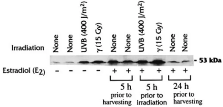
Estradiol is known to increase the level of p53 in the human mammary tumor cell line T47-D (30). Therefore, experimental conditions were optimized to allow us to distinguish the effect of UVB and estrogens on p53. When the analysis was made 5 h after the addition of estradiol, p53 was increased to a level equivalent to that observed with UVB alone or with the
Fig. 5. dhfr gene dosage in MTX resistant clones. Southern blot analysis of
MTX-resistant clones was performed to test for the presence of an amplified dhfr gene, using the gapdh gene as an internal control. The respective lanes correspond to: 1, wild-type MCF-7; 2, MTXR-spontaneous; 3, MTXR -E2clone 1; 4, MTXR-E2clone 2; 5, MTXR-2-OHE1clone 1; 6, MTXR-2-OHE1clone 2; 7, MTXR-4-OHE1clone 1; 8, MTXR-4-OHE1clone 2; 9, MTXR-16α-OHE1clone 1; 10, MTXR-16α-OHE1clone 2; 11, wild-type MCF-7.
Fig. 6. Induction of p53 and p21waf1/cip1proteins following UVB-irradiation. Wild-type MCF-7 cells were irradiated at the indicated dose of UVB-radiation and incubated for 15 h prior to harvest. Protein extraction, electrophoresis and immunoblotting are described in Materials and methods.
combination of UVB and estradiol (see Figure 7). The increase in p53 levels induced by estradiol was transient and the p53 level had essentially returned to normal 24 h after the addition of the hormone, which allowed us to determine the specific effect of UVB on p53 levels.
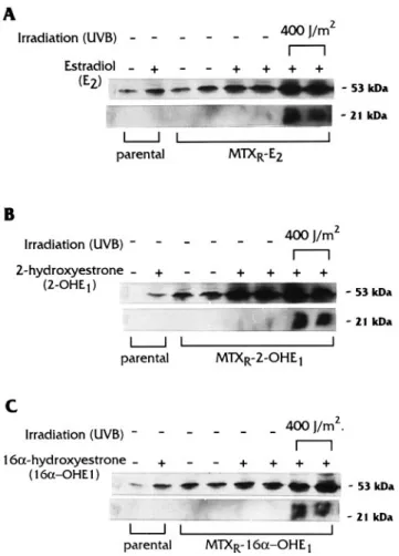
Figure 8 shows the levels of p53 and p21waf1/cip1obtained
following treatment with estrogens and UVB radiation on MTX-resistant clones induced by estradiol, 2-hydroxyestrone and 16α-hydroxyestrone, and on parental MCF-7 cells. Results obtained with the MTX resistant clones induced by 4-hydroxy-estrone were equivalent to those with the 2-hydroxy4-hydroxy-estrone (data not shown). In all MTX resistant clones studied, western blot analysis demonstrated that their p53 protein was active since they stimulated the transcription of p21waf1/cip1after UVB exposure, based on the increase in the p21waf1/cip1 protein.
However, there was an important difference in response to estrogen between the MTX-resistant clones and the parental MCF-7 cells. All four estrogens studied increased the level of p53 in the parental MCF-7 cells, with estradiol and 16α -hydroxyestrone having a greater effect. In the MTX resistant clones, the levels of p53 in the absence of estrogens were higher than the level measured in parental MCF-7 cells. For the MTX-resistant clones induced by estradiol and 16-hydroxyestrone, i.e. MTXR-E2 and MTXR-16α-OHE1, the
addition of their respective hormones increased the level of p53 only slightly (Figure 8A and C). On the other hand, a 10-fold enhancement was induced by 2-hydroxyestrone in the MTXR-2-OHE1clone. This enhanced level of p53 was
equiva-lent to the stimulation induced by UVB, but no activation of
p21waf1/cip1 transcription was measured when only the 2-hydroxyestrone was present, which suggests that the p53 accumulated in this MTX-resistant clone was transcriptionally inactive (Figure 8B).
Discussion
MCF-7 cells were exposed to these estrogens for 12 cell doublings, followed by an incubation in medium that contained
Fig. 7. Stimulation of p53 protein expression by treatment with estradiol
(E2) and/or DNA damaging agents. Cells were irradiated at the indicated dose of radiation in the absence or presence of estradiol, which had been added 5 h prior to irradiation. The effect of estradiol alone on p53 expression was verified by treating cells for 5 or 24 h prior to harvest. Cell treatments and protein extraction are described in Materials and methods.
Fig. 8. Modulation of p53 protein expression by estrogens and UVB
radiation. (A) Estradiol (E2); (B) 2-hydroxyestrone (2-OHE1); and (C) 16α-hydroxyestrone (16-OHE1) were used. The effect of each estrogen on p53 protein expression was tested on wild-type MCF-7 (parental) cells and also on MTX-resistant clones. Transcriptional transactivation of p21waf1/cip1 was observed following 400 J/m2of UVB radiation. Cell treatments and protein extraction are described in Materials and methods.
93LD50of MTX. Unlike other studies where MTX resistance was obtained after a step-wise selection in MTX (11,12), the event responsible for this resistance was induced during an exposure to the hormone alone. Under these in vitro conditions, the most efficient estrogens were the 4-hydroxyestrone and 16α-hydroxyestrone, which stimulated methotrexate resistance in MCF-7 cells by 88-fold. The 2-hydroxyestrone had an enhancement factor of 33-fold, whereas estradiol showed a slight effect with an enhancement factor of 3.2-fold. A similar small effect of estradiol has previously been reported (11). However, the effect of estradiol was not statistically significant (P5 0.07) in our study.
This enhancement of MTX-resistance cannot be the result of a difference in the number of cell divisions occurring during the exposure to the hormones, because a variation in cell number equivalent to only 0.27 cell divisions between the different series of Petri dishes was found at the end of the incubation with the estrogens.
After the incubation with estrogens, the MCF-7 cells were immediately transferred to Petri dishes that contained the selection medium. Therefore, it is possible that a significant amount of estrogen was still present in MCF-7 cells when MTX was added. Catecholestrones are rapidly methylated within a few hours by the enzyme catechol-O-methyltransferase to the inactive derivatives 2- and 4-methoxyestrone (31,32).
The 16α-hydroxyestrone is also short lived and binds cova-lently to amino groups contained in proteins (31,33,34). Since culture medium with fresh hormone was renewed only once, 5 days before the selection in MTX, it is reasonable to assume that these estrogens were already metabolized before selection in MTX. Only estradiol could still have been present at a significant concentration. However, estradiol showed only a slight effect with an enhancement factor for MTX resistance of 3.2-fold.
Alteration of MTX sensitivity can result from non-genotoxic or genotoxic effects. In 10T12cells transformed in vitro by
X-rays, estradiol can enhance the phenotype of genomic instabil-ity, as detected by the appearance of new genomic rearrange-ments in minisatellite sequences (19). To determine whether estrogen receptors might be involved in the development of methotrexate resistance, the pS2 gene was used as a reporter gene. It has been demonstrated that the induction of pS2 reaches a plateau after 3–6 days of incubation with estradiol at 10–8M with the MCF-7 cells (35). Under these conditions,
we have determined that the catechol metabolites, i.e. the 2-and 4-hydroxyestrone, did not increase the level of pS2 mRNA, which suggests that the development of MTX resistance can occur via a mechanism independent of the estrogen-receptor pathway. This result also suggests that 2- and 4-hydroxyestrone may induce MTX resistance in breast tumors that are estrogen-receptor negative.
Both estradiol and 16α-hydroxyestrone induced substantial increases in the level of pS2 mRNA. Thus, these estrogens may induce MTX resistance via a pathway that involves the estrogen receptor; however, the effect of 16α-hydroxyestrone may not be restricted to its action on the estrogen receptor. In short-term cultures, 16α-hydroxyestrone efficiently binds to the estrogen receptor and shows a degree of estrogenic activity that is almost as important as estradiol (8) when measured by stimulation of cell proliferation and elevated expression of progesterone receptors. However, with long-term incubations, 16α-hydroxyestrone forms covalent associations with estrogen receptors and proteins associated with DNA, which produces a marked decrease in its estrogenic activities. This irreversible binding has been suggested as one mechanism for malignant transformation in estrogen target tissue (33,34). Indeed, in the mouse mammary epithelial cell line C57/MG, 16α -hydroxyes-trone induced transformation (as detected by acquisition of anchorage-independent growth in soft-agar), hyperproliferation and DNA damage (5). In our Luria–Delbru¨ck assay, cells were incubated with the 16α-hydroxyestrone for 10 days. Therefore, it is possible that the induction of MTX resistance in MCF-7 cells by 16α-hydroxyestrone was a combination of pure estrogenic activity and genotoxic effects.
The metabolites of estradiol, 2- and 4-hydroxyestrone, can cause direct and indirect genotoxic effects (4). They are oxidized to semiquinones and then o-quinones that can undergo redox cycling mediated through cytochrome P450/P450 reduc-tase. This redox cycling produces free radicals that can induce oxidative damage to DNA (4). The 2- and 4-hydroxyestrone can also react directly with cellular nucleophiles such as protein and non-protein thiol groups and with DNA (4). DNA single strand breaks and 8-hydroxydeoxyguanosine, a mutagenic product of oxidative damage because of hydroxyl radical attack on DNA, were detected in vitro and in vivo (31). These mechanisms could be involved in the mutation of genes critical for the maintenance of the integrity of the genome (such as the tumor suppressor gene p53), which leads to the
accumulation of more DNA modifications. A second possibility is that the free radicals produced by the 2- and 4-hydroxyestrone could activate the antioxidant-responsive element (ARE) found in the promoter sequence of genes involved in the protection of DNA (36). The possible involvement of free radicals in the development MTX resistance is supported by the study of Hahn et al. (37) who demonstrated that after an exposure to 1000 cGy of ionizing radiation, drug resistance increased by as much as 1000-fold over unirradiated mouse EMT-6 cells. Furthermore, resistance to chemotherapeutic agents is observed in almost all lung cancers, an organ where oxidative stress is omnipresent.
An interesting observation made in our study was that the estrogen-induced MTX resistance was reversible, which is in agreement with previous studies of MTX resistance induced by other means. Instability of MTX resistance is not new. Flintoff et al. (38) reported that 12 out of 31 spontaneously MTX-resistant Chinese hamster ovary cell isolates had unstable phenotype, as well as some MTX-resistant clones induced by ethyl methanesulfone. Instability of the amplified DHFR gene was also observed in mouse sarcoma S-180 cells (39). Further-more, enhancement of MTX resistance induced by UV irradi-ation is also transient. The maximum enhancement was observed when 3T6 mouse cells were exposed to MTX at 12– 24 h after UV treatment (40). Different mechanisms of MTX resistance have been reported. Some are stable and are related to a mutation in genes coding for either the transmembrane transporter of MTX or the dhfr enzyme (41,42). MTX resistance can also occur via the amplification of the dhfr gene. This amplified gene can be present in double minute chromosomes or chromosomal DNA (43,44). Although this latter location is much more stable, it has been demonstrated that both forms of dhfr gene amplification can be lost during multiple cell divisions (44). Our results demonstrate that the exposure to estrogens was able to induce MTX resistance in a small fraction of the population of MCF-7 cells, which is consistent with either a gene mutation or an amplification event. However, the fact that the resistance was unstable suggests an amplifica-tion was involved.
Among the genes that are associated with MTX resistance and whose expression can be modified by estrogens, c-myc and the tumor suppressor gene p53 are particularly interesting. Overexpression of c-myc has been reported to favor the amplification of dhfr (44,45). Furthermore, it is known that estradiol can stimulate its expression (46). However, no correla-tion between this stimulacorrela-tion of c-myc and the amplificacorrela-tion of the dhfr gene has been reported.
In this study, we determined the status of the tumor sup-pressor gene p53 in the MTX resistant clones. The p53 protein is important in maintaining the stability of the genome, and some studies have reported an association between the appearance of MTX resistance and mutations in p53 (26,27). Our western blot analysis shows that p53 was still inducible in all MTX resistant clones studied, and was able to activate the transcription of p21waf1/cip1after UVB irradiation. However, the behavior of p53 was altered compared with the parental MCF-7 cells. The basal level of p53 was enhanced in all MTX clones. This level was further increased by 10-fold following the addition of 2-hydroxyestrone, but without stimulation of
p21waf1/cip1. These results suggest that the p53 was transcrip-tionally inactive, possibly as a result of a genomic modification induced during the incubation with the estrogens. If the p53 protein induced by 2-hydroxyestrone had been transcriptionally
competent, the resulting accumulation of the p21waf1/cip1protein
would have resulted in G1 cell-cycle arrest. However, the
2-hydroxyestrone had no inhibitory effects on cell growth. Accumulation of p53 is stimulated by reactive oxygen species (ROS) generated by ionizing radiation (47), but which are also produced by 2- and 4-hydroxyestrone. Therefore, a mutation in the defense system against oxidative stress could result in higher p53 accumulation in presence of the 2- and 4-hydroxyestrone.
In conclusion, this study demonstrated that estradiol metabol-ites 2-, 4- and 16α-hydroxyestrone enhance the development of MTX resistance in the breast carcinoma cell line MCF-7. The mechanisms involved still need to be elucidated and could implicate both non-genotoxic and genotoxic effects induced by estrogens.
Acknowledgements
This research project was funded by Medical Research Council of Canada and the Fonds pour la formation de chercheurs et l’aide a` la recherche. References
1. Wroblewski,D.H., Bhushan,A., Xuan,Y., Brinton,B.T., Tritton,T.R. and Hacker,M.P. (1996) Investigations on the mechanisms of methotrexate resistance in a cisplatin-resistant L1210 murine leukemia cell subline. Cancer Chemother. Pharmacol., 37, 337–342.
2. Haber,D.A., Beverley,S.M., Kiely,M.L. and Schimke,R.T. (1981) Properties of an altered dihydrofolate reductase encoded by amplified genes in cultured mouse fibroblasts. J. Biol. Chem., 256, 9501–9510.
3. Kaufman,R.J., Brown,P.C. and Schimke,R.T. (1979) Amplified dihydrofolate reductase genes in unstably methotrexate-resistant cells are associated with double min chromosomes. Proc. Natl Acad. Sci. USA, 76, 5669–5673.
4. Lieh,J.G. (1990) Genotoxic effects of estrogens. Mutat. Res., 238, 269–276. 5. Telang,N.T., Suto,A., Wong,G.Y., Osborne,M.P. and Bradlow,H.L. (1992) Induction by estrogen metabolite 16-hydroxyestrone of genotoxic damage and aberrant proliferation in mouse mammary epithelial cells. J. Natl Cancer Inst., 84, 634–638.
6. Bradlow,H.L., Hershcopf,R., Martucci,C. and Fishman,J. (1986) 16-Hydroxylation of estradiol: a possible risk marker for breast cancer. Ann. NY Acad. Sci., 464, 138–151.
7. Li,J.J., Gonzalez,A., Banerjee,S., Banerjee,S.K. and Li,S.A. (1993) Estrogen carcinogenesis in the hamster kidney: role of cytotoxicity and cell proliferation. Environ. Health Perspect., 101 (suppl. 5), 259–264. 8. Li,J.J., Li,S.A., Oberley,T.D. and Parsons,J.A. (1995) Carcinogenic
activities of various steroidal and nonsteroidal estrogens in the hamster kidney: relation to hormonal activity and cell proliferation. Cancer Res.,
55, 4347–4351.
9. Clarke,R., van den Berg,H.W. and Murphy,R.F. (1990) Reduction of the membrane fluidity of human breast cancer cells by tamoxifen and 17-estradiol. J. Natl Cancer Inst., 82, 1702–1705.
10. Clarke,R., van den Berg,H.W., Kennedy,D.G. and Murphy,R.F. (1985) Oestrogen receptor status and the response of human breast cancer cell lines to a combination of methotrexate and 17-oestradiol. Br. J. Cancer,
51, 365–369.
11. Cowan,K.H., Goldsmith,M.E., Levine,R.M., Aitken,S.C., Douglass,E., Clendeninn,N., Nienhuis,A.W. and Lippman,M.E. (1982) Dihydrofolate reductase gene amplification and possible rearrangement in estrogen-responsive methotrexate-resistant human breast cancer cells. J. Biol. Chem.,
257, 15079–15086.
12. Levine,R.M., Rubalcaba,E., Lippman,M.E. and Cowan,K.H. (1985) Effects of estrogen and tamoxifen on the regulation of dihydrofolate reductase gene expression in a human breast cancer cell line. Cancer Res., 45, 1644–1650.
13. Loeb,L.A. (1991) Mutator phenotype may be required for multistage carcinogenesis. Cancer Res., 51, 3075–3079.
14. Paquette,B. and Little,J.B. (1992) Genomic rearrangements in mouse C3H/ 10T12 cells transformed by X-rays, UV-C, and 3-methylcholanthene, detected by a DNA fingerprint assay. Cancer Res., 52, 5788–5793. 15. Paquette,B. and Little,J.B. (1994) In vivo enhancement of genomic
instability in minisatellite sequences of mouse C3H/10T12cells transformed in vitro by X-rays. Cancer Res., 54, 3173–3178.
16. Kronenberg,A. (1994) Radiation-induced genomic instability. Int. J. Radiat. Biol., 66, 603–609.
17. Russo,C.A., Weber,T.K., Volpe,C.M., Stoler,D.L., Petrelli,N.J., Rodriguez-Bigas,M., Burhans,W.C. and Anderson,G.R. (1995) An anoxia inducible endonuclease and enhanced DNA breakage as contributors to genomic instability in cancer. Cancer Res., 55, 1122–1128.
18. Stoler,D.L., Anderson,G.R., Russo,C.A., Spina,A.M. and Beerman,T.A. (1992) Anoxia-inducible endonuclease activity as a potential basis of the genomic instability of cancer cells. Cancer Res., 52, 4372–4378. 19. Paquette,B. (1996) Enhancement of genomic instability by 17beta-estradiol
in minisatellite sequences of X-ray-transformed mouse 10T12 cells. Carcinogenesis, 17, 1221–1225.
20. Tlsty,T.D., Margolin,B.H. and Lum,F. (1989) Differences in the rates of gene amplification in nontumorigenic and tumorigenic cell lines as measured by Luria–Delbruck fluctuation analysis. Proc. Natl Acad. Sci. USA, 86, 9441–9445.
21. Capizzi,R.L. and Jameson,J.W. (1973) A table for the estimation of the spontaneous mutation rate of cells in culture. Mutat. Res., 17, 147–148. 22. Chomczynski,P. and Sacchi,N. (1987) Single-step method of RNA isolation
by acid guanidinium thiocyanate–phenol–chloroform extraction. Anal. Biochem., 162, 156–159.
23. Miller,S.A., Dykes,D.D. and Polesky,H.F. (1988) A simple salting out procedure for extracting DNA from human nucleased cells. Nucleic Acids Res., 16, 1215.
24. Vojtesek,B., Bartek,J., Midgley,C.A. and Lane,D.P. (1992) An immunochemical analysis of the human nuclear phosphoprotein p53. New monoclonal antibodies and epitope mapping using recombinant p53. J. Immunol. Meth., 151, 237–244.
25. Brown,A.M.C., Jeltsch,J.-M., Roberts,M. and Chambon,P. (1984) Activation of pS2 gene transcription is a primary response to estrogen in the human breast cancer cell line MCF-7. Proc. Natl Acad. Sci. USA, 81, 6344–6348.
26. Go¨ker,E., Waltham,M., Kheradpour,A. et al. (1995) Amplification of the dihydrofolate reductase gene is a mechanism of acquired resistance to methotrexate in patients with acute lymphoblastic leukemia and is correlated with p53 gene mutations. Blood, 86, 677–684.
27. Lu¨cke-Huhle,C. (1994) Permissivity for methotrexate-induced DHFR gene amplification correlates with the metastatic potential of rat adenocarcinoma cells. Carcinogenesis, 15, 695–700.
28. Liu,M. and Pelling,J.C. (1995) UV-B/A irradiation of mouse keratinocytes results in p53-mediated WAF1/CIP1expression. Oncogene, 10, 1955–1960. 29. Zhan,Q., Fan,S., Smith,M.L., Bae,I., Yu,K., Alamo,I., O’Connor,P.M. and Fornace,A.J. (1996) Abrogation of p53 function affects gadd gene responses to DNA base-damaging agents and starvation. DNA Cell Biol.,
15, 805–815.
30. Hurd,C., Khattree,N., Alban,P., Nag,K., Jhanwar,S.C., Dinda,S. and Moudgil,V.K. (1995) Hormonal regulation of the p53 tumor suppressor protein in T47D human breast carcinoma cell line. J. Biol. Chem., 270, 28507–28510.
31. Yager,J.E. and Lieh,J.G. (1996) Molecular mechanisms of estrogen carcinogenesis. Annu. Rev. Pharmacol. Toxicol., 36, 203–232.
32. Schneider,J., Huh,M.M., Bradlow,H.L. and Fishman,J. (1984) Antiestrogen action of 2-hydroxyestrone on MCF-7 human breast cancer cells. J. Biol. Chem., 259, 4840–4845.
33. Miyairi,S., Ichikawa,T. and Nambara,T. (1991) Structure of the adduct of 16-hydroxyestrone with a primary amine: evidence for the Heyns rearrangement of steroidal D-ring alpha-hydroxyimines. Steroids, 56, 361–366.
34. Swaneck,G.E. and Fishman,J. (1988) Covalent binding of the endogenous estrogen 16-hydroxyestrone to estradiol receptor in human breast cancer cells: characterization and intranuclear localization. Proc. Natl Acad. Sci. USA, 85, 7831–7835.
35. Westley,B., May,F.E.B., Brown,A.M.C., Krust,A., Chambon,P., Lippman,M.E. and Rochefort,H. (1984) Effects of antiestrogens on the estrogen-regulated pS2 RNA and the 52- and 160-kilodalton proteins in MCF-7 cells and two tamoxifen-resistant sublines. J. Biol. Chem., 259, 10030–10035.
36. Prestera,T. and Talalay,P. (1995) Electrophile and antioxidant regulation of enzymes that detoxify carcinogens. Proc. Natl Acad. Sci. USA, 92, 8965–8969.
37. Hahn,P., Nevaldine,B. and Morgan,W.F. (1990) X-ray induction of methotrexate resistance due to dhfr gene amplification. Somat. Cell Mol. Genet., 16, 413–423.
38. Flintoff,W.F., Davidson,S.V. and Siminovitch,L. (1976) Isolation and partial characterization of three methotrexate-resistant phenotypes from Chinese hamster ovary cells. Somat. Cell Genet., 2, 245–261.
39. Kaufman,R.J., Brown,P.C. and Schimke,R.T. (1981) Loss and stabilization
of amplified dihydrofolate reductase genes in mouse sarcoma S-180 cell lines. Mol. Cell. Biol., 1, 1084–1093.
40. Tlsty,T.D., Brown,P.C. and Schimke,R.T. (1984) UV radiation facilitates methotrexate resistance and amplification of the dihydrofolate reductase gene in cultured 3T6 mouse cells. Mol. Cell. Biol., 4, 1050–1056. 41. Sirotnak,F.M., Moccio,D.M., Kelleher,L.E. and Goutas,L.J. (1981) Relative
frequency and kinetic properties of transport-defective phenotypes among methotrexate-resistant L1210 clonal cell lines derived in vivo. Cancer Res., 41, 4447–4452.
42. Haber,D.A., Beverley,S.M., Kiely,M.L. and Schimke,R.T. (1981) Properties of an altered dihydrofolate reductase encoded by amplified genes in cultured mouse fibroblasts. J. Biol. Chem., 256, 9501–9510.
43. Schimke,R.T. (1984) Gene amplification, drug resistance, and cancer. Cancer Res., 44, 1735–1742.
44. Mai,S., Hanley-Hyde,J. and Fluri,M. (1996) c-myc overexpression associated DHFR gene amplification in hamster, rat, mouse and human cell lines. Oncogene, 12, 277–288.
45. Lu¨cke-Huhle,C., Mai,S. and Moll,J. (1997) c-myc overexpression facilitates radiation-induced DHFR gene amplification. Int. J. Radiat. Biol., 71, 167–175.
46. Dubik,D. and Shiu,R.P.C. (1988) Transcriptional regulation of c-myc oncogene expression by estrogen in hormone-responsive human breast cancer cells. J. Biol. Chem., 263, 12705–12708.
47. Hain,J., Weller,E.M., Jung,T. and Burkart,W. (1996) Effects of ionizing-and UV B-radiation on proteins controlling cell cycle progression in human cells: comparison of the MCF-7 adenocarcinoma and the SCL-2 squamous cell carcinoma cell line. Int. J. Radiat. Biol., 70, 261–271. Received on November 13, 1998; revised on May 22, 1998; accepted on May 29, 1998