Experimental study on the role of lysophosphatidic acid
in mediating cytokine/chemokine production and on the
signaling pathways involved in inflammatory arthritis
Thèse
Weili Hui
Doctorat en microbiologie-immunologie
Philosophiae Doctor (PhD)
Québec, Canada
© Weili Hui, 2017
Experimental study on the role of lysophosphatidic acid
in mediating cytokine/chemokine production and on the
signaling pathways involved in inflammatory arthritis
Thèse
Weili Hui
Sous la direction de :
III
Résumé
La polyarthrite rhumatoïde (PR) est l’une des maladies auto-immunes qui atteint les articulations. Les symptômes principaux de la PR incluent une inflammation chronique de la synoviale dépendante de diverses cytokines et chimiokines inflammatoires, ainsi que de nombreux médiateurs de caractère lipidique, sécrétés par les cellules immunitaires et par les synoviocytes. L’acide lysophosphatidique (LPA) et son enzyme productrice, l’autotaxine (ATX) ont été détectés dans le liquide synovial. Les niveaux des ARNm de deux des six récepteurs du LPA, soit LPA1 et LPA3, sont aussi plus élevés dans les fibroblastes synoviaux des patients PR comparé à ceux d’arthrose ou suite avec une incubation avec le TNFα. Le LPA est aussi reconnu pour sa capacité à induire une production de cytokines et de chimiokines par des synoviocytes de type B (RAFLS) provenant de patients PR, ainsi que dans le modèle murin de poche d’air. D’ailleurs, la pré-incubation avec le facteur de nécrose tumorale alpha (TNFα) in vitro ou bien in vivo rehaussa cette sécrétion de cytokines et de chimiokines induite par la LPA. Dans le cadre de cette étude, nous avons démontré qu’une chimiokine appelée CXCL13 est impliquée dans le recrutement lymphocytaire induit par le LPA après une pré-incubation au TNFα, dans le modèle de poche d’air. Le mécanisme par lequel le LPA peut faire sécréter des cytokines et des chimiokines et par lequel la TNFα peut contribuer à une superproduction de cytokines et de chimiokines dépendante du LPA a aussi été investigué. Nos résultats indiquent que la p38MAPK, la cascade signalétique ERK-MSK-CREB, la Rho kinase, et la PI3K sont toutes des voies de signalisation stimulées par le LPA qui modulent une sécrétion de l’interleukine-8 (IL-8) chez les synoviocytes de type B. En revanche, nous avons remarqué que la sécrétion de l’IL-8 par les synoviocytes stimulés par le LPA devenait insensible aux inhibiteurs des dites kinases lorsque traités au préalable au TNFα. Il n’en demeure pas moins qu’une inhibition simultanée de la p38MAPK et d’ ERK a mené à une diminution
IV
efficace de la sécrétion de l’IL-6 et de l’IL-8. Cette étude a permis de mieux comprendre le mécanisme de la réponse inflammatoire déclenchée par le LPA et d’identifier les principales voies de signalisation intracellulaire contribuant à la sécrétion de cytokines et de chimiokines en présence ou en absence de TNFα. Le travail pourrait avoir des retombées majeures en ce qui concerne les stratégies pour développer des drogues ciblant l’axe ATX-LPA et les kinases mentionnées ci-dessus pour le traitement de la polyarthrite rhumatoïde.
V
Abstract
Rheumatoid Arthritis (RA) is one of the most severe inflammatory arthritides. The main features of RA include chronic inflammation in the synovium mediated by inflammatory cytokines and chemokines as well as lipid mediators, secreted by immune cells and synoviocytes. Lysophosphatidic acid (LPA), a monoacyl phospholipid mediator, and its producing enzyme autotaxin (ATX) were detected in synovial fluid. LPA1 and LPA3 receptor mRNA levels were also higher in synovium from RA patients than from normal individuals. LPA was previously reported to induce cytokine and chemokine production in RA fibroblast-like synoviocytes (RAFLS) and in a mouse air pouch model. In addition, TNFα pretreatment in vivo or in vitro enhanced this cytokine and chemokine secretion induced by LPA. In this study, we demonstrated that an LPA-induced chemokine named CXCL13 is also involved in LPA-induced leukocyte recruitment in the mouse air pouch model after TNFα pretreatment. The mechanism whereby LPA induces
cytokine/chemokine secretion and whereby TNFα pretreatment induces
cytokine/chemokine super-production mediated by LPA was also investigated in this study. Our data demonstrated that p38MAPK, the ERK-MSK-CREB axis, Rho kinase, and PI3K were all involved in the LPA signaling resulting in IL-8 secretion in RAFLS. However, we found that, after pretreatment with TNFα, LPA-induced IL-8 secretion became insensitive to inhibitors of those kinases mentioned above. Notwithstanding, blocking both the p38MAPK and ERK pathways could effectively decrease IL-8 and IL-6 secretion. This study allowed a deeper understanding of the mechanism of the LPA-induced inflammatory response, including the signal pathways regulating cytokine/chemokine secretion with or without the exacerbating effect of TNFα. The study may have important implications for the development of drugs targeting ATX-LPA axis and the aforementioned signaling pathways for the treatment of RA.
VI
Table des matières TABLE OF CONTENTS
RESUME ... III ABSTRACT ... V TABLE DES MATIERES ... VI LISTE DES TABLEAUX ... X LISTE DES FIGURES ... XI LISTE DES ABREVIATIONS ... XIII DÉDICACES ... XVII AVANT-PROPOS ET REMERCIEMENTS ... XVIII
CHAPTER 1 ... 1
INTRODUCTION ... 1
1.1LYSOPHOSPHATIDIC ACID ... 1
1.1.1 Lysophospholipids ... 1
1.1.2 History of LPA ... 1
1.1.3 The Structure of LPA Species ... 4
1.1.4 Metabolism of LPA ... 7
1.1.5 Carrier Proteins of LPA ... 16
1.1.6 LPA Receptors and Their Signaling ... 17
1.1.7 Other Proposed LPA Receptors ... 24
1.1.8 Intracellular signaling pathways- MAPK cascades ... 25
1.2RHEUMATOID ARTHRITIS ... 33
1.2.1 Symptoms and Features ... 33
1.2.2 Cytokines in RA ... 39
1.2.3 Chemokines in RA ... 48
1.2.4 Animal Models of RA ... 51
1.2.5 RA Treatment ... 54
1.2.6 Roles of LPA in RA ... 56
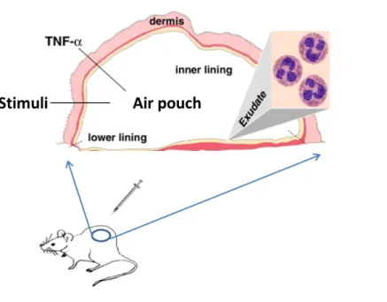
3EXPERIMENTAL MODELS USED IN THIS STUDY ... 59
3.1 The Murine Air Pouch Model ... 59
3.2 Fibroblast-like Synoviocytes from Rheumatoid Arthritis Patients ... 60
4GENERAL OBJECTIVES ... 61
4.1 Previous studies and unanswered questions (in vivo and in vitro) ... 61
4.2 Hypothesis ... 62
VII
CHAPTER 2 ... 64
2.1ABSTRACT ... 65
2.2INTRODUCTION ... 66
2.3MATERIALS AND METHODS ... 68
2.3.1 Materials ... 68
2.3.2 The Air Pouch Model ... 68
2.3.3 Flow Cytometry Analysis ... 69
2.3.4 Assessment of CXCL13 Secretion in the Air Pouch Lavage Fluids ... 69
2.3.5 Statistical Analysis ... 70
2.4RESULTS ... 71
2.4.1 LPA-Mediated Release of CXCL13 ... 71
2.4.2 LPA Recruits Various Leukocyte Subtypes into the Air Pouch ... 72
2.4.3 Effect of Blocking CXCL13 on LPA-Mediated CD3+ Cell Recruitment ... 73
2.5DISCUSSION ... 74 2.6FIGURES ... 77 2.7CONCLUSIONS ... 82 2.8REFERENCES ... 83 CHAPTER 3 ... 87 3.1ABSTRACT ... 88 3.2INTRODUCTION ... 89
3.3MATERIALS AND METHODS ... 92
3.3.1 Reagents ... 92
3.3.2 Cell Culture ... 93
3.3.3 Cell Treatment and Viability ... 93
3.3.4 Multiplex Immunoassay ... 93
3.3.5 Proteome Profiler™ Human Phospho-Kinase Array ... 93
3.3.7 Western Blotting ... 94
3.3.8 IL-8 and MCP-1 ELISA ... 95
3.3.9 Statistical Analysis ... 95
3.4RESULTS ... 96
3.4.1 LPA Induces Secretion of Multiple Inflammatory Cytokines/Chemokines in Human RAFLS ... 96
3.4.2 LPA Stimulates the Phosphorylation of Several Kinases and Transcription Factors in RAFLS ... 96
3.4.3 LPA Promotes the Phosphorylation of MSKs and CREB in RAFLS ... 97
3.4.4 MSKs are involved in LPA-Induced Chemokine Secretion in RAFLS ... 98
3.4.5 CREB is involved in LPA-Induced Chemokine Secretion in RAFLS ... 99
3.4.6 LPA-mediated Activation of CREB is Dependent on MSKs in RAFLS ... 99
3.4.7 The Impact of CREB Inhibitors on LPA-Induced Signaling and IL-8 Production by RAFLS ... 100
3.5DISCUSSION ... 101
VIII 3.7TABLE ... 110 3.8FIGURES ... 111 CHAPTER 4 ... 122 4.1ABSTRACT ... 123 4.2INTRODUCTION ... 124
4.3MATERIALS AND METHODS ... 126
4.3.1 Reagents ... 126
4.3.2 Cell Culture and Treatment ... 126
4.3.3 Analyses of Cytokine/Chemokine Production ... 127
4.3.4 Analyses of MSK Phosphorylation and CREB Phosphorylation ... 128
4.3.5 Statistical Analysis ... 128
4.4RESULTS ... 129
4.4.1 TNFα Up-Regulates MSK Phosphorylation Induced by LPA in RAFLS ... 129
4.4.2 Impact of Signaling on LPA-Mediated MSK Phosphorylation in Control- and TNFα-Primed RAFLS. ... 130
4.4.3 Inhibition of p38MAPK, ERK, Rho kinase, or MSK Activation Alone Does Not Regulate LPA-Mediated IL-8 Secretion in TNFα-Primed RAFLS. ... 131
4.4.4 Combination of Signaling Inhibitors Can Reduce LPA-Induced IL-8 Secretion after TNFα Priming132 4.5DISCUSSION AND CONCLUSION ... 134
4.6REFERENCES ... 139
4.7FIGURES ... 144
CHAPTER 5 ... 159
GENERAL DISCUSSION AND FUTURE DIRECTIONS ... 159
5.1OUR PRINCIPAL CONCLUSIONS BASED ON THIS STUDY ... 160
5.2DISCUSSION ... 161
5.2.1 Targeting LPA receptor mediated cell response ... 161
5.2.2 Indirect chemoattractant effect of LPA on leukocyte migration ... 162
5.2.3 Targeting intracellular signaling pathways in RA ... 163
5.2.4 The role of MSK and CREB in LPA-mediated cytokine and chemokine secretion ... 165
CHAPTER 6 ... 167
PERSPECTIVES ... 167
6.1THE ROLE OF LPA IN LYMPHOCYTE FUNCTION: LIMITATIONS OF THE MOUSE AIR POUCH MODEL AND OTHER SOLUTIONS ... 167
6.2HOW IS NF-ΚB INVOLVED IN LPA-INDUCED CYTOKINE/CHEMOKINE SECRETION WITH AND WITHOUT TNFΑ PRIMING? ... 168
6.3WHERE IS MSK ACTIVATION LOCATED, IN THE CYTOSOL OR INSIDE THE NUCLEUS? ... 168
6.4WHAT DO THE LPA RECEPTOR KNOCKOUT MICE LOOK LIKE IN ARTHRITIS MODELS? ... 169
6.5WHAT DO THE MSK1/2KO MICE LOOK LIKE IN ARTHRITIS MODELS? ... 170 6.6WHAT IS THE MECHANISM OF RAFLS PROLIFERATION?ARE SIGNALING PATHWAYS OTHER THAN MAPK
IX
SUCH AS HIPPO-YAP SIGNALING INVOLVED? ... 170
BIBLIOGRAPHIE ... 172 ANNEXES ... 195
X
Liste des tableaux Chapter 1
Table 1 Determination of LPA in different biological fluids Table 2 Phenotypes of LPA receptor knockout mice
Table 3 Characteristics of lysophospholipid receptors Table 4 The stages of RA
Table 5 Roles of some cytokines in the pathogenesis of autoimmune arthritis Table 6 Animal models of arthritis
Table 7 Studies showing the involvement of ATX-LPA in arthritis models
Chapter 3
Table 1 Effect of LPA on cytokine/chemokine secretion in human RAFLS with or without TNFα pre-treatment.
XI
Liste des figures Chapter 1
Figure 1. Lysophospholipid receptors and their intracellular signaling pathways
Figure 2. Chronology of the LPL field, LPLs and other lipid receptors, and overview of proximal LPL signaling features
Figure 3. Chemical structure of LPA species, analogs and cyclic derivatives Figure 4. LPA metabolism schematic
Figure 5. Domain organization and crystal structure of ATX
Figure 6. Illustration of the interaction between ATX, activated integrin and LPC Figure 7. Schematic overview of MAPK cascades
Figure 8. Stimuli and MAPK pathways that activate MSK Figure 9. The domain structure of CREB
Figure 10. Joint deformation of RA patients
Figure 11. Diagrammatic representation of synovial joint Figure 12. Pathophysiology of rheumatoid arthritis
Figure 13. Two forms of TNFα and TNFα mediated signaling
Figure 14. The cytokine network in lymphoid tissues and inflamed joints in autoimmune arthritis
Figure 15. Leukocyte trafficking into the inflamed synovium
Chapter 2
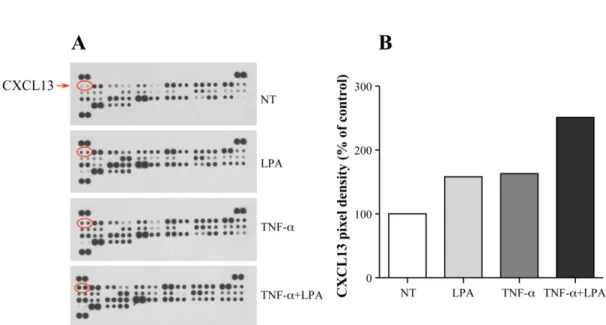
Figure 1. Effect of LPA on CXCL13 secretion in the murine air pouch with or without TNFα pretreatment
Figure 2. Effect of LPA and TNFα on CXCL13 secretion in the air pouch
Figure 3. LPA-induced leukocyte recruitment in untreated and TNFα-primed air pouches Figure 4. Effect of the CXCL13 neutralizing antibody on LPA-mediated lymphocyte recruitment into TNFα-primed air pouches
Chapter 3
Figure 1. Effect of LPA on kinases and kinase substrate phosphorylation in human RAFLS Figure 2. Effect of LPA on MSKs and CREB phosphorylation in human RAFLS
Figure 3. Effect of inhibition of MSKs on LPA-induced IL-8 secretion in human FLS Figure 4. Effect of silencing CREB on LPA-induced IL-8 secretion in human RAFLS Figure 5. Effect of silencing MSKs and of a MSK inhibitor on LPA-induced CREB phosphorylation
Figure 6. Effect of CREB inhibitor 217505 on LPA-induced IL-8 secretion and phosphorylation of MSKs and CREB
XII
Figure 7. Effect of CREB inhibitor KG-501 on LPA-induced IL-8 secretion and phosphorylation of MSKs and CREB
Figure 8. Schematic illustration of signal transduction pathways involved in LPA-induced IL-8 expression in human RAFLS
Chapter 4
Figure 1. Schematic illustration of the experimental design
Figure 2. Priming with TNFα enhances LPA-mediated MSK phosphorylation
Figure 3. Inhibition of ERK1/2 blocks LPA-mediated MSK1 Ser-376/MSK2 Ser-360 phosphorylation
Figure 4. Inhibition of ERK1/2 blocks LPA-mediated MSK1 Ser-212 phosphorylation Figure 5. Combinations of signaling inhibitors attenuate LPA-mediated MSK
phosphorylation
Figure 6. The sensitivity pattern of LPA-mediated IL-8 secretion to signaling inhibitors is altered after priming of RAFLS with TNFα
Figure 7. High concentration of Bay11-7082 totally blocks LPA-induced MSK phosphorylation but is highly cytotoxic for RAFLS
Figure 8. Combinations of signaling inhibitors differentially impact LPA-mediated IL-8, IL-6, IP-10, MCP-1 and RANTES secretion in TNFα-primed cells
Figure 9. Proposed signaling pathways involved in LPA-induced IL-8 secretion
Figure 10. Proposed signaling pathways involved in LPA-induced MSK phosphorylation after priming with TNFα
XIII
Liste des abréviations
AC adenylyl cyclase
ACKR Atypical chemokine receptor
acyl-CoA acyl coenzyme A, cofactor formed of pantothenate, cysteamine
and adenosine 3’-phosphate 5’-pyrophosphate
AGC group a kinase group named after the protein kinase A, G, and C families (PKA, PKC, PKG)
Akt Protein kinase B (PKB), a serine/threonine-specific protein
kinase
APC allophycocyanin
ApoM Apolipoprotein M a.k.a. Apom, component of low-and
high-density lipoproteins
ATF-1 activating transcription factor-1
ATX autotaxin, lysophospholipase D that produces LPA from LPC;
a.k.a. ENPP2, NPP2
B103 ASSAY bioassay on a rat central nervous system neuronal cell line B103
BSA bovine serum albumin
C/EBP-1 CCAAT/enhancer binding protein
CBP CREB-binding protein
CCP cyclic citrullinated peptide
CCR receptors for CC chemokines
CD The cluster of differentiation
CIA collagen-induced arthritis
COX-2 cyclo-oxygenase 2 or prostaglandin endoperoxide synthase
CREB cAMP response element-binding protein
CXCL chemokine (C-X-C motif) ligand
CXCL1/KC C-X-C chemokine motif ligand 1/Keratinocyte chemoattractant
CXCL10/IP-10 C-X-C chemokine motif ligand 10/Interferon gamma-induced
protein 10
CXCL8/IL-8 C-X-C chemokine motif ligand ligand 8/Interleukin 8
CXCR receptors for CXC chemokines
DAG diacylglycerol
DCs dendritic cells
DMARD disease modifying anti-rheumatic drug
DUSP Dual Specificity Phosphatase
EDG endothelial differentiation genes, family of receptors for LPA
XIV
eNOS endothelial nitric oxide synthase
ENPP nucleotide or ectonucleotide pyrophosphatase/
phosphodiesterase; a.k.a. Enpp; see ATX.
ERK extracellular signal-related kinase; a.k.a. p44/42
FAB-MS fast-atom bombardment mass spectrometry, a technique for the
analysis of protein sequence and structure
FABP3 fatty-acid binding protein 3
FBS fetal bovine serum
FLS fibroblast-like synoviocyte, also named synovial fibroblast
(SF)
Foxp3 forkhead box P3, a member of the forkhead transcription factor
family
FPP farnesyl (trimethyldodecatrienyl) pyrophosphate
GC-MS gas chromatography–mass spectrometry
GM-CSF granulocyte and macrophage colony-stimulating factor
GPAT glycerophosphate acyl transferase
GPCR G-protein-coupled seven-transmembrane-helix receptor
HDL high-density lipoprotein
HEV high-endothelial venule
HLA-DRB1,4 human major histocompatibility class II complex, locus DR, beta chain, variants 1 and 4
HMGN1 high mobility group nucleosome binding domain 1
HPTLC High performance thin layer chromatograph
HTLV human T-cell lymphotropic virus type 1; a.k.a. human T-cell
leukemia type 1
IFNgamma interferon gamma (type II interferon)
IKK The I kappa B kinase
IL-1 interleukin 1
IL-17RA the interleukin-17 receptor subunit
IL-1Ra The interleukin-1 receptor antagonist
JAK Janus kinase
JNK c-Jun N-terminal kinases
K/BxN Mice expressing both the T cell receptor (TCR) transgene KRN
and the MHC class II molecule A(g7) (develop severe inflammatory arthritis)
KRN mice Mice expressing the transgenic T cell receptor (TCR)
LCAT Lecithin–cholesterol acyltransferase
LC-MS liquid chromatography–mass spectrometry
XV
LPA lysophosphatidic acid, the subject of this thesis
LPAAT LPA acyl-transferase
LPC lysophosphatidylcholine, precursor of LPA
LPE lysophosphatidylethanolamine
LPG lysophosphatidylglycerol
LPI lysophosphatidylinositol
LPL lysophospholipid
LPP lipid phosphate phosphatase
LPS lipopolysaccharide
LPT lysophosphatidylthreonine
LTB4 leukotriene B4
lysoPS lysophosphatidylserine
MAG monoacyl glycerol
MAPK mitogen-activated protein kinase
MAPKAP-2 MAPK-activated protein kinase 2; a.k.a. MK2
MAPKK/MAP2K MAP kinase kinase (mitogen-activated protein kinase kinase)
MAPKKK/MAP3K MAP kinase kinase kinase
MCP-1/CCL2 monocyte chemoattractant protein 1/C-C motif chemokine
ligand 2
MEK MAP/ERK kinase; a.k.a. MKK, MAP2K, see MAP2K
MIP-2/CXCL2 macrophage inflammatory protein 2/C-X-C motif chemokine
ligand 2
MK2/3 MAPK-activated protein kinase 2/3
MMP matrix metalloproteinase
MSK mitogen- and stress-activated protein kinase; a.k.a. ribosomal
protein S6 kinase A5
NF-κB nuclear factor kappa-B, a transcription factor
NLD domain nuclease-like domain; a.k.a. NUC doamin
NPP nucleotide pyrophosphatase/phosphodiesterase
OA osteoarthritis
OPG osteoprotegerin
PA phosphatidic acid
PDE domain phosphodiesterase domain
PI3K phosphatidylinositol-4,5-bisphosphate 3-kinase
PKC Protein kinase C
PLA phospholipase A
PLC phospholipase C
PLD phospholipase D
XVI
RA rheumatoid arthritis
Rac protein monomeric GTPase, belongs to Rho family GTPases
RANK/RANKL receptor activator of nuclear factor κB ligand, a.k.a.
osteoprotegerin ligand (OPGL)
RANTES/CCL5 regulated on activation, normal T cell expressed and
secreted/Chemokine (C-C motif) ligand 5 Rho
kinase/ROCK/ROK
kinase for small G protein Ras homolog A (RhoA); a.k.a. Rock1, Rock2
RSK/S6K ribosomal s6 kinase
S1P sphingosine 1-phosphate
SKG mice a genetic model of RA due to altered signal transduction in T-cells
SMB domain somatomedin B-like domain
SPC sphingosylphosphatidylcholine
STAT signal transducer and activator of transcription protein
TAK1 transforming growth factor-β-activated kinase 1, a MAP3K
TAZ a transcriptional co-activator (with PDZ-binding motif)
Th17 type 17 T helper cell
TLC-GC thin-layer chromatography- gas chromatography
TLR Toll-like receptor
TNF tumor necrosis factor
TRAP tartrate-resistant acid phosphatase
Treg cells regulatory T cells
VEGF vascular endothelial growth factor
VLDL very-low-density lipoprotein
XVII
Dédicaces
Dedicated to my parents, my colleagues and all the friends who support me during my studies…
XVIII
Avant-propos et Remerciements
During my PhD studies, a lot of friends and colleagues in the lab gave me tremendous help and support, and I really appreciate it. As an international student from China, it is a challenge to adapt to the new environment of Quebec. This section is to thank everyone who gave me kind help in the lab and in my life.
First of all, I would like to give my thanks to the first two mentors in Quebec I met: my director Dr. Sylvain Bourgoin and my co-director Dr. Chenqi Zhao. Without them, I would not have had the chance to come to Canada to pursue my PhD. Dr. Bourgoin is very easygoing and nice to all his students and colleagues. He is very active in discussing interesting scientific questions and is always willing to help. He shows his humor all the time in public speaking and creates a good atmosphere around him. In our lab meetings, he makes me actively think to find the answers to his questions. Dr. Zhao is always very positive and kind, supporting me with every aspect of studies and life from the first day I arrived in Quebec, which left me a deep impression. She gave me a lot of mentorship and also some very practical advice in the techniques I used in my experiments. She is always very happy working in the lab and talks to everyone with a big smile, sometimes even telling some jokes to lighten up the day.
Second, I will give a brief introduction of all my closest colleagues all of whom gave me specific help and contributed to my thesis and my PhD studies:
Lynn Davis: Thanks for all the help and support with my experiments, improving my English and correcting my thesis. I will never forget your neat lab notebook writing, pretty frank smile, and healthy life style, which is a positive influence on my life!
Dr. Maria Fernandes: Thanks for your practical advice and chatting during my puzzled and confused days in the lab!
XIX
Dr. Paul Naccache: Thanks for your kindness in sparing me the time and effort of listening to the little problems in my studies and for giving me very meaningful guidance in my career and life!
Dr. Eric Boilard: Your story was a legend which gave me spiritual support, persuading me to stay on the road of science. By the way, thanks for your support at the beginning for the FACS analysis.
Dr. Fawzi Aoudjit: Thanks for all your support for helping me with the FACS analysis, courses and pre-doctoral exam!
Emmanuelle Rollet-Labelle: It is a joy to discuss science with you! I will remember your kind support and positive attitude toward life!
Finally, thanks to all the people who gave me such support in the research center—especially the axes of Rheumatology and Immunology, the assistants working in the animal facility, the FACS analysis platform and the Luminex platform. Thanks for the warm help from all the classmates and friends I met in the laboratory!
Contribution (in parentheses, estimation of my contributions to: experimental design; execution; data analysis; and writing) and publications:
Chapter 2
Hui W, Zhao C, Bourgoin SG. 2015. LPA promotes T cell recruitment through synthesis of CXCL13. Mediators Inflamm. 2015: 248492 (30%; 60%; 100%; 80%)
Chapter 3
Zhao C, Hui W, Fernandes MJ, Poubelle PE, Bourgoin SG. 2014. Lysophosphatidic acid-induced IL-8 secretion involves MSK1 and MSK2 mediated activation of CREB1 in human fibroblast-like synoviocytes. Biochem Pharmacol. Jul 1; 90(1):62-72. (10%; 50%; 40%; 10%)
XX
Hui W, Zhao C, Bourgoin SG. 2017. Tumor Necrosis Factor α governs the sensitivity of lysophosphatidic acid-induced cytokine/chemokine secretion to signaling inhibitors in synovial fibroblasts. (Manuscript in preparation, 30%; 60%; 70%; 60%)
Appendix
Bourgoin SG. Hui W. 2015. Role of mitogen- and stress-activated kinases in inflammatory arthritis. World J Pharmacol 4(4):265-273. (NA; NA; NA; 70%)
1
Chapter 1 Introduction 1.1 Lysophosphatidic Acid
1.1.1 Lysophospholipids
The term lysophospholipid (LPL) refers to any derivative of a phospholipid in which one acyl derivative is removed by hydrolysis. Lysophospholipid mediators, such as sphingosine 1-phosphate (S1P) and lysophosphatidic acid (LPA), can be synthesized from membrane phospholipids. Other LPLs include lysophosphatidylserine (lysoPS),
lysophosphatidylinositol (LPI), lysophosphatidylglycerol (LPG),
lysophosphatidylethanolamine (LPE), lysophosphatidylthreonine (LPT) and
lysophosphatidylcholine (LPC). Some of these have been found to be important signaling molecules mediating diverse effects by binding to and activating their cognate receptors [1-3] (Figure 1). The actions of those LPLs as lipid mediators including LysoPS, LPT, LPE and LPG have been reviewed in [3]. The history of LPLs and discovery of their receptors have been reviewed elsewhere (Figure 2). They will not be further discussed here as this is not the scope of this thesis. In this thesis, we focus on the study of LPA.
1.1.2 History of LPA
The discovery of LPA and its function date back to the 1950s and 1960s, when many lipid mediators with pharmacological activities were identified [4]. In 1954, Arneil and Dekanski detected an unknown vasopressor principle (named Arneil factor) in heparinized plasma (incubated at room temperature for clinical purposes) in patients or healthy subjects [5]. It was suggested to be LPC at first in 1960 [6], but was demonstrated later to be LPA produced from LPC by the action of a lysophospholipase D (lyso-PLD)-like activity in 1986 by the group of Tokumura [7]. LPA was first isolated and identified in 1978 as a vasopressor phospholipid in crude soybean lecithin
2
[8] and later was also identified in feline plasma incubated at body temperature [9]. Early reports showed that LPA was able to cause contraction of smooth muscle cells [10] and induce an increase in intracellular Ca2+ levels in 3T3 mouse embryonic fibroblast cells [11] and myeloma cells [12], indicating the existence of LPA receptors. In addition, LPA had been implicated as a bioactive lipid mediator with hormone- and growth factor-like activities [13]. Though many earlier reports demonstrated the involvement of putative LPA receptors [11, 14], the first high-affinity, cognate cell surface receptor for LPA was identified (LPA1) in 1996 [15] (Figure 2) by the group of Jerold Chunwhile they were identifying novel GPCR genes related with neurodevelopment.Until now, six LPA receptors have been identified [16-21], and an intracellular receptor of LPA was also identified (peroxisome proliferator-activated receptor gamma, PPARγ) [22]. Recent reviews have summarized the diverse roles of LPA in the past twenty years and updated nomenclature of LPA receptors [23-28]. As evidenced by the booming number of publications since the twentieth century, LPA has been implicated in various physiological and pathological states.
3
Figure 1. Lysophospholipid receptors and their intracellular signaling pathways (from [24])
4
Figure 2. Chronology of the LPL field, LPLs and other lipid receptors, and overview of proximal LPL signaling features ( From [29])
1.1.3 The Structure of LPA Species
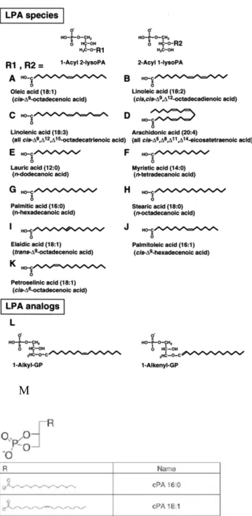
LPA can be generated naturally during platelet activation [30-32], thus platelets are a very important source of LPA species. Many other mammalian cell types are also known to produce LPA, such as adipocytes, neutrophils, fibroblasts, ovarian and prostate cancer cells, and neuronal cells [33, 34]. Many species of LPA are present in various biological fluids in different forms, due to the ester and ether linkages, the variety of fatty acid structure (fatty acid chain length and degree of saturation), and fatty acid linkage to sn-1 or sn-2 position of the glycerol backbone. The widely used species of LPA for signaling studies in the laboratory is 1-oleoyl-2-hydroxy-
5
polyunsaturated fatty esters, alkyl, alkenyl ether, as well as analogs 2, 3-cyclic phosphates [35] (Figure 3). The rank order of the acyl species of LPA in normal human plasma differs from the rank order in serum or in resting and thrombin-stimulated platelets, indicating that serum LPA and plasma LPA species have distinct precursors [31, 36]. The majority of serum or plasma LPA is in the form of acyl-LPA with palmitic (16:0), stearic (18:0), oleic (18:1), linoleic (18:2), and arachidonic acid (20:4) [36]. The 18:2 and 20:4 species make up 84% of the LPA found in serum [32].
6
M
Figure 3. Chemical structure of LPA species, analogs and cyclic derivatives
A-L, Chemical structures of LPA species and analogs; M, 2,3-cyclic derivatives of
7
1.1.4 Metabolism of LPA 1.1.4.1 Synthesis of LPA
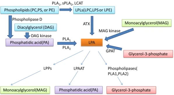
LPA is an important intermediate product in the biosynthesis of phospholipids and triacylglycerol, and as a signaling molecule. Thus far, several hypothetical pathways of LPA production have been postulated. LPA is present in blood with plasma concentration ranges from 0.1 to 1µM, and serum concentrations up to 10µM [38]. Serum LPA can be generated by platelet-dependent and platelet-independent pathways [39]. However, the mechanism by which LPA is produced in different biological fluids (saliva, seminal fluid, ascites, synovial fluid, follicular fluid) is still not very clear. The concentration of LPA in different biological fluids has been reviewed elsewhere (Table 1) [25, 40]. LPA can be produced: (1) intracellularly from glycerol-3-phosphate and acyl-CoA by a glycerophosphate acyl transferase (GPAT) [39, 41]; (2) from monoacylglycerol (MAG) by a monoacylglycerol kinase in mitochondria and microsomes [4, 42]; (3) from phosphatidic acid (PA) by PLA1- or PLA2- (phospholipase A) mediated hydrolysis [43, 44], which is the main mechanism in activated platelets (PA can be generated from hydrolysis of phospholipids by a phospholipase D (PLD) or from phosphorylation of diacylglycerol by a diacylglycerol kinase); (4) from lysophospholipids by lyso-PLD-mediated hydrolysis (autotaxin, ATX) [45] which is the primary source of extracellular LPA; (5) from oxidative modification of low density lipoprotein (LDL) [42]. Serum LPA can be produced mainly through pathway (3) and (4) [39] (Figure 4). The enzymes involved in these two pathways have been thoroughly discussed in the review of Aoki [39]. As pathway (1) is conserved in lower organisms without evidence of extracellular LPA production, it is not considered to be involved in extracellular LPA-dependent signaling events [39].
8
Figure 4. LPA metabolism schematic LCAT: Lecithincholesterol acyltransferases LPPs: lipid phosphate phosphatases
9
Table 1. Determination of LPA in different biological fluids. (Adapted from [25, 40])
10
1.1.4.2 The Enzyme Autotaxin
Autotaxin was first found to be secreted by melanoma cells and to be a chemoattractant for cancer cells [46]. Later autotaxin was proven to be the main enzyme responsible for LPA production which contributes to cancer cell migration (reviewed in [46, 47]).
(1) General
Autotaxin (ATX) is a member of the NPP (nucleotide
pyrophosphatase/phosphodiesterase) family, and is also referred to as ENPP2 (ectonucleotide pyrophosphatase/phosphodiesterase). Similar to NPP1/ENPP1 and NPP3/ENPP3, ATX is a multi-domain protein consisting of two N-terminal Cys-rich somatomedin B-like (SMB) domains, a central catalytic phosphodiesterase (PDE) domain and a C terminal nuclease-like domain (NLD) that is catalytically inactive (Figure 5A) [46]. ENPP1 and ENPP3 are membrane-bound enzymes which can be cleaved into soluble protein functionally hydrolyzing nucleotides, while ENPP2 is a secreted form [47]. Though ATX also hydrolyzes nucleotides, similar to ENPP1 and ENPP3, it shows 10-fold enhanced affinity for LPC than for nucleotides. LPC enhances the ATX-dependent cell migration [48], demonstrating that ATX could hydrolyze LPC to produce LPA as a cell response stimulus. Besides the catalytic function to produce LPA as a lysophospholipase D (lysoPLD), ATX is also reported to hydrolyze other LPLs, such as LPE, LPS, LPI [49] and SPC [50].
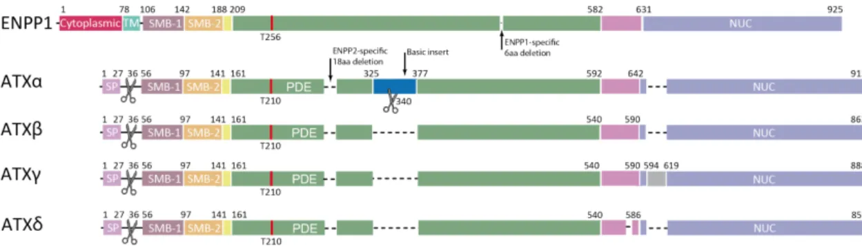
(2) Isoform
The alternative splicing of ATX mRNA results in distinct isoforms, of which α, β, γ, and δ have been identified with similar lysoPLD activities and substrate preferences [47](Figure 5B). ATXβ is the most widely expressed and is the predominant isoform that has been thoroughly investigated. ATXγ is brain specific. But whether distinct ATX isoforms are associated with specific (patho) physiological conditions is not clear. One report showed that ATXα but not ATXβ, by specific binding to heparin and
11
heparan sulfate, has increased lysoPLD activity toward LPC [51]. The demonstration of the crystal structure of ATX (Figure 5C) will allow us to better understand the interaction between ATX and its substrates, and protein-protein interactions, as well as the modulation of enzyme activity.
(3) Structure/Function Relationships
As shown in Figure 5C, ATX crystal structure studies have elucidated its domain organization and interaction, including interaction between the central catalytic PDE domain and SMB domains, as well as interaction between the PDE domain and the NLD which binds Ca2+ by an EF hand-like motif. ENPP1 and ATX/ENPP2 both harbor a shallow pocket for accommodating and binding the nucleotide for breaking the phosphodiester bond, which also accommodates LPLs (Figure 5C). Compared with ENPP1 or other phospholipases, ATX displays a distinct deep hydrophobic lipid-binding pocket (Figure 5C and Figure 6) unique for hydrolyzing LPLs and a tunnel through which LPA exits. The function of the deep pocket is confirmed by mutational and biochemical studies. This deep pocket is distinct from the autoinhibitory loop of secreted phospholipase A2 (sPLA2) which functions as an interfacial enzyme. The fourth feature of ATX is a SMB domain with integrin binding properties. In ATX, this domain interacts with the catalytic domain. The SMB domain of ATX has been found to bind β1 and β3 integrins in activated platelets and other cells for localizing LPA synthesis to the cell surface (Figure 6) [52]. As one example of this, ATX binds to integrins, then releases LPA which itself stimulates lymphocytes through their LPA receptors, enhancing their motility [53]. Some researchers suggest that ATX may interact with integrins to exert non-catalytic signaling functions like stimulating cell proliferation, which is independent of LPA signaling [54].
12
(4) Genetics
ATX showed physiological importance in embryonic development from the evidence that ATX knockout mice (Enpp2-/-) die before embryonic day 10 with clear vascular and neural tube defects [55]. Enpp2-/+ heterozygotes, survive until adulthood, but have only half the plasma LPA level of wild-type mice [56], and showed attenuated neuropathic pain [57]. Since the phenotypes of ATX knockout mice are more severe than those of any LPA receptor knockout mice (listed in Table 2), it is possible that other as yet unidentified LPA receptors may be involved in ATX function, or certain LPA receptors may have redundant functions, or ATX could function through other mechanisms unrelated to LPA signaling.
13
Figure 5A. Domain organization of NPP/ENPP family members (from [46])
Figure 5B. Different domain organizations of ATX isoforms compared with that of ENPP1.
14
Figure 5C. ATX crystal structure
15
Figure 6. Illustration of the interaction between ATX, activated integrin and LPC (From [52])
1.1.4.3 Degradation of LPA
As shown in Figure 4, LPA can be degraded through three distinct pathways: dephosphorylation into MAG by a family of lipid phosphate phosphatases (LPPs, including isoforms LPP1, LPP1a, LPP2, LPP3), which also dephosphorylate sphingosine-1-phosphate, phosphatidic acid and ceramide 1-phosphate in a non-specific manner; conversion back to PA by LPA acyltransferases (LPAAT, including five members named LPAATα, β, γ, δ, ε); hydrolysis into glycerol phosphate through LPA-specific phospholipases [42].
16
1.1.5 Carrier Proteins of LPA
Early studies related to LPA identified bovine or human serum albumin (BSA or HSA) [4, 42, 59], and liver fatty acid-binding protein [60] as endogenous LPA-binding proteins. Plasma gelsolin, the secreted form of gelsolin which is an intracellular actin binding protein, also binds LPA with nanomolar affinity [42]. In addition, FABP3 (fatty acid binding protein 3) has been identified as an intracellular LPA carrier protein in human coronary artery endothelial cells, regulating LPA-induced PPARγ activation in the nucleus [61]. Lipoprotein is known to be a lipid transportation complex. LDL is one of the major lipoproteins that plays a role in atherosclerosis [62]. In the same study, LPA is reported to be produced during oxidation of LDL, which actively initiates platelet activation. Phosphatidylcholine (PC), the major phospholipid component of VLDL [63], has been mentioned previously to generate LPA by two pathways through hydrolysis by PLA and PLD. High density lipoprotein (HDL) was identified as a carrier protein for several lysophospholipids including S1P, sphingosylphosphorylcholine (SPC) and lysosulfatide (LSF) [64]. S1P has been shown to bind to apolipoprotein M (ApoM) [65]. ApoM-bound S1P can activate S1P1 receptor on endothelial cells [66]. About 55% S1P in plasma is bound to HDL via ApoM and 35% to albumin [67]. Only ~10% of plasma S1P resides in other lipoproteins such as LDL and VLDL. It is possible that ApoM could function as a carrier protein of LPA species in a similar way as S1P, since there is LPA production during LDL oxidization and from VLDL. A novel approach was reported for measuring S1P and LPA binding to carrier proteins using monoclonal antibodies and the Kinetic Exclusion Assay [68], which will help to understand more about potential LPA carrier proteins and their role in controlling LPA levels. One research group found that a low concentration of albumin (15µM) could inhibit activation of LPA receptor, implying an important role for LPA carrier protein in LPA-mediated cell responses [69]. LPA in biological fluids may also form micelles
17
with proteins on the lipid droplet surface that can increase the stability of the micelles [70], which is important for LPA transportation and half-life in biological fluids.
1.1.6 LPA Receptors and Their Signaling
As an extracellular signaling molecule, LPA stimulates cellular responses through binding to the 7-transmembrane domain of G protein-coupled receptors (GPCRs) and activating downstream G proteins [24]. Early reports named the first three LPA receptors LPA1, LPA2, and LPA3 as EDG-2, EDG-4, and EDG-5 respectively (the endothelial differentiation, G-protein-coupled receptor gene family, EDG) (Table 3). The six LPA receptors and five sphingosine-1-phosphate (S1P) receptors constitute lysophospholipid receptors (LPL-R) [71]. GPCRs that bind specifically LPA have had their nomenclature updated recently: protein names LPA1 – LPA6, and italicized gene names LPAR1-LPAR6 (human) and Lpar1-Lpar6 (non-human) [24]. These GPCRs are believed to couple to one or more of the four classes of heterotrimeric G proteins (Gα12/13, Gαq/11, Gαi/o, and Gαs) [72], though signaling without this coupling may also be possible [73]. Activation of these receptors and G proteins in turn exerts a broad range of biological and pathological effects by activating various downstream signaling pathways (some of them are shown in Table 3) [25, 33, 74, 75].
18
19
1.1.6.1 LPA1
LPA1 (previous name: VZG-1/EDG-2/mrec1.3) was the first identified and the best characterized high affinity receptor for LPA [15]. The mouse cDNA of LPA1 encodes a 41-kDa protein consisting of 364 amino acids with 7 transmembrane domains, which is highly conserved in other mammalian (human and rat) Lpar1 genes [33]. It shares ~50-60% amino acid sequence identity with LPA2 and LPA3, and 30%, 32% and 37% amino acid identity with the cannabinoid receptors, melanocortin receptor and S1P1 receptor, respectively [76, 77]. The unique feature of LPA1 gene structure is that it contains five exons with one conserved intron (shared within Lpar1-3) located within the middle of transmembrane domain VI [76]. Of note, a variant of Lpar1 (mrec1.3) with a 18-amino acid deletion of the N terminus exists, but its biological significance is still unknown [78].
LPA1 is widely expressed in mouse and human tissues, particularly in the developing nervous system (reviewed in [23]). The gene expression chart file of LPA1 can be found at the BioGPS website (http://biogps.org/). After binding to LPA1, LPA signals through the G proteins Gi/o, Gq, and G12/13. In this way, cellular responses such as cell proliferation and cell migration can be initiated by LPA. These activated G proteins activate phospholipase C (PLC), mobilize Ca2+, activate the kinases MAPK, Akt, and Rho kinase, and induce binding to the serum-response element (SRE) (Table 3, Figure 1) (reviewed in [23, 33, 79]). One interesting function of LPA1 is that LPA mediates cell rounding in neuronal cells through G12/13 by Rho kinase activation [80], but mediates cell adhesion in other cell types at least in part through the same receptor [81]. Other cellular responses mediated by LPA1 have also been reviewed in [25].
Half of LPA1 gene knockout mice die perinatally [23, 82]. A defective sense of smell, smaller body size, a blunted snout, and brain defects including cortical thinning are those main phenotypes reported in LPA1 gene knockout mice [82, 83].
20
1.1.6.2 LPA2
LPA2 was identified through GenBank searches of orphan GPCR genes after LPA1 identification. The amino acid sequence and molecular weight of LPA2 are very similar to that of LPA1 (Table 3). Lpar2 mRNA expression is relatively restricted, with high expression levels in kidney, uterus, and testis; but lower expression in brain, heart, thymus, spleen and stomach compared with that of Lpar1 in mice [84]. Human LPAR2 mRNA is highly expressed in testis and leukocytes [25]. LPA2 has been reported to be involved in cancer development with aberrant expression in several cancer cell lines [85-87]. Moreover, LPAR2 gene expression was also reported in dendritic cells and unstimulated T cells (reviewed in [24]), which is different from LPAR1 that is predominantly expressed in stimulated T cells.
LPA2 was reported to couple to the same heterotrimeric G proteins as LPA1 (Figure 1 and Table 3), yielding similar cellular responses through the same downstream signaling pathway. LPA2 signaling is reported to be mainly involved in cell migration and survival, especially tumor metastasis [24, 88].
Lpar2−/− mice are viable and normal, which is different from Lpar1 knockout
mice. However, double-null mutants (Lpar1−/−/Lpar2−/−) present exacerbated frontal hematomas, and decreased response to LPA stimulation in primary fibroblasts and embryonic cortical neurons, suggesting the existence of redundant functions for LPA1 and LPA2 [89]. Lpar2−/− mice also have a defect in CD4+ T cell early migration through HEVs and in lymph nodes [90], indicating a role for LPA2 in T cell migration from the blood stream into lymphoid organs.
1.1.6.3 LPA3
LPA3 (EDG7) was also discovered and identified as LPA receptor using the same strategy as for LPA2. Compared with LPA1 and LPA2 which can be stimulated by LPA with both saturated and unsaturated fatty acids, LPA3 has a higher affinity for 2-acyl-LPA with unsaturated fatty acid chains (18:1, 18:2 and 18:3) when assessed in
21
Ca2+ mobilization assays [35, 91, 92]. LPA
3 gene expression is also more restricted compared with that of LPA1. Though human LPAR3 mRNA is highly expressed in heart, testis, prostate, and pancreas; and mouse Lpar3 mRNA is highly expressed in lung, kidney, uterus and testis [23, 93], the function of LPA3 was reported more related to reproduction through studies using gene knockout mice. One report showed that progesterone and estrogen cooperatively regulate LPA3 expression in mouse uteri [94]. Another report showed that female Lpar3−/− mice are viable but have a defect in embryo implantation and exhibit smaller litter size [95, 96], demonstrating a role in mammalian reproduction. LPA3 can couple to heterotrimeric G proteins Gαi/o and Gαq, but is unable to couple to Gα12/13 (Table 3), which is shown in LPAR overexpression systems: LPA3 does not mediate cell rounding in neuronal cells which requires signaling through Gα12/13 and Rho kinase [97]. Although the efficacy and potency of LPA varies from LPA1 to LPA3, these three receptors could mediate LPA-dependent inositol phosphate production, PLC activation, modulation of adenylyl cyclase, Ca2+ mobilization, MAPK activation, and arachidonic acid release through multiple G proteins [97] (Table 3, Figure 1). In addition, LPA3 is involved in promoting erythropoiesis and megakaryopoiesis [98, 99], which contrasts with LPA2 that was found to suppress erythropoiesis and megakaryopoiesis using pharmacological blockers and knockdown experiments in vitro.
1.1.6.4 LPA4
Analysis of the expressed sequence tag (EST) database successfully identified the orphan receptor p2y9/GPR23 [100, 101], which was later reclassified and renamed LPA4 after ligand screening data demonstrated specific LPA binding [18]. Distinct from LPA1, LPA2 and LPA3, which belong to the Edg family, LPA4 shares more homology with the P2Y purinergic receptor named p2y9. However, LPA4 shows no nucleotide or nucleoside ligand binding affinity [18, 100]. LPA4 has more specific binding affinity for
22
1-oleoyl-LPA (18:1-LPA) compared to other LPA species [18]. Human LPAR4 mRNA is highly expressed in the ovaries compared to other tissues [18, 23]. In mouse,
Lpar4 mRNA is mainly expressed in bone marrow stromal cells and in mesenchymal
stem cells (data available in BioGPS gene expression database: http://www.biogps.org) [102]. LPA4 mediates morphological changes including neurite retraction, cell rounding and stress fiber formation through the Gα12/13 and Rho/Rho-kinase pathways (the same observation was reported in LPA1-, LPA2-, and LPA5-expressing cells), and promotes intracellular cAMP accumulation through Gαs (Fig. 4) [23, 103, 104]. Notably, Gαs-coupling is only reported for LPA4 signaling but not for LPA1-3 signaling (Fig. 4) [103, 104]. In contrast to LPA1-LPA3 that stimulate cell motility, LPA4 suppresses LPA-dependent cell migration and invasion in fibroblasts and colon cancer cells [105].
LPA4-deficient mice display no apparent abnormalities [105], except increased trabecular bone volume and thickness [106]. LPA4 deficiency in a human mesenchymal stem cell line demonstrated the role of LPA4 in the inhibition of osteoblastic differentiation [106]. LPA1 and LPA4 have opposite effects on bone metabolism, where LPA1 hinders bone resorption and LPA4 promotes bone resorption [107], indicating that both receptors are involved in the regulation of osteoblast and osteoclast functions.
Lpar4-/- mice showed partial embryonic lethality due to the abnormal development of
blood and lymphatic vessels [108]. One report showed that LPA4-deficient mice also exhibit a delay in the recovery of hematopoietic stem cell numbers in the bone marrow and spleen, and were highly sensitive to myelosuppression [102], suggesting a role in hematopoiesis. Another report showed that in LPA4 knockout mice, lymphocytes accumulated in the high endothelial venule (HEV) endothelial cell layer and there was a mild reduction of lymphocyte transmigration across HEVs, indicating a role for LPA4 in regulating lymphocyte extravasation across HEV and entry into the peripheral lymph node [109].
23
1.1.6.5 LPA5
LPA5 (GPR92) was identified in 2006 [19, 21], and is structurally closer to LPA4 (35% homology), and dissimilar to LPA1-3 (~22% homology) [21]. LPA5 exhibits a higher affinity (~6.4 nM) than that of LPA4 (45 nM) for 1-oleoyl 18:1-LPA [18, 19]. Oh
et al. in 2008 [110] suggested that two other naturally occurring ligands, farnesyl
pyrophosphate (FPP) and N-arachidonoylglycine (NAGly), are more potent LPA5 agonists than LPA 18:1. However, the data of Williams et al. in 2009 [111] showed that LPA18:1 is a more potent ligand of LPA5 than farnesyl phosphate analogs (EC50 = 8.9 ± 0.7nM for LPA 18:1; 40 ± 15nM for FPP; 49 ± 13nM for farnesyl monophosphate), thus confirming LPA5 as a member of the non-Edg receptor family. LPA5 mRNA is widely expressed in mouse tissues, including placenta, brain, gut, spleen, and dorsal root ganglion neurons, and is highly expressed in heart and gastrointestinal tract [19, 112]. Recently, Lpar5 expression was also reported in the early embryonic mouse brain, thereby suggesting a role in brain development [25]. In humans, LPAR5 mRNA is highly expressed in the spleen, moderately expressed in the small intestine, heart, and placenta, and to a lesser degree in the colon, and liver [23]. LPA5 and LPA4, are the most abundant LPA receptor mRNA transcripts in human platelets [69, 113].
Cell lines expressing LPA5 show that LPA induces neurite retraction, stress fiber formation, and receptor internalization through Gα12/13; increased intracellular calcium levels and cAMP accumulation through Gq [21]. LPA5 expressed by CD8 T cells was reported to suppress antigen receptor signaling, cell activation and proliferation in vitro and in vivo [114]. Interestingly, LPA5 also negatively regulates BCR signaling, B cell activation and immune response via a Gα12/13-Arhgef1 pathway [115]. LPA5 is found to be highly expressed in human mast cells and microglia cells [116]. Inhibition of LPA5 by its small molecular inhibitor (specific non-lipid LPA5 antagonist) prevents the activation of these cells [116, 117], indicating its role in chronic neuroinflammation. LPA5 knock-out mice were less sensitive to pain, were more hyperactive at night, and
24
were less anxious and less socially connected, implying a role for LPA5 in pain reception and normal brain function [112, 118]. Recent reports showed that LPA5 is also expressed in an increased level inmousemelanoma cells [119]. In vitro and in vivo data from gene silencing and gene knockout demonstrate that LPA5 inhibits melanoma cell invasion, whereas LPA2 stimulates cell invasion [114, 119-122].
1.1.6.6 LPA6
LPA6 (P2Y5) is a member of the P2Y receptor family. LPA6 plays a role in hair growth and hair follicle development as a mutation in LPA6 was found in a family with hypotrichosis simplex, a disease with familial hair loss [20, 123]. A method detecting GPCR activation called TGF-α shedding assay, identified LPA6 as an LPA receptor [124]. LPA6 has some preference for 2-acyl-LPA vs. 1-acyl-LPA, and requires high concentrations of LPA to mediate cell responses [125]. LPA6 stimulates cAMP accumulation and morphological change through G13-Rho signaling (Figure 1).
LPA6 knockdown inhibited pancreatic cancer cell and endothelial cell motility, as well as cancer cell invasiveness, which is an observation opposite to LPA5 knockdown [122]. LPA6 KO mice also showed lymphocyte accumulation in HEV endothelial cell layers, but did not have a defect in lymphocyte extravasation, indicating a different role for LPA6 in regulating lymphocyte extravasation across HEV and entry into the peripheral lymph nodes [109].
1.1.7 Other Proposed LPA Receptors
LPA was also reported to be an agonist of other GPCRs including GPR87 [69, 126], GPR35 [127] and GPR45/PSP24 [128], as well as nuclear hormone PPARγ receptors [22] in cell lines overexpressing these receptors. However, these receptors bind many other ligands such as uridine diphosphate-glucose and cysteinyl-leukotrienes for GPR87 [129], and kynurenic acid for GPR35 [130]. It is still unclear whether these
25
receptors are physiologically related LPA receptors. Further investigation of their physiological significance is proposed in the future by some researchers [131].
1.1.8 Intracellular signaling pathways- MAPK cascades
The mitogen-activated protein kinases (MAPKs) are one of the best-known signal transduction families involved in LPA signaling. MAPKs are critical for regulating cellular processes such as growth, differentiation and apoptosis [132]. Stimuli that are capable of activating MAPKs include growth factors, cytokines, neurotransmitters and hormones. These signaling molecules transmit their signals either through receptor tyrosine kinases, G-protein coupled receptors or hormone receptors. They function as effector kinases of MAPK cascades composed of three kinases: The above-mentioned environmental stimuli transmit the signals through their receptors to activate a MAP kinase kinase kinase (MAPKKK/MAP3K), leading to activation of a MAP kinase kinase (MAPKK/MAP2K), and finally MAPK that modulates a cellular response by phosphorylation of protein substrates (Figure 7). The dominant multifunctional effector of this MAPK cascade is the MAPK element. Mammalian MAPK subgroups include: ERK1/2; ERK3/4; JNK1/2/3; p38 proteins (α/β/δ/γ); ERK5, ERK8 (ERK7 is the rat homolog) and NLK (Nemo-Like Kinase, Serine/threonine protein kinase).
26
Figure 7. Schematic overview of MAPK cascades (From [133])
As an evolutionally conserved kinase, extracellular-signal-regulated kinase (ERK) phosphorylates various substrates (enzymes, transcription factors and cytoskeletal proteins). ERK signaling is well known to play a crucial role in cell growth, proliferation, differentiation, migration and survival [133]. Differences in the duration (sustained or transient activation), magnitude (strong or weak activation) and subcellular compartmentalization (cytoplasmic or nuclear activation) of ERK activity determine signaling specificity [134]. In addition, several proteins were found to act as spatial, temporal or strength-controlling regulators of ERK activity (such as Sef, PEA-15 and KSR) [134]. ERK1 and ERK2 (ERK1/2) are known to be activated by the upstream MAP/ERK kinase 1 (MEK1) and MEK2 (MEK1/2, MAPKK family members) [135]. First, MAPKKK-mediated phosphorylation leads to MEK1/2 activation; activated MEK1/2 then phosphorylates threonine and tyrosine residues of ERK1/2
27
(ERK activation). Activated ERK1/2 then phosphorylates many substrates (transcription factors, such as Elk1 and c-Myc; and protein kinases, such as RSK and MSK) [136]. Subsequently, expression of immediate early genes, such as c-Fos, is induced [137]. ERK1 gene knockout mice are viable and fertile [138] but ERK2 knockout is embryonic lethal [139]. B-cell-specific double ERK1 and ERK2 null mice have defect in B cell development [140], while T cell development defect is observed in either ERK1 or ERK2 T cell-specific deletion [141], indicating a pivotal role of ERK in lymphocyte development.
The p38 MAPK cascade is activated by a broad spectrum of cellular stress stimuli, such as UV-C or ionizing radiation, heat or oxidative stress, hypoxia, PAMPs, heavy metals and some antibiotics [136, 142]. It is also activated by a variety of physiological signaling molecules, including pro-inflammatory cytokines and retinoic acid [143]. These stimuli employ several distinct MAPKKKs to activate MKK3 and MKK6 (MAPKKs), which finally activates the p38 family members p38α, p38β, p38γ and p38δ. These four kinases share approximately 60% amino acid sequence similarity. They also differ highly in their expression patterns, substrate specificities and sensitivities to inhibitors [144]. MKK3, 4 and 6 are kinases known to activate p38 MAPKs [145]. The p38MAPK pathway regulates cytokine synthesis and several inhibitors of p38MAPK have been evaluated in clinical trials for the treatment of autoimmune and inflammatory diseases [146-148].
Methods to identify physiological substrates for p38α and p38β include using specific inhibitors such as SB203580 and SB202190, as well as using mice deficient for each p38 MAPK. Pharmacological inhibitors, siRNA and shRNA are widely used and effective tools for elucidating these signaling pathways. Knockout of p38α in mice results in embryonic lethality, whereas p38β, p38γ and p38δ and double p38γ/p38δ knockout mice are viable and fertile [144]. p38α and p38β are able to phosphorylate transcription factors directly or activate transcription factors through phosphorylating
28
other protein kinases, other structural proteins or metabolism-related enzymes (reviewed in [144]).
The mitogen and stress-activated kinases-1 and -2 (MSK1 and MSK2) were discovered as protein kinases that mediate signal transduction by MAPK cascades [149]. MSK1 and MSK2 are activated by ERK1/2 and the p38 family (Figure 8) [150]. MSKs contain several different domains, motifs and phosphorylation sites (See Appendix Figure 1). MSK phosphorylation ultimately activate the N-terminal kinase domain (reviewed in [142]). In stage 1, MSK is locked in its inactive state due to autoinhibitory elements and lack of activating phosphorylation. In stage 2, active ERK1/2 or p38 bound to the MAPK docking site in MSK, phosphorylates Ser360, Thr581, and Thr700. Phosphorylation of these site activates the C-terminal domain. In stage 3, the activated C-terminal domain phosphorylates Ser212, Ser376, and Ser381. This phosphorylation leads to N-terminal domain activation, then phosphorylation of other auto-phosphorylation sites in MSK and phosphorylation of the downstream substrates of MSK. Well validated MSK substrates include CREB, ATF-1, Histone H3, HMGN1, NF-κBp65 and RARα1 (Retinoic acid receptor). MSK1 and MSK2 knockout mice are viable and fertile in pathogen-free conditions [151]. MSK1 and MSK2 double knockout mice were found to be hypersensitive to lipopolysaccharide (LPS)-induced endotoxic shock [152]. MSK1 and MSK2 are believed to down-regulate Toll-like receptor-driven inflammation, especially by limiting the production of proinflammatory cytokines and promoting DUSP1 expression (MAPK phosphatase DUSP1 deactivates p38) in response to LPS in macrophages, implying a role for MSKs in regulating innate immunity. MSKs are also implicated in neurodegeneration and synaptic plasticity [153]. The impact of MSKs in RA has been summarized and highlighted in the appendix.
CREB (cAMP response element-binding protein) is a transcription factor capable of binding DNA via the bZIP domain (Basic Leucine Zipper Domain) and regulating gene expression. CREB is comprised of several conserved domains, including the bZIP, Q1, KID and Q2/CAD domains (Figure 9) [142]. The KID domain of CREB is the
29
critical domain for the regulation of CREB activity. The KID domain contains several sites (Ser 133, Ser129, Ser142) for posttranslational modification. Ser 133 phosphorylation has been the most extensively studied and was considered to be critical for the regulation of CREB activity. CREB can be activated downstream of a number signaling pathways, including cAMP, Ca2+, and MAPK-dependent pathways [154]. These pathways all promote the phosphorylation of CREB on Ser133. Three groups of MAPK activated kinases have been linked to CREB phosphorylation: RSKs, MSKs, and MK2/3 [155]. MSKs seem to be the major MAPK activated CREB kinases in cells. Studies using a specific MSK inhibitor SB747651A [156], MSK1/2 double knockout mice [151] and knockin mice with mutation of Ser133 in CREB have confirmed a role for MSK in phosphorylating CREB [157]. In macrophages, TLR agonist LPS induces CREB phosphorylation also via MSK1 and MSK2 [152].
Rho kinase (also named ROCK/ROK) is a serine/threonine kinase that was identified as one effector of Rho, a small GTPase [158]. It plays a dominant role in regulating cytoskeletal reorganization downstream of GPCR. Rho kinase also plays various roles in regulating cell migration, morphology, proliferation, and gene expression. The Rho kinase family has two members: Rho kinase α and β (alternative names ROCK2 and ROCK1, respectively). Both are composed of a catalytic domain (N-domain), a coiled coil domain with Rho binding domain (middle-domain), and a pleckstrin homology (PH) domain (C-domain) [159]. ROCK1 and ROCK2 knock-out mice die in early postnatal life [159]. Double heterozygous offspring created by crossing ROCK1+/- with ROCK2+/- mice are fertile, but their numbers are less than expected by Mendelian inheritance, showing some synthetic lethality [160]. Y-27632 is a broadly used inhibitor of Rho kinase. It selectively inhibits Rho kinase in an ATP-competitive manner. Studies on the structure of the Rho-kinase-Y-27632 complex showed the mechanism of action of this inhibitor: induced-fit conformational change of this complex increased the binding of Y-27632 with the phosphate loop of Rho kinase [161]. The substrates of Rho kinase include myosin light chain, ERM
30
(Ezrin/Radixin/Moesin) domain proteins, MAP-2/Tau, Calponin, endophilin, LIM-kinase, eNOS, etc.[162]. Some recent studies indicate a role of Rho kinase on cytokine [163] and chemokine [164, 165] regulation in immune responses. A previous study in our lab found that Y-27632 could inhibit p38MAPK to decrease cytokine/chemokine secretion. It would be interesting to investigate the involvement of Rho kinase in regulating actin organization and cytokine/chemokine expression simultaneously.
PI3Ks (phosphoinositide 3-kinases) are capable of converting its substrate phosphatidylinositol (3, 4)-bisphosphate (PIP2) to phosphatidylinositol (3, 4, 5)-trisphosphate (PIP3) by adding a phosphate at the 3-OH group of the inositol ring [166]. Receptor tyrosine kinases and GPCRs can activate PI3Ks, stimuli of which are growth factors and cytokines. Activated PI3Ks then generate phospholipids to activate AKT and other downstream effector pathways, mediating cell survival, proliferation and differentiation. Based on their structure, PI3Ks are divided into three classes: Class I (A and B), Class II and Class III [167]. The only difference between Class IA and Class IB PI3Ks in the structures situates in adaptor/regulatory subunit. Class IA catalytic subunits include three isoforms p110α, β and δ (110–120 kDa), these constitutively bind the adaptor subunit p85 (in one of the two isoforms, α and β), which contains the lipid substrate binding activity [168]. Class IB consists of a catalytic subunit p110γ and a regulatory subunit p101 or p84 [168, 169]. Class II PI3Ks (PI3K-C2α, β or γ) are large (170–210 kDa) proteins that share similar catalytic domain with Class I PI3Ks (45–50% similarity) and also have distinct domains: a coiled-coil domain, a proline-rich domain, and a Phox domain [170, 171]. Class II PI3Ks preferentially phosphorylate PI and PIP, while Class I PI3Ks are able to phosphorylate PI, PIP, or PIP2 [170]. Class III PI3K can only phosphorylate PI and is capable of forming a heterodimer with Vps15 (vacuolar protein sorting-associated protein) bound to the intracellular membrane [167]. Potential downstream effectors of PI3K include p70S6 kinase, Akt kinase, RAC, GSK3 and protein kinase C [167, 168]. Wortmannin,
31
discovered as a fungal metabolite with anti-inflammatory activities, is a relatively more selective PI3K inhibitor [172]. The IC50 values for Class I and Class III PI3K are in the range of 1-10 nM, for Class II PI3Ks, in the range of 50-450nM [170]. Some PI3K inhibitors have been processed through clinical phases for treatment of cancer and inflammation.
As mentioned above, LPA receptors may mediate cell responses through a MAPK cascade or even Rho kinase or PI3K signaling. Depending on the cell type, regulation of LPA-induced IL-8 expression depends on the Gi protein, p38MAPK, ERK1/2, JNK, PKCδ, phosphoinositide-3-kinase and/or Rho, as well as on activation of several transcription factors, such as AP-1 (c-Jun) and NF-κB [173-175]. In this thesis, we will focus on the role of LPA in cytokine/chemokine secretion and the involved signaling pathways, in the context of rheumatoid arthritis.
32
Figure 8. Stimuli and MAPK pathways that activate MSK
33
1.2 Rheumatoid Arthritis
1.2.1 Symptoms and Features
Rheumatoid arthritis (RA) is a chronic autoimmune disease. The predominant symptoms of RA are progressive, symmetrically distributed joint deformation, ankylosis and pain. It mainly affects the wrist and small joints of the hand (Figure 10), as well as other peripheral joints in the late phase. RA is more frequent in women than men [176]. There is ~1% of the population affected by RA in the world. As an autoimmune disease, it features the existence of autoantibodies (named rheumatoid factor for RA diagnosis) against the Fc portion of IgM or IgG, and autoantibodies against cyclic citrullinated peptides [177]. One of the main features of RA is the constantly and gradually worsening inflammation of peripheral joints and adjacent tissues resulting in the destruction of cartilage and bone. Another typical feature of the RA synovium is hyperplasia of synovial lining cells along with infiltration of leukocytes, forming a “pannus”. The “pannus” covers and invades the articular surface, releasing pro-inflammatory mediators and matrix degrading enzymes, that contribute to cartilage damage and bone loss (Figure 11 and 12). Though the mechanism of synovial hyperplasia is not fully explained, the roles of the various immune cells and inflammatory mediators involved have been thoroughly investigated in past decades. Angiogenesis in the synovial membrane is another feature of RA in the early phase [178]. The pathologic process of RA in different phases has been reported and summarized in Table 4.
34
Figure 10. Joint deformation of RA patients (From: http://www.physio-pedia.com/RA )
Figure 11. Diagrammatic representation of synovial joint
35
Figure 12. Pathophysiology of rheumatoid arthritis (From [180])
![Figure 2. Chronology of the LPL field, LPLs and other lipid receptors, and overview of proximal LPL signaling features ( From [29])](https://thumb-eu.123doks.com/thumbv2/123doknet/6259547.163143/24.892.189.755.119.575/figure-chronology-field-receptors-overview-proximal-signaling-features.webp)
![Figure 6. Illustration of the interaction between ATX, activated integrin and LPC (From [52])](https://thumb-eu.123doks.com/thumbv2/123doknet/6259547.163143/35.892.125.775.113.566/figure-illustration-interaction-atx-activated-integrin-lpc.webp)
![Figure 14. The cytokine network in lymphoid tissues and inflamed joints in autoimmune arthritis (pathogenic, pro-inflammatory fashion is in red; a protective, anti-inflammatory fashion is in blue, from [196])](https://thumb-eu.123doks.com/thumbv2/123doknet/6259547.163143/66.892.154.748.117.478/cytokine-inflamed-autoimmune-arthritis-pathogenic-inflammatory-protective-inflammatory.webp)
![Table 5. Roles of some cytokines in the pathogenesis of autoimmune arthritis ( From [166])](https://thumb-eu.123doks.com/thumbv2/123doknet/6259547.163143/67.892.132.754.122.940/table-roles-cytokines-pathogenesis-autoimmune-arthritis.webp)
![Table 7. Studies showing the involvement of ATX-LPA in arthritis models ( From [228])](https://thumb-eu.123doks.com/thumbv2/123doknet/6259547.163143/78.892.142.432.97.1048/table-studies-showing-involvement-atx-lpa-arthritis-models.webp)