Université de MontréaÏ
Expression et fonction des gènes du groupe Potycontb (PcG) dans l’hématopoïèse normale et leucémique
par Julie Lessard
Programme de biologie moléculaire, Université de Montréal faculté des études supérieures
Thèse présentée à la Faculté des études supérieures En vue de l’obtention du grade de doctorat
En biologie moléculaire
Avril, 2003
—s \IÇ
a
I)
Université
de Montréal
Direction des bibliothèques
AVIS
L’auteur a autorisé l’Université de Montréal à reproduire et diffuser, en totalité ou en partie, par quelque moyen que ce soit et sur quelque support que ce soit, et exclusivement à des fins non lucratives d’enseignement et de recherche, des copies de ce mémoire ou de cette thèse.
L’auteur et les coauteurs le cas échéant conservent la propriété du droit d’auteur et des droits moraux qui protègent ce document. Ni la thèse ou le mémoire, ni des extraits substantiels de ce document, ne doivent être imprimés ou autrement reproduits sans l’autorisation de l’auteur.
Afin de se conformer à la Loi canadienne sur la protection des renseignements personnels, quelques formulaires secondaires, coordonnées ou signatures intégrées au texte ont pu être enlevés de ce document. Bien que cela ait pu affecter la pagination, il n’y a aucun contenu manquant.
NOTICE
The author of this thesis or dissertation has granted a nonexclusive license allowing Université de Montréal to reproduce and publish the document, in part or in whole, and in any format, solely for noncommercial educational and research purposes.
The author and co-authors if applicable retain copyright ownership and moral rights in this document. Neither the whole thesis or dissertation, nor substantial extracts from it, may be printed or otherwise reproduced without the author’s permission.
In compliance with the Canadian Privacy Act some supporting forms, contact information or signatures may have been removed from the document. While this may affect the document page count, it does flot represent any loss of content from the document.
Université de Montréal Faculté des études supérieures
Cette thèse intitulée
Expression et fonction des gènes du groupe Potycomb (PcG) dans l’hématopoïèse normale et leucémique
présentée par
Julie Lessard
a été évaluée par un jury composé des personnes suivantes
Dr Sylvain Meloche Président-rapporteur Dr Guy Sauvageau Directeur de thèse Dr Trang Hoang Membre du Jury Dr John Dick Examinateur externe Université de Toronto Dr H-J Olney
Représentant du doyen de la FES
111
RESUME
ENFRANÇAIS ET MOTS CLES FRANÇAIS
Des études présentées dans cette thèse ont permis de démontrer que le gène du Groupe Polycomb (FcG,) Bnii-] est un déterminant génétique essentiel à l’activité proliférative des cellules souches hérnatopoïétiques (CSHs) foetales et adultes. Appuyant cette observation, l’hornozygotie pour un allèle nul du gène Bmi-1 conduit à
une anémie aplasique progressive et létale chez la souris. De plus, ce travail démontre que la fonction du gène Bmi-] est maintenue au niveau des cellules souches leucémiques (CSH-Ls). Cette importante observation renforce la notion d’une structure dans la hiérarchie leucémique où Bmi-1 définit la “prirnitivité” et suggère que la fonction des cellules souches (qu’elles soient normales ou leucémiques) est régulée par un groupe commun de déterminants géniques. Il devient alors impératif de vérifier si la fonction de Bmi-] s’étend aussi aux cellules souches normales et cancéreuses provenant d’autres types tissulaires. Une étude d’expression détaillée de gènes de la famille Polvcomb (FcG) dans des populations purifiées de cellules de moelles osseuses humaines a révélé une curieuse dissociation entre le patron d’expression du gène Bmi-1 (restreint aux cellules souches hématopoïétiques) et celui des autres protéines Potycoinb (PcG) avec lesquelles Bmi-l interagit physiquement. Dans le but de définir la composition du complexe contenant Bmi-1 au niveau des CSHs, nous avons utilisé une approche de double-hybride chez la levure. Ceci a conduit à l’identification de huit nouveaux co facteurs de Bmi-l spécifiques aux CSHs, incluant une molécule à doigts de zinc qui semble représenter un médiateur clé de la fonction pro-proliférative de Bmi-1 au niveau des CSHs. Par ailleurs, nous avons démontré que le gène Polycomb (PcG) eed possède une activité antagoniste à Bmi-l dans la régulation de la prolifération des CSHs. Les souris hétérozygotes pour un aflèle nul du gène eed (eed3354j développent des anomalies myelo- et lyrnpho-prolifératives sévères, indiquant qu’Eed est impliqué dans la régulation négative de la prolifération des progéniteurs de la moelle osseuse. En conclusion, le travail présenté dans cette thèse démontre que l’activité proliférative de la cellule souche hématopoïétique est régulée par la contribution relative d’un complexe PcG contenant Bmi-l stimulant leur prolifération et d’un complexe contenant Eed, réprimant cette activité.
Mots clés: Gènes du Groupe FoÏycomb (Pc, cellule souche hématopoïétique (CSH), cellule souche leucémique (CSH-L), hématopoïèse, Bmi-1, eed.
RESUME
ENANGLAIS ET MOTS CLES ANGLAIS
The studies presented in this thesis establish that the Folycomb Group (PcG,) gene
3mi-J is a key genetic determinant of the proliferative capacity of fetal and aduit hemopoietic stem ceils (HSCs). Consistent with this, hornozygosity for a nuli allele of
3mi-J in mice leads to progressive and lethal aplastic anemia by early adulthood. This work also shows that the function of Bmi-J is preserved in leukemic stem celis (L HSCs), providing the first molecular basis for the concept that stem ceil function (whether normal or neoplastic) is regulated by common regulatory genes. These findings reinforce the notion of a structure in the leukemic hierarchy where 3m i-1 defines “stemness”. Detennining whether this function of Bmi-J extends to other types of normal and “neoplastic stem celis” is eagerly awaited. A detailed expression analysis of selected members of the PcG gene family in purified subpopulations of hurnan bone marrow ceils revealed a curious dissociation of the expression profile ofBmi-1 (mostly restricted to the HSC cornpartrnent) and that of the other known PcG proteins which physically interact with Bmi- 1. In an attempt to define the biochemical nature of the Brni-1-containing complex in HSCs, a yeast-two-hybrid screen was performed using an expression !ibrary enriched for primitive hernopoietic ceïls. This approach lcd to the identification of eight novel stem ceil-specific co-factors of Bmi-l, including a zinc finger molecule which appears to represent a key mediator of Brni-1 -induced HSC proliferation. Conversely, it was established that the PcG gene eed performs an antagonistic function to Bmi-J in the regulation ofHSC proliferation. Heterozygosity for a nuil allele of eed (eed3354j leads to severe myelo- and lympho-proliferative defects and lyrnphoid turnor development in mice, indicating that Eed is involved in the negative regulation of the pool size of early bone manow progenitor celis. Together, the work presented in this thesis reveals that the proliferative tone ofthe HSC is intrinsically regulated by the relative contribution of a pro-proliferative (Bmi-Ï-containing) and an anti-prou ferative (Eed-containing) PcG gene comp lex.
Key words: Polycomb Group (FcG) genes, hernopoietic stem ce!! (HSC), leukemic stem ceil (L-HSC), hemopoiesis, Bini-], Eed.
V
TABLE DES MATIÈRES
RÉSUMÉ EN FRANÇAIS ET MOTS CLÉS FRANÇAIS III
RÉSUMÉ EN ANGLAIS ET MOTS CLÉS ANGLAIS
w
TABLE DES MATIÈRES V
LISTE DES TABLEAUX X
LISTE DES FIGURES XI
LISTE DES SIGLES ET ABRÉVIATIONS
xvi
LA DÉDICACE XXII
REMERCIEMENTS XXIII
CHAPITRE 1 1
INTRODUCTION: SECTION 1 I
Molecular Genetics of Hemopoietic Stem Celis 1
1.1.1 Genetic determinants of early hernopoiesis 3
1 .1.2 Seif-renewal: a key property of the hernopoietic stem celJ9 5
1.1.3 Intrinsic regulators ofHSC seif-renewal 8
1.1.4 Extrinsic regulators ofHSC seif-renewal 10
1.1.4.1 Regulation ofgerm Une stem cet! (GSC) sef-reitewat in Drosophita... 10
1.1.4.2 Positionat hformation aitd the regutation ofstem cet! setjrenewal: a
conserved inechanism? 11
1.1.5 Cellular organization ofnormal and leukernic hemopoiesis: a common function
forstemcelis 13
References 15
INTRODUCTION: SECTION 2 24
Po!ycoinb Group (PcG) Genes as Epigenetic Regulators of Normal and Leukemic
Hemopoiesis 24
Sections
1.2.1 Definition, evolutionary conservation and organization ofPcG proteins into
distinct multimeric complexes 26
Evohttioitaiy conservation ofFcG complexes 3]
Synergy between E$C-E(’4) and PRCJ complexes 32 1.2.2. PcG-rnediated regulation ofHSC/progenitor seif-renewal and proliferation...33
1.2.3. Regulation ofsenescence: the PcG and INK4aJARF connection 38 1.2.4. FcG-mediated repression: a link with the basal transcription and chrornatin
assembly/condensation at rnitosis7 40
1.2.5. Epigenetic modifications ofhistone tails by PcG proteins convey stable
inheritance ofthe silenced state: a role in hemopoiesis7 43
Methylation 44
Histone cleacetvlation 47
1.2.6. Role ofEed/Enxl in patemal X chromosome inactivation in trophoblast stem
celis: an epigenetic phenornenon 48
1.2.7. PcG genes in regulation oflyrnphoid ceil proliferation and differentiation 50 1.2.8. Deregulated expression ofPcG genes in solid tumors 53
Acknowledgements 54
References 57
CHAPITRE 2 $9
ARTICLE $9
Stage-Specîfïc Expression of Polycontb Group Genes in Human Bone Marrow Celis $9
Abstract 91
Introduction 92
vii
Resuits. 101
Expression ofknown and novel Pc genes in ‘D34htmtan bonemarrow ceils 101 Quantitative andilysis of Pc-G gene expression infitnctionally distinct
subpopuÏations ofhumait boite marrow celis 103
Several Pc-G genes are expressed as nitdttpÏe alternative transcrpts in primarv
ceÏÏs and in tetikemic ceÏÏ liii es 109
Discussion 111
Acknowledgrnents 1 14
Refereiices 115
CHAPITRE 3 122
ARTICLE 122
functional Antagonism of the Po!ycomb-Group Genes eed and Bmil in Hemopoietic
Ccli Proliferation 122
Abstract 124
Introduction 125
Resuits 127
Expression of eed anciBmil in boite marrow cells 127
eed is a negative regidator of boitemari-ow progen itor cellprohferation 129 Lymphoid and niyeÏoid ]tvper—protiJratioit iii older eed ntutant niice 131 eed aitd Bntil Itave oppositefuitctioits in regutating hemopoietic cell
prohferation 137
ink4a and Hox gene expressioit are itot altered
ut
eed mutant mice 141Discussion 144
functioitaÏ alttagoitisnt betweeit eed and3mil 144 EecÏ, a PcGprotein witlt tuntor suppressing activity 146 A geitetic hierarcÏty ofPcGfitnction in hentopoiesis 146 Downstreant ntediators ofeed and Bmi] fiinction 148
Materials and Methods.149
Acknowledgrnents 153
References 154
CHAPITRE 4 160
ThePotycomb Group PcG) Geneembryoizic ectoderm devetopineitt (eed) is a
Negative Regutator of Bone Marrow Progenitor Ceil Prolïferation and Suppresses
Radiation-Induced Lymphomagenesis in Mice 160
References 172
CHAPITRE 5 174
ARTICLE 174
Bmi-1 Determines the Proliferative Capacity of Normal and Leukaemic Stem CelIs 174
Abstract 176
Introduction 176
Resuits 179
function ofBmi-1 in stem andprogenitor ceils 1 79
-7- . +/+
Generation ofteukenua in Bmt-1 and Bim-] fL celis 182 Bini-T7- AMLs do not repopidate secondaiy reciients 185 In vitro propel-ties ofBini-T7- leukaemic ceÏts 187
Derivatio,t oJclonesfrom B,ni-F7-AMLs 187
Bmi-1’ HPC’s are weakty teukaemogenic 191
Rescue ofBmi-] HPCs AML-inducing capacity 191
Discussion 194
Methods 197
References 199
ix
CHAPITRE 6.203 Identification of Novel Co-factors of Bmi-l in Hemopoietic Stem Celis 203 E4F-1, a potential mediator ofBmi-1 function in HSCs 20$ BHIP-1: a novel Brni-1 interacting partner in HSCs 216 Bmi-1 interacts with the thyroidhormonereceptor (TR) coactivator protein Trip23O
27 Human Vacuolar protein sorting 11 (hVPS1 1) specifically associates with Brni-1 in
yeast 219
BHIP-2: a KRAB and Zinc finger dornain-containing protein that associates with
Bmi- in yeast 220
References 222
CHAPITRE 7 225
Conclusions, Perspectives and Future Directions 225
“Epigenetics” and the regulation ofliernopoiesis 227 Epigenetic regulation ofcell fate by the Polycoinb grott’p (PcG) genes 227 FcG function in regulating hemopoiesis: a role for the hox cluster genes9 229 The proliferation ofhernopoietic progenitor celis is regulated by the combined action
ofEed and Bmi-1-containing PcG protein complexes 230 3mi-J as a regulator of embryonic and aduit HSC function 232 3mi-J determines the proliferative activity of normal and leukemic hernopoietic stem
celis 235
Bmi-1: a common regulator of stem ceil function9 23$
LISTE DES TABLEAUX
Table I PoÏycomb (PcG) and Eiihancers of Trithorax and PoÏycoinb (ETP) Groups
ofgenes 55
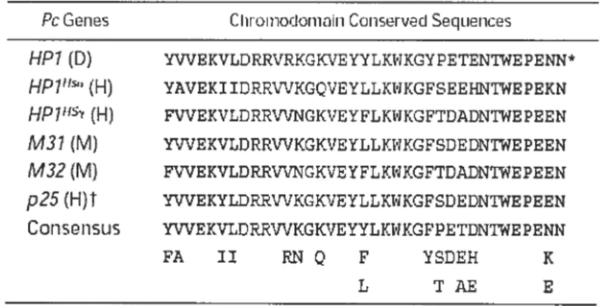
Table II Alignrnent of the chrornodomains of the Pc genes and generation of a consensus sequence to design the degenerate oligonucleotides used in these
studies 102
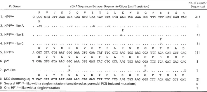
Table III Pc gene sequences obtained from cDNA isolated from purified CD34 burnan bone marrow ceils with degenerate oligonucleotides designed based on conserved sequences ofPc genes 103
xi
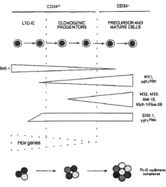
LISTE DES FIGURES
Fig. 1.1 Schematic representation indicating the position ofessential function of some transcription factors known to be active in hernopoiesis 5
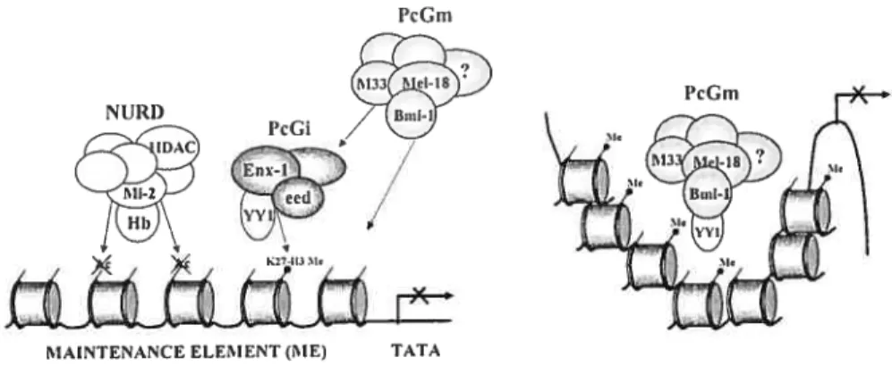
Fig. 1.2 Schematic representation ofthe functional interaction between the PcGe and PcGrn complexes in regulating gene expression through epigenetic
modification of chrornati n structure 32
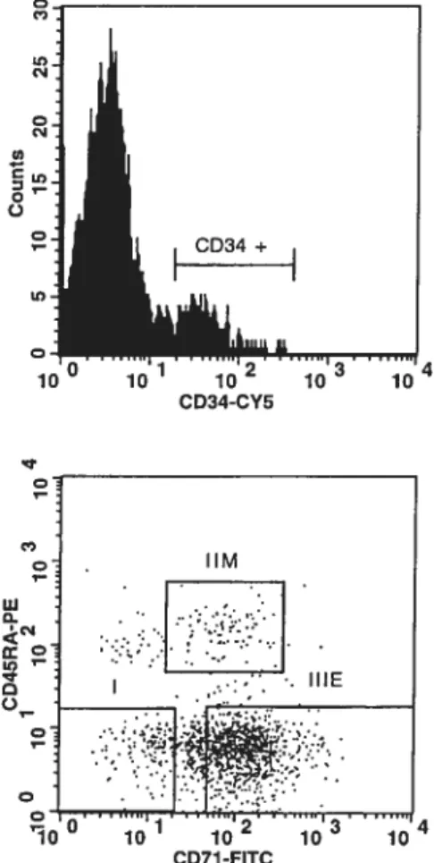
Fig. 2.1 FACS profiles ofthe CD34 subpopulations isolated from donor no. 1 104
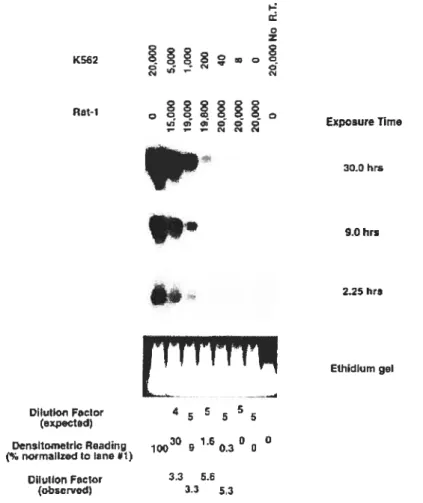
Fig. 2.2 Representative amplification ofrnRNA by quantitative RT-PCR 105
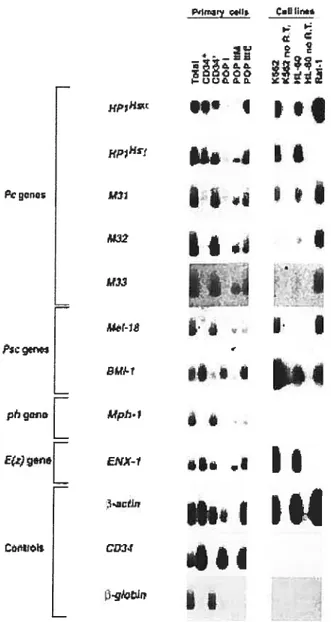
Fig. 2.3 Expression ofmarnrnalian Pc-G genes in purified bone marrow CD34
subpopulations 10$
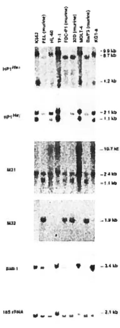
Fig. 2.4 Northern blot analysis showing the expression of selected members
ofthe Pc-G family in human and murine hematopoietic celi unes 110
Fig. 2.5 Sumrnary ofPc-G gene expression pattens observed in different purified
subpopulations ofhuman bone manow celis 114
Fig. 3.1 Expression of eed and Bmi] PcG genes in purified subpopulations
ofrnurine bone marrow celis and hernopoietic celi unes 128
Fig. 3.2 Eed is a negative regulator of the proliferative activity ofbone marrow
myeloid an lymphoid progenitor ceils 130
Fig. 3.3 Cytopathological (A) and cytofluorometric (B) analyses ofhernatopoietic ceils isolated from older control and eed3354 mutant mice 134
Fig. 3.4 Myelo-and lyrnphoproïiferation occur in old eed3354 mutant mice.136
Fig. 3.5 Monoclonal B-cell tumor in a 3-montl;-old eed’989’989 mutant mice 137
fig. 3.6 Hernopoietic parameters of3mil mutant and eed/Bmil double
mutantmice analyzed at 12-16 weeks of age 140
Fig. 3.7 Expression0f16JK4a, 1319ARF and seÏected Hox genes in various
hemopoietic organs of eed mutant mice together with an eed’989’989 mutant mice presenting a monoclonal B-ceIl Iymphoma 143
Fig. 3.8 $chernatic representation ofEed and Brnil mediated effects in ernbryonic
versus hemopoietic development in mice 149
Fig. 4.1 Ethylnitrosourea (ENU)-induced point mutations at the
eed locus in mouse 164
Fig. 4.2 Survival curves showing an increased incidence ofirradiation-induced (600 RADs) lyrnphornas in eed’98911989 and eed3354 mutant mice
when compared to control littennates 165
Fig. 4.3 Representative examples ofT-cell lymphomas arising in ilTadiated
eect’939’989 mutant mice 165
fig. 4.4 Irradiation-9nduced lymphomas in eed’98911989 rnice are of monoclonal
or oligoclonal origin 166
Fig. 4.5 Representative examples ofB-and T-cell abnonnalities arising in eed3354 irradiated mice, as analyzed by fluorometric activated
xiii
Fig. 4.6 Irradiation-induced lymphornas in eecÏ3354 mice are monoclonal or
oligoclonal 16$
Fig. 4.7 Decreased methylation levels at the lysine 27 (K27)residue ofhistone H3 (H3-K27) in eed’98911989 MMLV-induced T-cell lyrnphomas when
compared to that isolated from control littermates 169
Fig. 4.8 Structural aberrations at 1 lq are among the most common aberrations
involved in lymphoproliferative disorders in humans 170
Fig. 4.9 Schematic representation ofthe Eed-containing PcGe complex in
mammalian celis 170
Fig. 5.1 Bmi-1 is expressed in CD34 leukaernic ceils 17$
Fig. 5.2 Bmi-] regulates the proliferative potential ofernbryonic day (E) 14.5
fetal liver (FL)-derived haematopoietic ceils 181
Fig. 5.3 Bini-I is dispensable for the generation of AML in primary recipients 184
fig. 5.4 Bini1 is essential for the “transplantabiÏity” ofAML into
secondary hosts 1 $6
Fig. 5.5 Characterization of control and 3mi-1leukaemic ceils in vitro 1 $9
Fig. 5.6 Loss of expression of several CKIs in Bmi-F’ Hoxa9-Meis HPCs 190
Fig. 5.7 Bmi-1 rescues the weak leukaemogenic potential ofBmi-T’ HPCs 193
Fig. 5.8 Bini-I regulates the proliferative capacity ofstem and progenitor celis,
Fïg. 5.9 Expression of selected genes inBmi-]and Bmi-THoxa9-Meis] highly proliferative clones overexpressing or not Bmi-] 1 96
Fig. 6.1 Yeast transformation and screening protocol 207
Fig. 6.2 Bmi-1-interacting clones identified in our yeast-two-hybrid (Y2H) screen..207
Fig. 6.3 120E4Fand REDprotein are co-expressed together withBnii-]
in primitive bone marrow celis and hemopoietic ce!! unes 210
Fig. 6.4 Bmi-1 specifically associates with E4F-1 in yeast 211
Fig. 6.5 Brni-1 and E4F-1 specifically coimmunoprecipitate in hurnan 293T ceils ...212
Fig. 6.6 Immunoprecipitation of an endogenous Brni- 1 and E4f- 1 complex
in human ceils 212
Fig. 6.7 Direct physical association between Brni-1 and E4F-1 213
Fig. 6.8 Mapping the minimal interaction dornains of E4F- 1 and Bmi- 1
using a panel ofdeletion mutants 213
Fig. 6.9 The HTHTHT dornain ofBmi-1 is essential for its interaction
with E4F-1 in yeast 214
Fig. 6.10 A domain comprising the zinc linger 3 to 6 ofE4F-1 is essential
for its ability to interact withBrni-l 215
xv
Fig. 6.12 Amino acids sequence ofthe human BHJP-1 protein.217
Fig. 6.13 Schernatic representations ofthe human Trip23O protein and
the Trip23O clone identified in our yeast-two-hybrid screen 218
Fig. 6.14 Trz23O is expressed at low leveÏs in purifled subpopulations
ofmurine bone rnaiiow ceils 219
Fig. 6.15 Arnino acid sequence ofthe human Vacuolar protein
sorting il (hVPS li) protein 219
LISTE DES SIGLES ET ABRÉVIATIONS
Abréviation Signifïcatfon
3-AT 3-amino-1,2,4-triazoÏe
a.a. Amino acid
AD Activation domain
Ade Adenine
AIP Anterior-posterior
Ag Antigen
AGM Aortic-gonadal-mesonephros ALL Acute lyrnphoblastic leukemia AML Acute myeloid leukernia ANT-C Antemiapedia complex
Arg3 Arginine 3
Asx AdditionaÏ sex cornbs
ATM Ataxia Telangiectasia Mutated ATP Adenosine triphosphate
3-CLL B-cell chronic lymphocytic leukemia BCR B-cell receptor
BFU-E Burst-forrning-unit-erythroid
BHIP-l Bmi-1 Hemopoietic Interacting Protein-1
BM Bone rnarrow
f3-ME Beta-mercaptoethanol
Brin Brahma
BSA Bovine serum alburnin BX-C Bithorax complex
C Cysteine
C2H2 Zinc finger domain ofthe C2H2 subtype Ccf Centrosornal and chrornosornal factor
xvii
CDS Coding sequence
CFC Colony-forming celi
CFU-GEMM Colony-forrning-unit-granulocyte-erythroid-rnonocyte-rnegakaryocyte CFU-GM Colony-forrning-unit-granulocyte-rnacrophage
CFU-S2 Colony-forrning-units in spleen day 12 CTHR Canadian Institute ofHealth Research CKI Cyclin-dependent kinase inhibitor CtBP C-terminal binding protein
CTRL Control
D Aspartic acid
DN Double negative
DNA Deoxyribonucleic acid
DP Dotible positive
E Glutamic acid
E(Pc) Enhancer ofPolycomb E(z) Enhancer of Zeste
Eed Ernbryonic ectoderm development EGFP Enhanced green fluorescent protein ENU Ethylnitrosourea
Epo Erythropoietin
Esc Extra Sex Combs
EST Expressed sequence tag
ETP Enhancers of Trithorax and Polycomb Eu Immunoglobin heavy chain enhancer FACS Fluorescence activated celI sorting
FCS Fetal calfserum
FITC Fluorescein isothiocyanate
FL Fetal liver
GC Germinal center
GEF GTP/GDP exchange factor GTF General transcription factor
H Histidine
H3 Histone 3
HA Hemaggiutin protein
Hb Hunchback
HDAC Histone deacetylase
hGM-CSF Human granulocyte-macrophage colony-sti mulating factor
His Histidine
HMTase Histone methyÏtransferase
HOM-C Homeotic
Hox Homeobox
HPC Highly proliferative clone HPP-CFC High proliferative potential-CFC HRS Hodgkin’s disease Reed-Sternberg HSC Hemopoietic stem ceil
HTHTHT Helix-tum-helix-tum-helix dornain hVPS1 1 Human Vacuolar protein sorting 11 i.d. Identification
IgH Immunoglobulin heavy chain
IL Interleukin
IP Immunoprecipitation
IRCM Institut de Recherches Cliniques de Montréal
K Lysine
K4 Lysine4
K9 Lysine 9
K27 Lysine 27
KRAB Kruppel-associated box
L Lyrnphnode
L-blast Leukernic blast
LCBQ Leukernic Celi Bank ofQuebec LD Lirniting dilution
xix
L-HSC Leukemic hernopoietic stem celi
Li Liver
Lib Library
Lin Lineage-specific
LN Lymph noUe
LOH Loss ofheterozygosity LP Lyrnphoproliferation
LPP-CFC Low proliferative potential-CFC LTC-IC Long-tenu culture-initiating celi LTRC Long-terni repopulating celi Lys 12 Lysine 12
Lys27 Lysine 27
Mabs Monoclonal antibodies
MBLR Mel-18 and Bmi-1-like RING finger MCL Mantie celi lymphoma
M-CSF Macrophage colony-stirnuÏating factor
MDa Mega Dalton
MDR Multi-Drug Resistance ME Maintenance elernent MEC Mammary epithelial celi MEF Mouse embryonic fibroblast MLL Mixed lineage leukemia
MMLV Moloney murine leukemia virus MNU N-rnethyl-N-nitrosourea
MoAbs Monoclonal antibodies MPD Myelo-proliferative disease
MRC Medical Research Council of Canada MSCV Murine Stem Celi Virus
Mxc Multi sex combs
MZ Mantie zone
NC Nucleated celi N.D. Not deterrnined
NED No evidence ofdisease NHL Non-Hodgkin’ slyrnphorna
NLS Nuclear localization signal No R.T. No reverse transcription NSCLC Non-small celi lung cancer
NuRD Nucleosome remodeling and histone deacetylation
PBL Peripheral Nood
PBS Phosphate-buffered sait
Pc Polycomb
PcG Polycomb Group
PcGe PcG complex making the epigenetic mark PcGm PcG complex involved in the maintenance
PcI Polycomblike
Pco Polycombeotic
PE Phycoerythrin
PEV Position effect variegation
Ph Polyhorneotic
PI Propidium iodine
PLZf PromyeÏocytic leukernia zinc finger pRb Retinoblastoma tumor-suppressor protein PRCI Polycornb repressive complex 1
PRE Polycomb response element PS Para-aortic splanchnoplcura Psc Posterior sex combs
R Arginine
Rb Retinoblastoma protein
RNA Ribonucleic acid
RNAi RNA interference
xxi
S Spleen
Sce Sex combs extra
5cm Sex comb on midleg
SDS Sodium dodecyl sulfate
Ser Serine Shh Sonic hedgehog SP Single positive SP Side population SPL Spleen SPF Specific pathogen-free
Sxc Super sex combs
T Thymus
Thy Thymus
TBP TATA-binding-protein TCR T-cell receptor
TCRf3 T-cell receptor
f3
TdT Terminal deoxynucleotidyl transferase TNT Transcribed and translated
T-PLL T-cell prolymphocytic leukemia TR Thyroid honrione receptor
Trp Tryptophane
TrxG Trithorax Group
TS Trophoblast stem
Tx Transplanted
VSV Vesicular stomatiti s virus WBC White blood ceil
\VW-IC Whitlock-Witte-initi ating celi
Xeed Xenopus eed
Y2H Yeast-two-hybrid
Zn Zinc linger domain
LA DÉDICACE
xxiii
REMERCIEMENTS
Je voudrais remercier mon directeur de thèse, Dr Guy Sauvageau, pour son encouragement, esprit critique et originalité examplaires. Je lui sais gré de la confiance qu’il m’a accordée ainsi que son enthousiasme contagieux pour la science. Mon séjour dans son laboratoire aura eu, sans contredit, un impact majeur sur le développement de ma personnalité scientifique.
Je voudrais aussi remercier Sherry Niessen pour son amitié et contribution scientifique majeure à mon travail. Je remercie aussi Amélie Faubert et Martin Sauvageau pour leur collaboration à plusieurs de mes projets, Simon Girard et Nadine Mayotte pour leur excellent support technique, ainsi que Louise Leblanc pour son professionnalisme dans la préparation de la thèse et de certains manuscrits. Enfin, je voudrais exprimer ma gratitude à tous les membres du laboratoire (présents et passés) pour leur support, tant au plan humain que scientifique. Leur complicité et camaraderie auront grandement contribué à faire de mon passage à l’Institut un séjour inoubliable. Je remercie aussi les Instituts de Recherche en Santé du Canada (IRSC) pour leur support financier tout au long de mes études.
Finalement, je voudrais remercier tout spécialement mes parents et amis personnels pour leur affection et continuel support.
INTRODUCTION: SECTION 1
7
The present Ph.D. thesis, consisting of 7 chapters, describes the expression, function and mechanisms of action of selected members of the PcG gene farnily in normal and leukemic hernopoiesis.
Chapter 1 is a literature review which is subdivised into two sections. The first section describes the genetic determinants of early hemopoiesis, the intrinsic and extrinsic regulators of hemopoietic stem ccli (HSC) seif-renewai, and the rnechanisms regulating stem celi homeostasis in model systems such as the DrosophiÏa germ une. The next section summarizes the role of selected members of the Polycomb Group (PcG,) gene farnily in the regulation of normal and teukernic hernopoiesis. A particular emphasis is given to the epigenetic mechanisrns underlying Polycomb Group (PcG) gene function across species. This last section bas been recently published as a review Article. It was written by Julie Lessard under the supervision ofDr Guy Sauvageau.
1.1.1 Genetic determinants ofearly hemopoiesis
Hernatopoiesis is an ordered developmental program of differentiation and proliferation, leading to the generation of mature blood celis of multiple distinctive lineages from totipotent hernopoietic stem celis (HSCs). Regulation of this highly complex process occurs at multiple levels, and can be simplistically viewed as the combined effects of extemal influences (composed of boti; humoral factors and ceil-ceil or ceil-matrix interactions) and intracellular signaling events, ensuring transcriptional factor regulation and consequent changes in gene expression programs.
Until recently, very littie was known about the genetic mechanisrns that bring about the intrinsic processes of lineage comrnitment and subsequent lineage-speci fie differentiation of early hemopoietic celis. Accumulating evidence, from a number of recent studies, is now pointing to transcription factors suc!; as ScL/tal-1 (stem celi leukernia hemopoietic transcription factor), rbtn2/LMO2 (aiso known as ttg-2), AML-J (aÏso Ïrnowit as RUNX1/3fA2 and PEBF2B), GATA-2 and Ikaros as key genetic detenninants of early hemopoiesis. Depicted in figure 1 are the positions of essential function for these transcription factors in the hernopoietic hierachy. The b-HLH domain containing tal-l/SCL and the LIM domain-containing rbtn2/LMO2 proteins (which have been shown to interact in vivo and form a heterocomplex) 1,2 act very ear!y in ontogeny, as mice deficient for either ofthese gene products lack ail lineages ofboth primitive and definitive hemopoiesis A role for the runt homology domain protein AML-1 in the initiation of definitive hernopoiesis lias been demonstrated, as AML-1 miii mice present normal primitive hernopoiesis but completely lack definitive hernopoiesis .
The TEL (transÏocaction-Ets-Ïeukemia or ETV6) gene was shown to be required specifically for hemopoiesis of ail lineages in the bone maiow 6 Further studies will be necessary to establish whether this reflects an inability of TEL HSC/progenitors to migrate to the bone rnarrow or, more likely, to respond appropriately and/or survive within the bone manow microenviromnent. Absence of the zinc finger protein GATA-2 in mice appears to impair the proliferation capacity of early hemopoietic cells .
Mice that lack functional c-myb protein have a phenotype similar to that ofthe GATA-2 nuli mice, with
4
the exception of the rnegakaryocyte lineage which appears to develop norrnally in the absence of c-myb
.
Both GATA-2 and c-myb are nornially expressed in primitive hemopoietic ceÏÏs and then down-reguÏated as these celis differentiate 9,10• forced overexpression of GATA-2 and c-mvb in progenitor celis prornotes their proliferation and blocks differentiation 10,1 1• Homozygous nuli mice for tue ets family member PU] lack ceils of the granulocytic, monocytic and B celi lineages —the celis in which PU] is norrnally expressed 12 Interestingly, a separate une of PU] nuli mice (derived using a different targeting vector) has a more severe phenotype with additional defects in the T and erythroid lineages .
Finally, the Ikaros gene encodes six alternative transcripts that are differentially expressed in lymphoid ceils and whose products regulate the expression of a number of lineage specific genes ‘. Mice lacking Ikaros function displayed a complete absence of ail celis of the lyrnphoid lineage (T, B and NX), whereas both progenitor and mature celis of the myeloid and erythroid lineage where increased 15
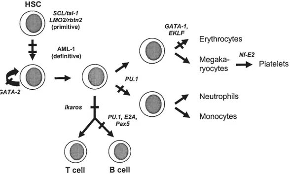
Fig. 1.1 Schematic representation indicating the position of essential function of sorne transcription factors known to be active in hemopoiesis. Positioning of each gene product is based on the earliest block observed in hernopoiesis resulting from its absence. Adapted from Shivdasani R A. and Orkin 5H. (1996) Blood 87: 4025-4039.
Other factors appear to be more lineage specific in action such as GATA-], EKLF, Nf E2, Pax5 and E2A, as their absence affects onÏy one hernopoietic lineage 16-22 For example, absence of GATA-1 blocks hemopoietic differentiation at the proerythroblast stage followed by apoptosis of these celis, suggesting that the functional role of GATA 1 is to penriit survival and maturation of erythroid progenitor ceils by preventing apoptosis 23
1.1.2 Self-renewal: a key property of the hemopoietic stem celi?
The hemopoietic stem ce!! (HSC) can be operationa!f y defined as a !ong-term repopulating ce!! with both lyrnphoid (T and B) and rnye!oid potentia! 24 The first evidence for the existence of sucli a ce!!-type cornes from experirnents by Ray Owen and
HSC GI SCUta!-1 LMOZfrbtn2 (primitive) AML-1 (definitive) —‘ Pu.1 GA TA-1, EKLF
Air
-K Erythrocytes Nf-E2 .—‘ Platelets Ne utroph ils Monocytes T celi B celi6
colleagues, in 1945, which showed that bovine fraternal twins, sharing a single placenta and blood circulation, retained production of blood ceils genetically defined to be from both throughout their Iife 25 Twenty years later, elegant experiments by Tili, McCulloch, Wu, Becker, Sirnonovitch and colleagues demonstrated that aduit bone marrow contained single celis that had the ability to forrn macroscopic nodules of myeloerythroid celis on the spleen, $ to 12 days after intravenous injection into myelo ablated recipients 26-28 These spleen-colony-forrning-units (CfU-S) were shown to be clonaI 29 and, in rnany cases, couÏd generate similar colonies upon transplantation into secondary recipients 28 As they shared several characteristics attributed to HSCs (including high proliferative potentiaÏ, multipotentiality and seÏf-renewaÏ abiÏity), CFU-S were initially considered to be HSCs 2$ The validity ofthe CFU-S assay to detect HSCs with long-terni repopulating potential was questioned after the discovery that some of these ceils were capable of only unilineage differentiation and/or lacked the ability to self-renew (functional heterogeneity). Although most ofthe ceils possessed the ability to differentiate into the erythrocyte and myeloid lineages, their lymphoid potential rernained controversial 30-32 is now clear that most CFU-S cells in the aduit bone rnarrow are cornmitted myeloid progenitors which can be physically separated from more primitive cells with long-term lympho-rnyeloid repopulating potential Although the CFU-S assay played a key role in the development of concepts of primitive hemopoietic ccli organization and regulation, its inability to analyze pure stem ceils meant that rnost oftheir functions were irnplied rather than directiy analyzed.
The first attempts at purifying the HSC came from the school ofTill and McCullogh 33,40 and Van Bekkum in the Netherlands 41 From this work, it lias becorne possible to routinely identify and isolate highly purified murine and hurnan HSCs based mainly on characteristic cell surface proteins that are either present (Sca-1 and c-kit) or absent (using rnarkers of lineage cornrnitted cells such as CD38, Mac-l and CD8) (for review
42)
Despite the progress that has been made in identifying and obtaining enriched HSC populations, analysis
of
the population dynamics and cdl cycle kinetics of HSCsremains difficuit. One of the most intriguing properties of aduit HSCs is a robust maintenance of the dynamic equilibrium between seif-renewal and differentiation ‘n• Under homeostatic conditions in vivo, most HSCs are quiescent, as demonstrated by their relative resistance to killing by the cytotoxic dnig 5-fluorouracil (5-FU) when compared to comrnitted progenitor celis When they enter cycle, HSCs can divide asymrnetrically or symmetrically, resulting in different HSC fates. Stem ccli maintenance divisions give rise to one daugbter HSC with essentially identical biological properties (a process referred to as seif-renewal) and one committed daughter ceil. The comrnitted daughter celi enters a transient state of rapid cellular proliferation and, upon exhaustion of its proliferative potential, withdraws from the ce!! cycle and progressively acquires the specialized characteristics of one of the 10 mature blood celi lineages ta process known as differentiation). Although the relative influence of intrinsic versus extrinsic factors on HSC seif-renewal remains to be detenriined, it bas been easier to identify the environmental factors having a negative impact on this process than those that enhance it. Thus, rnost in vivo culture conditions defined to date lead to depletion of the HSC pool by favoring symmetric divisions (generation of two daughter differentiated celis) and concomitant expansion ofcornmitted progenitor populations
Several sttidies using retroviral marking of HSCs have dernonstrated the ability ofHSCs to undergo seif-renewa! divisions 46-49 A!though most of these studies failed to accurately quantify the magnitude of seif-renewa! events, considerable evidence suggests that this property is not unlimited. First, following bone marrow transplantation, the HSC pool is flot found to regenerate to ievels higher than 10% of normal pre-transplantation values, despite a complete regeneration of bone rnaiow
cellularity and progenitor ce!! numbers 5054 Some investigators have suggested the involvement of negative feedback rnechanisrns irnposed in vivo by more mature ceils as a possible mechanism that could prematurely inhibit HSC expansion following transplantation .
Alternatively, this loss of long-tenu repopulating ability may result from damage to the recipient’s microenvironment inflicted by the conditioning regimen (i.e. irradiation). However, experirnents performed in the anemic (WWV) recipient mouse strain, which possess a normal microenvironment but poorly competitive
$
hernopoietic celis due to a mutation in the c-kit ligand receptor56, rather suggest that this defect is intrinsic to the transplanted ceils themseives 51,57•
A major concem is that nearly ail HSC assays assessing seif-renewal rely on the genel-ation of functionalÏy mature celÏs, and therefore provide a retrospective rather than a current view of potential HSC attributes. In a transplantation setting, the accuiacy of the HSC readout relies on the efficiency of the transplanted ceils to home and engraft to the speciaiized niches of the bone marrow rnicroenvironment .
The heterogeneity of the HSC cornpartment further complicates the interpretation of such experimental designs. Age-related and strain-specific 59,60 differences in HSC numbers and/or competitive abilities have been reported (for review 61) Moreover, a functionai deciine in the proliferative potential of FISCs derived from the fetal liver, umbilical cord (at birth) and aduit bone marrow indicates ontogeny-related differences in HSC function
54,62-64
Whether this heterogeneity represents true intrinsic quantitative and/or qualitative differences in HSC properties, or in the expression of this potentiai due to stochastic events, remains unclear. So, a question remains: can HSCs tntly seif-renew or does cell division impair their qualitative (biological) properties?
1.1.3 Intrinsic regulators of HSC seif-ren ewal
While the concept of “true HSC seifrenewai” remains controversial, genetic programmes regulating the self-renewal and di fferentiati on outcomes of early hemopoietic cell divisions have been described. Recent attention bas focused on bemopoietic cytokines (see next section) 65-69 and cell intrinsic pathways whose activation bas caused sorne HSC expansion ex vivo, with the ultimate goal of durable in
vivo engraftrnent. Enforced expression of the P glycoprotein purnp genes MDR] and ABCG2 in murine bone marrow cells led to the expansion of side population cells with retained in vivo repopulation ability70• Activation of retinoic acid receptor signaling by addition of all-trans retinoic acid resulted in retention of long-term repopulating activity of cultured hemopoietic stem cells 71 Constitutive Notch signaling in purified Sca-l Lin c-kit bone rnaiiow cells led to the immortalization of blast-like ceils that retained
pluripotency and Ïong-terrn repopulating potential 72• In particular, retroviral overexpression of HOXB4 in mouse bone marrow celis significantly enhanced the rate of HSC seif-renewal (potentially tip to 1000-fold net increase of transduced HSCs) in
both prirnary and secondary recipients 0f note, this stem-ceil specific proliferative
effect of HOXB4 occurs without impairing normal differentiation or inducing cellular transformation. A role for the Wnt signaling pathway in seif-renewal of HSCs has also recently been demonstrated 76• Overexpression of activated 3-catenin expands the pool of HSCs in long-terni cultures by both phenotype and ftmction. Ectopic expression of
axin or afrizzled ligand-binding domain, inhibitors of the Wnt signaling pathway, leads to inhibition of HSC growth in vitro and reduced reconstitution in vivo. Furthermore, activation ofWnt signaling in HSCs induces increased expression ofNotchl and Hoxb4, suggesting a molecular hierarchy of regulation of HSC developrnent. Purified Wnt3a protein also induces seif-renewal of repopulating celis (HSCs?), signifying its potential role in tissue engineering Serial transplantation studies demonstrated a critical role for the transcription-regulating/chrornatin modifying CREB-binding protein (CBP), but flot its paralogous protein p300, in maintaining an adequate pool of murine HSCs through seif-renewal 78• The cyclin-dependent kinase inhibitor (CKI) p21 also appears to be an essential component of the molecular switch governing the entry of the hernopoietic stem celis in cycle as in its absence, increased cdl cycling leads to stem ceil exhaustion
.
Most importantly, the PoÏycoinb group gene Bmi-] is absolutely required for the rnaintenance/self-renewal ofboth fetal and aduil HSCs (see below)80’81.
It was recently demonstrated that the horneoprotein Nanog is required for maintenance of pluripotency in mouse epiblast and embryonic stem (ES) cells 82,83• Despite very different genetic programs in vivo, tinder normal circtirnstances, both ernbryonic (ES) and hemopoietic stem cells share fundamental common properties: rnultipotency and the ability to self-renew. Insights into flic regulation of Nanog- as well as other regulators of self-renewal/multipotency such as Oct4-, FoxD3-, Sox2- and Stat3-directed transcription pathways and the network of crosstalk between these factors might contribute to a better understanding of stem ceil behavior in other renewing tissues such as blood. However, an important caveat is that ES celis truly self-renew whereas HSCs
10
may not. This raises the hypothesis that distinct molecular programmes may be involved in regulating this process.
1.1.4 Extrinsic regutators of HSC seif-renewal
1.1.4.1 Regulation of germ une stem celi (GSC) seif-renewal in Drosophita
Further defining the molecular mechanisms controlling stem ceil function is crucial to the future use of stem celis in regenerative medicine as well as understanding the processes of aging, turnor formation and degeneration. As mentioned above, a ftindamental characteristic of adult stem ceils is their capacity to either divide asyrnrnetrically or symmetrically. Thus, general mechanisrns may exist to balance self renewal capacity with differentiation.
The Drosoph lia germ une represents an excellent system to study adult stem ceils at the cellular and molecular level and their relation with their microenvironrnent. li the female, at the anterior end of cadi ovariole of an ovary (or gennarium) rest two or three gerrn-line stem celis (GSCs) that originate from primordial genn stem ceils (PGCs). These primitive ceils are located in a niche which is composed of three differentiated somatic ce!! types: terminal filament (TF) cells, cap celis and inner germarium sheath (IGS) celis. This microenvironment is instrumental in regulating the expansion of tic gemi une stem ceil pool. Anterior PGCs adjacent to Tf/cap celis give rise to two daughters that both contact Tf/cap ceils and will eventually develop into GSCs (symmetric division). The remaining PGCs (not in physical contact with Tf/cap ceils)
directly differentiate into mature oocytes (asymmetric division) 8490• In die male germ une, PGCs are also selected to become GSCs based on their juxtaposition to a cluster of somatid/support cells (called the hub) located at the apical tip of die testis. Upon ce!! division, the daughter ccli maintaining contact with the hub retains stein ceil identity, whereas the celi displayed away from the hub initiates differentiation into a gonialblast
91,92
Thus, the orientationlposition of genn line stem ceil divisions in the niche seems to be criticalin regulating tic expansion of tic germinal stem ccli pool (detenninistic fate).
of transcription (JAK-STAT) signal transduction pathway is an important intrinsic regulator of GSC seif-renewal in the male gerrn une Similarly, piwi, the Drosoph lia counterpart of human HIWI, is aiso required for the seif-renewal ability of G$Cs, but is absolutely dispensabie for their differentiation into committed daughter celis Decapentapiegic (Dpp,) (the Drosoph lia homoiog of human boue moiphogeneticprotein
2/4), is expressed in anterior somatic ceils of the gonad and is essential for PGC proliferation. PGCs mutant for thick veins, an essential dpp receptor, ate impaired in
their ability to clonally populate a niche, further suggesting that dpp is one of the extrinsic mitotic signais that promote flic clonai expansion of GSCs in a niche 90• In the male germ une, the asymmetric outcome of stem ceil divisions is specified, extrinsicaily, through direct interaction with niche ceils expressing the ligand Unpaired (Upd) ‘°. DE-cadherin-mediatcd ccii adhesion was aiso shown to be essential for anchoring ovarian GCSs to their niches and stimulating their proliferation Loss of function of the Drosoph lia Epidermal growth factor receptor (EGFR,) in sornatic ceils increases the
number of GSCs in the male genu une, suggesting that the EGFR gene is aiso an extrinsic (but negative) reguiator of GSC proliferation 96•
1.1.4.2 Positional information and the regulation of stem celi seif-renewal: a conserved mechanism?
Positionai cues from the microenvironrnent seem to be critical in regulating stem celi fate decisions in severai other organisms and developmentai systems. In the C. elegans gonadal ami, mitotic gerrn une stem ceils (GSCs) reside distaliy, while differentiating gametes progressiveiy move towards the proximal end. Sornatic distal tip ceils (DTCs) located at the extrernity of the gonad constitute a niche that is essential for promoting GSC proliferation and preventing meiosis (for review 97)• In the developing brain, mammaiian neural stem celis (NSCs) are known to be located in close contact with the ventricular zone surface, whereas differentiated post-mitotic neurons progressively move
12
towards the dorsal zone (for review, 9$)• Interestingly, it has been suggested that NSCs dividing along the ventricular surface give rise to two stem celis, while the perpendicular division generates one stem ceil and one differentiated neuronal celi .
The small intestine is cornposed of ciliated villi, each surrounded by crypts, embedded in the intestinal wall for protection. Each crypt is composed of about 250 simple epithelial ceils that include the stein ceil compartrnent for replenishing the villi. The multipotent stem ceils are iocated near or at the base of each crypt To maintain horneostasis, slow cycling stem celis are converted to rapidly but transiently proÏiferating celis that move to the rnidsegment and subsequently differentiate into functionally mature and specialized ceils (i.e. either absorptive enterocytes, mucus-secreting gobÏet or enteroendocrine ceils). Similarly, the epidermis is composed of a single inner (basal) layer of dividing sternlprogenitor celis, which periodically withdraw from the celi cycle, commit to differentiate terminally, and move outward toward the skin surface ‘°‘.
The bone marrow aiso provides aduit HSCs with a rich, but complex, milieu (or niche) composed of many ccli types including macrophages, adipocytes, fibrobiasts and mesenchyrnal celis. Bone rnarrow strornal factors that positively impact on HSC maintenance, propagation and homing include soluble and membrane-bound stem celi
factor (SCF), soluble Sonic Hedgehog, the fibrobiast growth factors fGf-1 and fGF-2, the a-chemokine strornal-1 ccii derived factor-l (SDF-1) and a slew of extra ceilutar matrix (ECM) moiecules, ail having the abiiity to specifically interact with various receptors on the HSC surface (for review 102) On the other hand, purified primitive
(Sca-1 Lin c-kif’) bone rnarrow ceils are severely compromised in their short and long
term multilineage reconstituting ability when activated by TNF-alpha or through Fas 103 or when engineered to overexpress f 1t3 104 providing molecular evidence for extrinsic
negative regulators of HS C seif-renewal.
Despite major efforts to characterize the bone rnarrow niche, in vitro studies indicate that HSCs still survive better when cuitured with bone marrow strorna than when placed in a defined medium supplernented with characterized bone manow components. This is
fibrob!asts to display their optimal survival and proliferative capacity 105 Thus, the orientation of the stem ceil division plane in the niche rnight represent a general and evolutionariÏy conserved mechanism invoÏved in regulating both germinal and somatic stem celi horneostasis. Locating and analyzing the stem celi niches and identifying the molecuÏes that orchestrate these environmental positional cues are of major importance in stem ce!! biology.
1.1.5 Cellular organization of normal aiid leukemic hemopoiesis: a common function for stem ceils
There is strong support for the idea that cancer is a stem-celI disease 106 The simi!arity in the hierarchical organization of malignant and normal tissues is best characterized in the hernopoietic system. Hurnan acute myeloid leukemia (AML) originates from a rare population of primitive cel!s (CD34 CD38) high!y enriched in hemopoietic stem cells (HSCs) 107 Most leukemic ceils (blasts) are limited in their proliferative capacity and must be constant!y replenished by rare, se!f-renewing “leukemic stem ce!ls” (L-HSCs). So, like the normal hemopoietic system, leukemia seems to be organized as a hierarchy that originates from a stem-ce!! pool which rnost likely retains remnants of the nonrial deve!oprnental program.
It has also been proposed that the initial, cancer-causing (“transforming”) niutations occur in the seif-renewing stem ceil pool, rather than in a!ready committed precursors. In this view, fewer mutations would be required to generate fu!!y malignant ce!!s if they were to originate from already seif-renewing stem ce!!s, as opposed to cornmitted progenitors with !ow pro!iferative potential. Thus, two important findings have recent!y emerged from studies of stem ce!! biology and carcinogenesis: 1-) in the process of neoplastic transformation, the genetic events responsible for di sease progression must occur in a stem ce!!, un!ess one of the mutations would penriit se!f-renewal in a downstream comrnitted progenitor; 2) within the cancer or leukemia, on!y a subset ofthe cells that make up the tumor mass are tumorigenic —the “cancer stem celis” These ideas predict similarities in the rno!ecular programmes of norma! and cancer/AML stem ceils.
14
The genetic rnechanisms regulating seif-renewal of the HSCs may be more generally applicable to other regenerating tissue systems. Recent findings implicated the Notch
72,109,110
\Vnt and Shh 111-113 signaling pathways in prornoting stein ce!! seif-renewal in a variety of different epithelia in addition to HSCs. 1nterestingly, mutations of these pathways have been associated with a number of hurnan neoplasia, including colon carcinoma and epidermal tumors 114,115 (Wnt), rnedulloblastoma and basal cali
carcinorna 116,117 (Shh), and T-ce!! leukernias 11$(Notch).
As uncontrolled stem ce!l seif-renewal represents the basis of cancer, the identification of stern-celÏ specific genes, especiaÏly those involved in the deregulation of their self renewa! capacity is critical. The goal ofrny PhD thesis was specifically to identify such a gene. To achieve this, I used a candidate gene approach and decided to focus rny studies on the PoÏycomb (PcG,) fami!y of genes because: 1-) the PcG genes are upstream transcriptional regulators of the homeotic (Hox, genes in skeletal precursor ce!!s 2-) the Hox genes are important regu!ators of hemopoietic deveÏoprnent; 3-) HOXB4 is a critical regulator ofHSC self-renewal; its retroviral overexpression in mouse bone marrow cells leads to 1000-fo!d net increase of transduced HSCs in both primary and secondary recipients N; 4-) in the rnid-90’s, expression of some FcG genes in hurnan and murine
hernopoietic ceil unes had already been reported; 5-) van Lohuizen and co!!eagues had identified the Polycoinb Group (PcG,) gene Bmi-] as an essential regulator of the
proliferative activity of boue manow mye!oid and !ymphoid progenitors 119 suggesting
a putative roTe for this gene in the regu!ation of HSC behavior. The work presented in
this PhD thesis was intended at verifying whether selected members of the Polycomb group (PcG) gene family 1-) were expressed in the hernopoietic tissue; and 2-) rnight be involved in regulating critical aspects ofHSC function.
The next section xviii provide a detaiied overview of the Folycomb (FcG) group genes, with special emphasis on their implication in the regulation of hernopoietic ce!! developrnent.
Referen ces
1. Valge-Archer,V.E. etctt. The LIM protein RBTN2 and the basic helix-loop-helix protein TAL1 are presentin a complex inerythroid celis. Proc. NatÏ. Acad. Sci.
U. S. A 91, 8617-8621 (1994).
2. Wadman,I. et aï. Specific in vivo association between the bHLH and LIM proteins irnplicated inhuman T ceil leukemia. EMBOI 13, 483 1-4839 (1994). 3. Porcher,C. et aï. The T celi leukemia oncoprotein SCL/tal-1 is essential for
development of ail hematopoietic lineages. Ccli 26, 47-57 (1996).
4. Warren,A.J. et ai. The oncogenic cysteine-rich LIM domain protein rbtn2 is essentiai for erythroid development. Ccii 78, 45-57 (1994).
5. Okuda,T., van Deursen,J., Hiebert,S.W., GrosveÏd,G. & Downing,J.R. AIVIL1, the target of multiple chromosomal transiocations in human leukemia, is essential for normal fetai liver hernatopoiesis. Ccli 84, 32 1-330 (1996).
6. Wang,LC. et ctt. The TEL/ETV6 gene is required specificaliy for hematopoiesis
inthe bone marTow. Genes Dcv. 12, 23 92-2402 (1998).
7. Tsai,F.Y. et aï. An early haematopoietic defect in mice lacking the transcription factor GATA-2. Nature 371, 22 1-226 (1994).
8. Mucenski,M.L. et aï. A functional c-myb gene is required for normal murine
fetal hepatic hematopoiesis. Ccii 65, 677-689 (1991).
9. Thompson,M.A. & Ramsay,R.G. Myb: an old oncoprotein with new roies. Bioessays 17, 341-350 (1995).
10. Yamamoto,M. et al. Activity and tissue-specific expression ofthe transcription factor NF-E1 multigene famlly. Genes Dcv. 4, 1650-1662 (1990).
11. Briegel,K. et al. Ectopic expression of a conditionai GATA-2/estrogen receptor chimera arrests erythroid differentiation in a hormone-dependent manner. Genes Dcv. 7, 1097-1109 (1993).
12. McKercher,S.R. et aï. Targeted disniption ofthePU. 1 gene resuitsinmultiple hematopoietic abnormalities. EMBOI 15, 5647-5658 (1996).
13. Scott,E.W., Sirnon,M.C., Anastasi,J. & Singh,H. Requirement of transcription factor PU.1 in the development of multiple hematopoietic lineages. Science 265, 1573-1577 (1994).
16
14. Georgopoulos,K., Moore,D.D. & Derfler,B. Ikaros, an early Iyrnphoid-specific transcription factor and a putative rnediator for T cel! comrnitment. Science 258, $0$-812 (1992).
15. Georgopoulos,K. et aï. The Ikaros gene is required for the development of ail lymphoid lineages. CeÏÏ 79, 143-156 (1994).
16. Nuez,B., Michalovich,D., Bygrave,A., PÏoemacher,R. & GrosveÏd,f. Defective
haernatopoiesis in fetal liver resulting from inactivation of the EKLf gene. Nctture 375, 316-318 (1995).
17. Perkins,A.C., Sharpe,A.H. & Orkin,S.H. Lethal beta-thalassaemia inmice
lacking the erythroid CACCC-transcription factor EKLF. Natitre375, 318-322 (1995).
18. Pevny,L. et aï. Erythroid differentiation in chimaeric mice blocked by a targeted mutation in the gene for transcription factor GATA-1. Nature349, 25 7-260 (1991).
19. Shivdasani,RA. et aï. Transcription factor NF-E2 is required for platelet formation independent of the actions of thrombopoietinlMGDF in megakaryocyte development. Ccli 81, 695-704 (1995).
20. Urbanek,P., Wang,Z.Q., Fetka,I., Wagner,E.F. & Busslinger,M. Complete block ofearly B celi differentiation and a!tered patterning of the posterior midbrain in mice lacking PaxS/BSAP. CeÏt 79, 901-912 (1994).
21. Weiss,M.J., Keller,G. & Orkin,S.H. Novel insights into erythroid development reveaied through in vitro differentiation of GATA- 1 embryonic stem celis. Genes Dcv. 8, 1184-1197 (1994).
22. Zhuang,Y., Soriano,P. & Weintraub,H. The helix-loop-helix gene E2A is required for B ce!! formation. Ccii 79, 275-884 (1994).
23. Weiss,M.J. & Orkin,S.H. Transcription factor GATA-1 penriits survival and maturation of erythroid precursors by preventing apoptosis. Froc. Nati. Acad. Sci. U. S. A 92, 9623-9627 (1995).
24. Orlic,D. & Bodine,D.M. What defines a pluripotent hematopoietic stem ceil (PHSC): vi11 the reai PHSCplease stand up! Blood 84, 3991-3994 (1994). 25. Owen,R.D. Immunologenetic consequences of vascular anastomoses between
bovine twins. Science 102, 400. 1945. 1945.
26. Till,J.E.M.E.A. A direct measurernent of the radiation sensitivity of normai mouse bone rnalTow celis. RctcÏiat. Res. 14, 1419-1430 (1961).
27. Becker,A., McCulloch,E. & Tili,J. Cytologicai demonstration ofthe clonai nature of spleen colonies derived from transplanted mouse rnaiow ceils. Nature 197, 452-454 (1963).
2$. Sirninovitch,L., McCulloch,E.A. & Till,J.E. The distribution ofcolony-fonning ceils among spleen colonies. J C’eÏl. Coinp. PhysioÏ. 62, 327-336 (1963).
29. Wu,A.M., Till,J.E., Sirninovitch,L. & McCulloch,E.A. Cytological evidence for a relationship between normal hernotopoietic coÏony-fonning ceils and celis of the lyrnphoid system. I Exp. lied. 127, 455-464 (196$).
30. Wu,A.M., Sirninovitch,L., Till,J.E. & McCulloch,E.A. Evidence for a
relationship between mouse hernopoietic stem ceils and ceils forming colonies in culture. Proc. Nati. Acad. Sci. U. S. A 59, 1209-12 15 (196$).
31. Lala,P.K. & Johnson,G.R. Monoclonal origin of B lymphocyte colony-forming ceils in spleen colonies forrned by multipotential hernopoietic stem ceils. I Exp. Med. 148, 146$-1477 (197$).
32. Lepault,F., Ezine,S. & Gagnerault,M.C. T- and B-lymphocyte differentiation potentials of spleen colony-fonning ceils. Btood 81, 950-955 (1993).
33. Worton,R.G., McCulloch,E.A. & Till,J.E. Physical separation ofhemopoietic stem celis differing in their capacity for seif-renewal. I Exp. Med. 130, 91-103 (1969).
34. Jones,R.J., Celano,P., Sharkis,S.I. & Sensenbrenner,L.L. Two phases of
engraftrnent established by serial bone marrow transplantation in mice. Blood 73, 397-401 (1989).
35. Jones,R.J., Wagner,J.E., Celano,P., Zicha,M.S. & Sharkis,S.J. Separation of pluripotent haematopoietic stem celis from spleen colony-forming ceils. Nature 347, 18$-189 (1990).
36. Mulder,A.H. & Visser,J.W. Separation and functional analysis ofbone manow ceils separated by rhodarnine-123 fluorescence. Exp. Hematot. 15, 99-104 (1987).
37. Spangrude,G.J. et aï. Mouse hematopoietic stem celis. BÏood 78, 1395-1402 (1991).
38. van der Loo,J.C., van den,B.C., Baert,M.R., Wagernaker,G. & Ploemacher,R.E. Stable multilineage hematopoietic chirnerisrn in alpha-thalassemic mice induced by a bone marrow subpopulation that exciudes the majority ofday-12 spleen colony-forming units. Bïood 83, 1769-1777 (1994).
18
39. Visser,J.W. & de Vries,P. Isolation of spÏeen-colony fonriing celis (CFU-s) using wheat gerrn aggiutinin and rhodamine 123 labeling. BÏood CeÏÏs 14, 369-384 (198$).
40. Worton,R.G., McCulloch,E.A. & Till,J.E. Physical separation of hemopoietic stem ceils from celis forming colonies in culture. I CellFhvsioÏ 74, 171-182 (1969).
41. van Bekkum,D.W., van den Engh,G.J., Wagernaker,G., Bol,S.J. & Visser,J.W. Structural identity of the pluripotential hernopoietic stem ccli. BÏood CelÏs 5, 143-159 (1979).
42. Weissrnan,I.L. The road ended up at stem ceils. ImmtmoÏ. Rev. 185, 159-174 (2002).
43. Morrison,S.J., Shah,N.M. & Anderson,D.J. Regulatoiy mechanisrns in stem ccli biology. Ceti $8, 287-29$ (1997).
44. Hodgson,G.S. & Bradley,T.R. Properties ofhaernatopoietic stem celis surviving 5-fluorouracil treatment: evidence for a pre-CFU-S ccli? Nature 281, 38 1-382 (1979).
45. Lemer,C. & Harrison,D.E. 5-Fluorouracil spares hemopoietic stem ceils responsible for long-term repopulation. Exp. Hematol. 18, 114-118 (1990). 46. Fraser,C.C., Szilvassy,S.J., Eaves,C.J. & Humphries,R.K. Proliferation of
totipotent hernatopoietic stem celis in vitro with retention oflong-term competitive in vivo reconstituting ability. Froc. Natt. Acad. $ci. U. S. A 89,
1968-1972 (1992).
47. Jordan,C.T. & Lernischka,I.R. Clonai and systemic analysis ofiong-terrn hernatopoiesis in the motise. Genes Dcv. 4, 220-232 (1990).
48. Kelier,G. & Snodgrass,R. Life span ofrnuitipotential hematopoietic stem ceils in vivo. I Exp. MecÏ. 171, 1407-1418 (1990).
49. Lemischka,I.R., Rauiet,D.H. & Mulligan,RC. Developrnental potential and dynarnic behavior ofhernatopoietic stem ceils. CeÏÏ 45, 9 17-927 (1986). 50. Haiiison,D.E., Astle,C.M. & Delaittre,J.A. Loss ofproliferative capacity in
immunohemopoietic stem ceiis caused by seriai transplantation rather than aging. I Exp. Med. 147, 1526-1531 (1978).
51. Harrison,D.E. & Astie,C.M. Loss of stem ccli repopuiating ability upon
transplantation. Effects of donor age, ccii number, and transplantation procedure. J Exp. Mccl. 156, 1767-1779 (1982).
53. Mauch,P. & HelÏman,S. Loss ofhematopoietic stem ccli sel f-renewal after bone marrow transplantation. BÏoocl 74, $72-$75 (1989).
54. Pawliuk,R., Eaves,C. & Humphries,R.K. Evidence ofboth ontogeny and transplant dose-regulated expansion ofhematopoietic stem celis in vivo. BÏood 88, 2852-2858 (1996).
55. Iscove,N.N. & Nawa,K. Hematopoietic stem cdils expand during serial transplantation in vivo without apparent exhaustion. Cuir. Biol. 7, 805-808 (1997).
56. Chabot,B., Stephenson,D.A., Chaprnan,V.M., Besmer,P. & Bemstein,A. The proto-oncogene c-kit encoding a transmembrane tyrosine kinase receptor maps to the mouse W locus. Nature 335, $8-89 (1988).
57. Gardner,R.V., Astle,C.M. & Hamson,D.E. The decrease in iong-term maiiow repopulating capacity seen after transplantation is not the result of irradiation induced strornal injury. Exp. Hematol. 16, 49-54 (198$).
58. Benveniste,P., Cantin,C., Hyam,D. & Iscove,N.N. Hernatopoietic stem celis engraft in mice with absolute efficiency. Nat. hnmunoÏ. 4, 70$-713 (2003). 59. Van Zant,G., Thompson,B.P. & Chen,J.J. Differentiation of chimeric bone
marrow in vivo reveals genotype-restricted contributions to hematopoiesis.Exp. Hematot. 19, 941-949 (1991).
60. Phillips,R.L., Reinhart,A.J. & Van Zant,G. Genetic control ofmurine
hematopoietic stem ccli pool sizes and cycling kinetics. Froc. Nati. Acad. Sci. U.
S. A 89, 11607-11611 (1992).
61. Geiger,H. & Van Zant,G. The aging oflyrnpho-hernatopoietic stem celis. Nat. inimunol. 3, 329-333 (2002).
62. Harrison,D.E., Zhong,R.K., Jordan,C.T., Lemischka,I.R. & Astle,C.M. Relative to adult malTow, fetal liver repopulates nearly five tirnes more effectiveiy long
tem than short-term. Exp. Hematol. 25, 293-297 (1997).
63. Rebel,V.I. et aï. A comparison oflong-term repopuiating hematopoietic stem ceils in fetal liver and aduitbonemarrow from the mouse. Exp. HematoÏ. 24, 63$-64$ (1996).
64. Rebel,V.I., Miiler,C.L., Eaves,C.J. & Lansdorp,P.M. The repopulation potential of fetal iiver hematopoietic stem cells in mice exceeds that of their liver adult
20
65. Zandstra,P.W., Connealiy,E., Petzer,A.L., Piret,J.M. & Eaves,C.J. Cytokine manipulation of primitive hurnan hernatopoietic ce!! seif-renewal. Froc. NatÏ. Acad. Sel. U S. A 94, 4698-4703 (1997).
66. Piacibeiio,W. et al. Extensive amplification and seif-renewai ofhurnan primitive hematopoietic stem ceils from cord biood. BÏood 89, 2644-2653 (1997).
67. Traycoff,C.M., Cometta,K., Yoder,M.C., Davidson,A. & Srour,E.F. Ex vivo expansion of murine hematopoietic progenitor celis generates classes o f expanded cells possessing different levels ofbone marrow repopulating potentiai. Exp. HematoÏ. 24, 299-306 (1996).
6$. Matsunaga,T., Hirayarna,F., Yonernura,Y., MuITay,R. & Ogawa,M. Negative regulation by interleukin-3 (IL-3) ofmouse early B-cell progenitors and stem ceils in culture: transduction ofthe negative signais by betac and betalL-3 proteins of IL-3 receptor and absence of negative regulation by granulocyte macrophage coiony-stimulating factor. BÏood 92, 901-907 (199$).
69. Ema,H., Takano,H., Sudo,K. & Nakauchi,H. In vitro seif-renewal division of hernatopoietic stem celis. J. Exp. MecI. 192, 1281-128$ (2000).
70. Bunting,K.D., Zhou,S., Lu,T. & Sorrentino,B.P. Enforced P-glycoprotein pump function in murine bone marrow ceils resuits in expansion of side population stem celis in vitro and repopulating cells in vivo. Blood96, 902-909 (2000). 71. Purton,LE., Bernstein,I.D. & Collins,S.J. Ali-trans retinoic acid enhances the
long-term repopuiating activity of cultured hematopoietic stem ceils. Blood 95, 470-477 (2000).
72. Vanutrn-Finney,B. et al. P luripotent, cytokine-dep endent, hematopoietic stem cells are imrnortaiized by constitutive Notchi signaling. Nat. Med. 6, 1278-12$ 1 (2000).
73. Antonchuk,J., Sauvageau,G. & Humphries,RK. HOXB4-induced expansion of adult hernatopoietic stem celis ex vivo. Celi 109, 39-45 (2002).
74. Sauvageau,G. et cil. Overexpression ofHOXB4 in hematopoietic celis causes the selective expansion ofmoreprimitive populations in vitro and in vivo. Genes Dey. 9, 1753-1765 (1995).
75. Thorsteinsdottir,U., $auvageau,G. & Hurnphries,R.K. Enhanced in vivo regenerative potentiai ofHOXB4-transduced hematopoietic stem ceiis with regulation oftheir pool size. BÏood 94, 2605-2612 (1999).
76. Reya,T. et aï. A role for Wnt signaliing in self-renewal ofhaematopoietic stem ceils. Nature 423, 409-414 (2003).
77. Wiiiert,K. et ctl. Wnt proteins are lipid-rnodified and can act as stem ce!! growth factors. Nature 423, 44$-452 (2003).
78. Rebel,V.I. et cxl. Distinct roles for CREB-binding protein and p300 in
hematopoietic stem ccli seif-renewal. Froc. Natt. Acad. Sci. U. S. A 99, 14789-14794 (2002).
79. Cheng,L et cil. Hernatopoietic stem ceil quiescence maintained by p2lcipl/waft. Science 287, 1804-1808 (2000).
80. Park,I.K. et aï. Bmi-1 is required for maintenance ofaduit seif-renewing haematopoietic stem ceils. Natttre 423, 302-305 (2003).
$1. Lessard,J. & Sauvageau,G. Bmi-1 detemiines the proliferative capacity of normal and ieukaemic stem celis. Nature 423, 255-260 (2003).
$2. Mitsui,K. et aï. The Homeoprotein Nanog Is Required for Maintenance of Pluripotency in Mouse Epibiast and ES Ceils. Ccli 113, 63 1-642 (2003). $3. Chambers,I. et aï. Functionai expression cioning ofnanog, a piuripotency
sustaining factor in embryonic stem celis. Ceil 113, 643-655 (2003).
$4. Cox,D.N. et aï. A nove! class ofevoiutionariiy conserved genes defined by piwi are essentiai for stem ccli seif-renewai. Genes Dey. 12, 37 15-3727 (1998).
$5. Cox,D.N., Chao,A. & Lin,H. piwi encodes a nucleoplasmic factor whose activity modulates the number and division rate of genriline stem ceils. DeveÏopntent 127, 503-5 14 (2000).
$6. King,F.J. & Lin,H. Somatic signaling mediated by fs(1)Yb is essential for gerniline stem ccli maintenance during Drosophila oogenesis. Development 126,
1833-1844 (1999).
$7. King,F.J., Szakmary,A., Cox,D.N. & Lin,H. Yb modulates the divisions ofboth germiine and somatic stem cetis through piwi- and hh-mediated mechanisrns in the Drosophila ovary. Mol. Ccli 7, 497-5 08 (2001).
8$. Xie,T. & Spradling,A.C. decapentaplegic is essential for the maintenance and division ofgermline stem ceils in the Drosophiia ovary. Ccli 94, 25 1-260 (1998). 89. Xie,T. & SpradÏing,A.C. A niche maintaining germ une stem ceilsin the
Drosophila ovary. Science 290, 328-330 (2000).
90. Zhu,C.H. & Xie,T. Clonai expansion ofovarian germiine stem celis during niche formation in Drosophila. Deveïopinent 130, 2579-2588 (2003).
7,)
91. Hardy,R.W., Tokuyasu,K.T., Linds!ey,D.L. & Garavito,M. The germinal proliferation center inthe testis ofDrosophila melanogaster. I Uttrastruct. Res. 69, 180-190 (1979).
92. Gonczy,P. & DiNardo,S. The gerrn une regulates somatic cyst ce!! proliferation and fate during Drosophila spermatogenesis. Development 122, 2437-2447 (1996).
93. Kiger,A.A., Jones,D.L., Schulz,C., Rogers,M.B. & Fuller,M.T. Stem celi self renewal specified by JAK-STAT activation in response to a support ce!! cue. Science 294, 2542-2545 (2001).
94. Tulina,N. & Matunis,E. Control of stem ccl! seif-renewal in Drosophila spermatogenesis by JAK-STAT signaling. Science 294, 2546-2549 (2001). 95. Song,X. & Xie,T. DE-cadherin-mediated ceil adhesion is essential for
maintaining somatic stem ceils in the Drosophila ovary. Froc. Nati. Acad. Sci. U. S. A 99, 14813-14818 (2002).
96. Duchek,P. & Rorth,P. Guidance of cel! migration by EGF receptor signaling during Drosophila oogenesis. Science 291, 13 1-133 (2001).
97. Kirnble,J.E. & White,J.G. On the control ofgerm celi developrnent in Caenorhabditis e!egans. Dcv. Biol. 81, 208-219 (1981).
98. Anderson,D.J. Stem ceils and pattem formation in thenervous system: the possib!e versus the actual. Neuron 30, 19-35 (2001).
99. Chemi,A. & McConnell,S.K. Cleavage orientation and the asymmetric
inheritance ofNotchl immunoreactivityinmamma!ian neurogenesis. C’ett $2, 631-641 (1995).
100. Loeffler,M., Birke,A., Winton,D. & Potten,C. Sornatic mutation, rnonoclonality and stochastic models of stem ceil organization in the intestinal crypt. I Theor. Biot. 160, 471-491 (1993).
101. Fuchs,E. & Segre,J.A. Stem celis: a new lease onlife. Ccli 100, 143-155 (2000). 102. Whetton,A.D. & Graharn,G.J.Horning andmobi!ization in the stem ce!! niche.
Trencis Ccli Biot. 9,233-238(1999).
103. Bryder,D. et al. Se!f-renewal ofrnu!tipotent !ong-term repopu!ating
hematopoietic stem ceils is negatively regulated by Fas and tumor necrosis factor receptor activation. I Exp. lied. 194, 941-952 (2001).
104. Adolfsson,J. et ai. Upregu!ation ofF!t3 expression within the bonernaiow Lin(