1 Lancellotti P, et al. Heart 2017;0:1–7. doi:10.1136/heartjnl-2017-311682
Stress echocardiography in patients with native
valvular heart disease
Patrizio Lancellotti,
1,2Raluca Dulgheru,
1Yun Yun Go,
1Tadafumi Sugimoto,
1Stella Marchetta,
1Cécile Oury,
1Madalina Garbi
3AbStrAct
Valve stress echocardiography (VSE) can be performed as exercise stress echocardiography (ESE) or dobutamine stress echocardiography (DSE) depending on the patient’s clinical status, severity and type of valve disease. ESE combines exercise testing with two-dimensional grey scale and Doppler echocardiography during exercise. Thus, it provides objective assessment of symptomatic status (exercise test), as well as exercise-induced changes of a series of echocardiographic parameters (different depending on the valve disease type), which yield prognostic information in individual patients and help in a better treatment planning. DSE is useful in symptomatic patients with low-gradient aortic stenosis. It clarifies its severity and helps in assessing surgical risk in patients with severe disease and systolic dysfunction. It can be also used to test valve haemodynamics in asymptomatic patients with significant mitral stenosis unable to perform an exercise test or to test the left ventricle response, namely to test viability, in patients with ischaemic secondary mitral regurgitation. VSE has taught us that history taking, clinical examination and resting echocardiography give an ’incomplete picture’ of the disease in patients presenting with a severe valve disease. Therefore, its use should be encouraged in such patients.
IntroductIon
Functional testing at its inception was used to assess symptoms during exertion, and it has evolved to answer several clinical questions with the help of stress echocardiography.1 Valve stress
echocardi-ography (VSE) helps refine diagnosis of severity, prognosis and management planning based on changes in various parameters induced by exercise or by dobutamine.2 3 The indications for VSE have
expanded in parallel with the growing evidence for early intervention benefit in valvular heart disease.
Currently, there is extensive evidence that VSE can benefit decision-making for a wide range of patients being followed up in valve clinics or those with newly diagnosed valvular heart diseases.4
Consequently, the recently published European Association of Cardiovascular Imaging and the American Society of Echocardiography joint recommendations for the use of stress echocardi-ography in non-ischaemic heart disease address the use of stress echocardiography in valvular heart disease.5 VSE is recommended for the following
clinical scenarios: corroborating the severity of valve disease with symptoms prior to intervention, detecting severe valve disease with left ventricular systolic dysfunction or other signs of haemodynamic compromise.4 For diagnosis of severe valve disease
with symptoms, VSE is performed in two catego-ries of patients: asymptomatic patients with severe valve disease with the aim to detect symptoms and symptomatic patients with non-severe valve disease with the aim to regrade valve disease severity. For diagnosis of severe valve disease with left ventric-ular systolic dysfunction, VSE is also performed in two categories of patients: patients with low-flow aortic stenosis with the aim to clarify aortic stenosis severity and patients with asymptomatic severe valve disease with the aim to detect subclinical left ventricular systolic dysfunction.6 7 Furthermore,
other signs of haemodynamic compromise may be demonstrated with VSE in asymptomatic patients with severe valve disease.8 9 A simplified
classifi-cation of VSE indiclassifi-cations groups them into three categories: severe valve disease without symptoms, non-severe valve disease with symptoms and valve disease with low flow. A more detailed and compre-hensive classification of VSE indications assigns them to the respective valve disease.
Methodology
Stress echocardiography with both dobutamine and exercise can be used for the assessment of valvular heart disease, depending on indication and on the aim of the test. Exercise testing can be performed with a treadmill; however, the postexercise rather than during exercise image acquisition in this case may result in peak exercise-related phenomena being underestimated or missed, and low work-load-related phenomena cannot be assessed. Ideally, VSE should be performed with supine bicycle exercise to allow imaging throughout the test and particularly at low workload and at peak effort. Imaging at low workload allows assessment of contractile reserve and changes in global longitu-dinal strain, both helping to detect subclinical left ventricular systolic dysfunction. Furthermore, the increase in systolic pulmonary artery pressure from low workload has higher specificity for a patho-logical response as haemodynamic consequence of severe valve disease.
Dobutamine stress is the only currently recom-mended VSE modality for the assessment of low-flow, low-gradient aortic stenosis with reduced left ventricular ejection fraction.10 11
Dobuta-mine stress can be used as an alternative to exer-cise in low-flow, low-gradient aortic stenosis with preserved left ventricular ejection fraction and in the assessment of mitral stenosis severity. Dobuta-mine stress does not allow assessment of systolic pulmonary artery pressure and the assessment of mitral regurgitation severity.
review
to cite: Lancellotti P, Dulgheru R, Go YY, et al. Heart Published Online First: [please include Day Month Year]. doi:10.1136/ heartjnl-2017-311682 1Departments of Cardiology, Heart Valve Clinic, University of Liège Hospital, GIGA Cardiovascular Sciences, CHU Sart Tilman, Liege, Belgium 2Gruppo Villa Maria Care and Research, Anthea Hospital, Bari, Italy
3King’s Health Partners, King’s College Hospital NHS Foundation Trust, London, UK correspondence to Professor Patrizio Lancellotti, Domaine Universitaire du Sart Tilman, Batiment B35 Department of Cardiology, University Hospital, Université de Liège, CHU du Sart Tilman 4000 Liège, Belgium; plancellotti@ chu. ulg. ac. be Received 6 September 2017 Revised 30 October 2017 Accepted 15 November 2017
Heart Online First, published on December 7, 2017 as 10.1136/heartjnl-2017-311682
group.bmj.comon January 23, 2018 - Published by
http://heart.bmj.com/
Exercise echocardiography allows the assessment of exer-cise tolerance and symptoms together with the assessment of haemodynamic parameters under physiological stress. The exercise tolerance is compared with age-matched, sex-matched and weight-matched normals.12 Symptoms of shortness of
breath are witnessed and can be occasionally found to be due to cardiac asthma—interstitial pulmonary oedema resulting in prolonged expiration and expiratory wheeze. Being symp-toms-limited, the exercise test is usually terminated before development of alveolar pulmonary oedema with wet lungs; on the contrary, alveolar pulmonary oedema may be occa-sionally induced by dobutamine stress echo in the assessment of low-flow, low-gradient aortic stenosis or mitral stenosis. Exercise echocardiography allows the assessment of systolic pulmonary artery pressure response, of mitral regurgitation severity and of severe mitral regurgitation consequences. Exercise echocardiography allows the assessment of the true left ventricular contractile reserve, as a physiological phenom-enon, rather than the left ventricular inotropic reserve, defined as the increase in ejection fraction in response to intravenous dobutamine infusion. Exercise echocardiography is used in the assessment of severe valve disease with no symptoms but also non-severe valve disease with symptoms and paradoxical low-flow aortic stenosis.
Parameters assessed depend on the VSE indication; thus, a specific protocol is used in each case, predefining necessary images and their order of acquisition appropriate for the aim of the test (figure 1).
clInIcAl ApplIcAtIonS
The VSE clinical application depends on the valve disease in question and on the indication for the test (tables 1 and 2,
figures 2–5).
In case of asymptomatic severe valve disease, the test should be always performed with exercise stress to detect symptoms and haemodynamic consequences of the valve disease in phys-iological circumstances, reproducible in daily life. Further-more, dobutamine stress is contraindicated in severe valve disease, particularly in high-gradient severe aortic stenosis. The administration of dobutamine in severe aortic stenosis can result in cardiogenic shock due to dobutamine-induced peripheral vasodilation with consequent systemic hypotension and concomitant acute pulmonary oedema. Exercise testing to detect symptoms is well established and recommended in all valve disease management guidelines, because symptoms are a class I indication for intervention, although having a subjective nature; furthermore, sedentary patients do not give themselves the opportunity to experience symptoms, and many active Figure 1 Schematic representation of different parameters that may be acquired during exercise stress echocardiography in each type of
asymptomatic severe heart valve disease. AR, aortic regurgitation; AS, aortic stenosis; Ch, chamber; E, early diastolic wave velocity; ESE, exercise stress echocardiography; LV, left ventricle; MG, mean pressure gradient; MR, mitral regurgitations; MS, mitral stenosis; PISA, proximal isovelocity surface area; SV, stroke volume; TAPSE, tricuspid annular plane systolic excursion; TTG, transtricuspid pressure gradient.
review
patients instinctively slow down their pace to avoid symptoms. In severe valve disease, given that the valve disease severity is already established, VSE image acquisition should begin with left ventricular views, tricuspid regurgitation for systolic pulmonary artery pressure estimation and other parameters suggestive of decompensation, for example exertion-induced mitral regurgitation in patients with aortic valve disease.
In case of symptoms but non-severe valve disease, the test performed with exercise stress allows corroboration of symp-toms with exertion-induced changes. However, dobutamine can be used to assess inducible ischaemia as reason for symp-toms or to regrade low-gradient aortic stenosis severity. Echo-cardiography is essential for diagnosis in this case, and the VSE image acquisition should begin with parameters of valve disease severity.
In case of low-flow aortic stenosis, exercise is recommended only if the ejection fraction is preserved and the patient is asymptomatic by history taking. The VSE image acquisition should begin with parameters of valve disease severity and flow reserve, which is the degree of blood flow increase in response to maximal stimulation, with left ventricular views acquired subsequently.
AortIc StenoSIS
Asymptomatic severe aortic stenosis
Severe aortic stenosis is not an indication for intervention in the absence of symptoms if the left ventricular ejection fraction is preserved. Exercise testing to detect symptoms is long well estab-lished in the follow-up of severe aortic stenosis in valve clinics for timing of intervention. The addition of echocardiography provides diagnostic and prognostic parameters.13 Classically,
during treadmill exercise testing, ST depression on ECG and drop in systolic blood pressure are indications for intervention.14 15 The
ST depression on ECG translates into induced regional myocardial hypokinesia during VSE. Left ventricular ejection fraction drop on exertion and inducible mitral regurgitation are also echocar-diographic signs of haemodynamic compromise. Reduced global longitudinal strain at rest and on exertion suggests subclinical left ventricular systolic dysfunction.16 17 Furthermore, mean gradient
increase by more than 20 mm Hg is a sign of adverse prognosis, suggestive of possible decompensation during follow-up.8 14 Symptoms despite non-severe aortic stenosis
In patients with non-severe aortic stenosis based on current classification criteria, symptoms should trigger stress testing to table 1 Stress echocardiographic parameters in native valvular heart disease
Symptom type of stress
echocardiographic parameters with
guideline recommendations1 2 echocardiographic parameters with clinical recommendations3
Severe high-gradient AS Asymptomatic ESE – Flow reserve/contractile reserve Ex-induced PAP >60 mm Hg Ex-MG increase >20 mm Hg1 Severe low-gradient AS with preserved LVEF Asymptomatic ESE – MG and AVA during exercise Ex-induced PAP >60 mm Hg Symptomatic DSE – MG and AVA at DES Severe low-gradient AS with reduced LVEF – DSE MG and AVA at DES,1 2 flow reserve/
contractile reserve1 Flow reserve/contractile reserve Severe AR Asymptomatic ESE – Flow reserve/contractile reserve
Ex-induced PAP >60 mm Hg Severe primary MR Asymptomatic ESE – Ex-induced PAP >60 mm Hg1
Flow reserve/contractile reserve Ex-increase in EROA >10 mm2
Haemodynamically significant MS Asymptomatic ESE or DSE Ex-MG increase >15 mm Hg for ESE2 Ex-MG increase >15 mm Hg for ESE or >18 mm Hg for DES
Ex-induced PAP >60 mm Hg for ESE AR, aortic regurgitation; AS, aortic stenosis; DSE, dobutamine stress echocardiography; EROA, effective regurgitant orifice area; ESE, exercise stress echocardiography; Ex, exercise; LVEF, left ventricular ejection fraction; MG, mean pressure gradient; MR, mitral regurgitations; MS, mitral stenosis; PAP, systolic pulmonary artery pressure.
table 2 Comparison of exercise versus dobutamine stress echocardiography
upright/treadmill Semisupine bicycle dobutamine
Does not allow imaging at intermediate level Allows for imaging at intermediate and peak levels Allows for imaging at intermediate and peak levels Postpeak/postexercise imaging (within 90 s after
termination)
Postpeak/postexercise imaging (recovery period) Postpeak/postdobutamine imaging (recovery period)
– Dynamic LV function assessment, E/e’ assessment (at low levels)
Dynamic LV function assessment, E/e’ assessment (at low levels)
– Changes in valve disease severity (gradients across stenotic valve, regurgitant volume or effective regurgitant orifice area for mitral regurgitation)
Changes in valve disease severity (gradients across stenotic valve)
– Slope of change in sPAP –
– Biphasic wall motion changes Biphasic wall motion changes
– Ischaemic threshold Ischaemic threshold
Change in loading condition due to change in posture – –
Potential drop in heart rate – –
E, early diastolic wave velocity; LV, left ventricle; sPAP, systolic pulmonary artery pressure.
group.bmj.com
on January 23, 2018 - Published by
http://heart.bmj.com/
regrade aortic stenosis severity based on stress-induced changes of echocardiographic parameters and to detect signs of haemo-dynamic significance described for severe aortic stenosis.6 7 classical low-flow, low-gradient aortic stenosis
Low-flow, low-gradient aortic stenosis with reduced left ventricular ejection fraction needs low-dose dobutamine VSE to assess the severity of aortic stenosis and the existence of flow reserve. At rest, because of low transvalvular flow as a result of reduced left ventricular ejection fraction, the echocardiographic assessment of aortic stenosis severity gives contradictory findings with calculated functional aortic valve area in the severe range and gradients in the moderate range. The diagnosis of aortic stenosis severity necessitates assess-ment of calculated functional aortic valve area and gradi-ents in conditions of increased transvalvular flow as a result of dobutamine.18 19 Gradients increase with no or minimal
increase in calculated functional aortic valve area suggest severe aortic stenosis. The assessment of aortic stenosis severity depends on the existence of flow reserve. In case of limited flow reserve, the projected aortic valve area can be used for diagnosis of severity.19 In case of no flow reserve, the
assessment of aortic valve calcification with CT may help with the diagnosis.20 Caution is required in case of induced
isch-aemia with consequent drop in left ventricular ejection frac-tion; interpretation of findings at this stage as representing a higher flow state would lead to an erroneous diagnosis of non-severe aortic stenosis and lack of contractile reserve.21 To
avoid missing the peak increase in flow, continuous acquisi-tion is recommended.
low-flow, low-gradient aortic stenosis with preserved left ventricular ejection fraction
Low-flow, low-gradient aortic stenosis with preserved left ventricular ejection fraction may benefit from dobutamine or exercise VSE to assess the severity of aortic stenosis, although caution ought to be exercised in patients with small hypertro-phic hearts.18 22 An increase in gradients with minimal or no
increase in calculated functional valve area during dobutamine stress points towards severe aortic stenosis physiology. Exercise testing allows concomitant assessment of exercise tolerance and symptoms. Often the test with dobutamine or exercise is termi-nated early due to heart failure symptoms as a result of diastolic Figure 2 Summary of different valve stress echocardiography modalities and parameters to assess according to each type of heart valve disease. AR, aortic regurgitation; AS, aortic stenosis; AVA, aortic valve area; DSE, dobutamine stress echocardiography; EROA, effective regurgitant orifice area; ESE, exercise stress echocardiography; Ex, exercise; Ex-MG, mean pressure gradient during exercise; GLS, global longitudinal strain; LVEDD, left ventricle end diastolic diameter; LVEF, left ventricular ejection fraction; LVESD, left ventricle end systolic diameter; LVESI, left ventricle end systolic index; MR, mitral regurgitation; MS, mitral stenosis; MVA, mitral valve area; PAP, systolic pulmonary artery pressure; RV, regurgitant volume; sPAP, systolic pulmonary artery pressure.
review
dysfunction and intolerance of sinus tachycardia or development of atrial fibrillation.
AortIc regurgItAtIon
In asymptomatic severe aortic regurgitation, exercise testing to detect symptoms is crucial because of the high mortality they
portent.23 In both severe and non-severe aortic regurgitation,
with or without symptoms, exercise VSE allows assessment of subclinical systolic dysfunction based on contractile reserve and on the global longitudinal strain at rest and at low workload exercise.24 25 Furthermore, it allows assessment of inducible
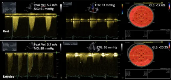
isch-aemia and of signs of left ventricular decompensation—ejection Figure 3 Exercise stress echocardiography in an asymptomatic patient with severe AS. Rest echocardiographic evaluation confirms the presence of normal-flow, high-gradient severe AS in a patient with preserved left ventricular ejection fraction (>50%) and LV global longitudinal strain, and no pulmonary arterial hypertension (systolic pulmonary artery pressure <50 mm Hg). The patient stopped the test prematurely for leg pain (peak heart rate 102 beats per minute). Several parameters of poor outcome were identified during exercise: an increase in mean transaortic pressure gradient by 19 mm Hg, a rapid increase in LV filling pressure (as assessed by E/e’) and the development of pulmonary arterial hypertension, despite preserved LV contractile reserve (increase in global longitudinal strain (GLS)). AS, aortic stenosis; E, early diastolic wave velocity; LV, left ventricle; MG, mean transaortic pressure gradient; TTG, transtricuspid pressure gradient; Vel, velocity.
Figure 4 Exercise stress echocardiography in an asymptomatic patient with degenerative mitral regurgitation. At exercise the degree of mitral regurgitation increased moderately while the global longitudinal strain decreased, identifying the absence of contractile reserve. EROA, effective regurgitant orifice area; GLS, global longitudinal strain.
group.bmj.com
on January 23, 2018 - Published by
http://heart.bmj.com/
fraction drop and development of mitral regurgitation.26 The
assessment of inducible ischaemia can be also performed with dobutamine.
VSE has no role in the assessment of aortic regurgitation severity; the aortic regurgitation appears less severe with tachy-cardia and consequent shortening of diastole.
MItrAl StenoSIS
The assessment of mitral stenosis severity and haemody-namic consequences based on stress echocardiography is long well established.27 VSE has changed the way mitral stenosis is
perceived and managed because mitral stenosis with valve area in the range of moderate is often demonstrated to have severe stress-induced rise in mean gradient and systolic pulmonary artery pressure28; this is mainly the case for rheumatic mitral
stenosis, characterised by lack of valve compliance to flow. VSE is recommended to assess asymptomatic mitral stenosis prior to major surgery or planned pregnancy to predict haemodynamic reserve or likelihood of decompensation with increase in flow.29
Current guidelines recommend VSE for management planning to inform decision-making for balloon valvotomy in asymptom-atic patient, and on the other hand proceed to intervention in symptomatic patients.11 12
Supine bicycle exercise VSE allows assessment of systolic pulmonary pressure rise at low workload and assessment of mean gradient rise at low workload and peak.30 Dobutamine
VSE allows assessment of mean gradient rise.31 The threshold
for severe rise in mean gradient is 15 mm Hg for exercise and 18 mm Hg for dobutamine VSE.
MItrAl regurgItAtIon primary mitral regurgitation
The role of supine bicycle exercise VSE in the assessment of asymptomatic severe primary mitral regurgitation is well estab-lished, and the test is recommended for surgical timing by the European Society of Cardiology (ESC)/European Association for Cardio-Thoracic Surgery (EACTS) valve disease management guidelines.11 The test is essential for an early repair strategy in
degenerative mitral regurgitation, detecting symptoms, subclin-ical left ventricular systolic dysfunction or pathologsubclin-ical systolic pulmonary artery pressure rise.32 33 Following a complete
assess-ment at rest, images are acquired at low workload and at peak for systolic pulmonary artery pressure assessment and for assess-ment of the left ventricle. A rise in systolic pulmonary artery pressure to over 60 mm Hg is a harbinger of poor outcomes.34–37
The use of supine bicycle exercise VSE for the assessment of primary mitral regurgitation severity in symptomatic patients with moderate regurgitation at rest is underpinned by robust evidence.38–40 Primary mitral regurgitation, regardless of
aeti-ology, can have a dynamic component, increasing on exertion and thus explaining the symptoms. The assessment of mitral regurgitation severity is mainly based on images acquired at low workload and intermediate level of exercise because mitral regurgitation quantification becomes more difficult at heart rate >115 beats per minute.
Secondary mitral regurgitation
Supine bicycle exercise VSE can be used for the assessment of secondary mitral regurgitation of ischaemic aetiology or due to non-ischaemic dilated cardiomyopathy. VSE can demonstrate an increase in secondary mitral regurgitation severity during exertion in patients with exertional symptoms not explained by the severity of mitral regurgitation and left ventricular systolic dysfunction at rest.41 However, often the severity of secondary
mitral regurgitation decreases with exertion in patients with non-ischaemic dilated cardiomyopathy because of contractile recruitment; the decrease in severity confirms the secondary nature of the regurgitation and a good prognosis because of the existence of contractile reserve. In patients with coronary disease, an increase in mitral regurgitation severity during VSE suggests need for mitral valve repair at the time of surgical revascularisation.
concluSIon
The use of VSE is a fruitful complementary technique to assess patients with valve disease and adds important missing pieces to the puzzle. In our view, it enhances diagnostic accuracy and enables a better treatment planning. Exercise stress echocardi-ography is safe and should be encouraged especially in heart valve clinics to understand the complex response of the left ventricle and valve haemodynamics during exercise in asymp-tomatic patients. Dobutamine stress echocardiography has an undeniable role in severity assessment and risk stratification of patients with low-flow, low-gradient aortic stenosis and reduced left ventricular ejection fraction. It proved useful in the severity assessment of symptomatic patients with low-gradient aortic stenosis and preserved ejection fraction. Larger scale studies on the role of VSE in each type of severe valve disease are needed.
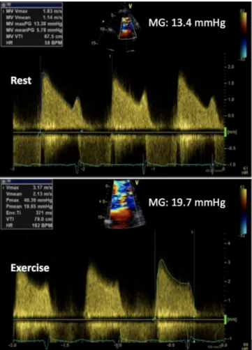
Figure 5 Exercise stress echocardiography in an asymptomatic patient with mitral stenosis and significant increase in mean mitral pressure gradient (MG).
review
contributors PL and MG wrote the first draft. TS, YYG, RD, CO, SM provided critical review and the figures (practical cases).
competing interests CO is Senior Research Associate at the belgian Funds for Scientific Research (FRS-FNRS).
provenance and peer review Commissioned; externally peer reviewed. © Article author(s) (or their employer(s) unless otherwise stated in the text of the article) 2017. All rights reserved. No commercial use is permitted unless otherwise expressly granted.
reFerenceS
1 Lancellotti P, Magne J. Stress echocardiography in regurgitant valve disease. Circ Cardiovasc Imaging 2013;6:840–9.
2 Wu WC, Aziz GF, Sadaniantz A. The use of stress echocardiography in the assessment of mitral valvular disease. Echocardiography 2004;21:451–8.
3 Piérard LA, Lancellotti P. Stress testing in valve disease. Heart 2007;93:766–72. 4 Garbi M, Chambers J, Vannan MA, et al. Valve stress echocardiography: a practical
guide for referral, procedure, reporting, and clinical implementation of results from the HAVEC group. JACC Cardiovasc Imaging 2015;8:724–36.
5 Lancellotti P, Pellikka PA, Budts W, et al. The clinical use of stress echocardiography in non-ischaemic heart disease: recommendations from the European Association of Cardiovascular Imaging and the American Society of Echocardiography. Eur Heart J Cardiovasc Imaging 2016;17:1191–229.
6 Rafique AM, Biner S, Ray I, et al. Meta-analysis of prognostic value of stress testing in patients with asymptomatic severe aortic stenosis. Am J Cardiol 2009;104:972–7. 7 Das P, Rimington H, Chambers J. Exercise testing to stratify risk in aortic stenosis. Eur
Heart J 2005;26:1309–13.
8 Lancellotti P, Lebois F, Simon M, et al. Prognostic importance of quantitative exercise Doppler echocardiography in asymptomatic valvular aortic stenosis. Circulation
2005;112(9 Suppl):I377–82.
9 Maréchaux S, Hachicha Z, Bellouin A, et al. Usefulness of exercise-stress
echocardiography for risk stratification of true asymptomatic patients with aortic valve stenosis. Eur Heart J 2010;31:1390–7.
10 Vahanian A, Alfieri O, Andreotti F, et al. Guidelines on the management of valvular heart disease (version 2012). Eur Heart J 2012;33:2451–96.
11 Nishimura RA, Otto CM, Bonow RO, et al. AHA/ACC guideline for the management of patients with valvular heart disease: executive summary: a report of the American College of Cardiology/American Heart Association Task Force on Practice Guidelines.
J Am Coll Cardiol 2014;2014:2438–88.
12 Masri A, Goodman AL, Barr T, et al. predictors of long-term outcomes in asymptomatic patients with severe aortic stenosis and preserved left ventricular systolic function undergoing exercise echocardiography. Circ Cardiovasc Imaging 2016;9:e004689. 13 Leurent G, Donal E, de Place C, et al. Argument for a Doppler echocardiography
during exercise in assessing asymptomatic patients with severe aortic stenosis. Eur J Echocardiogr 2009;10:69–73.
14 Alborino D, Hoffmann JL, Fournet PC, et al. Value of exercise testing to evaluate the indication for surgery in asymptomatic patients with valvular aortic stenosis. J Heart Valve Dis 2002;11:204–9.
15 Amato MC, Moffa PJ, Werner KE, et al. Treatment decision in asymptomatic aortic valve stenosis: role of exercise testing. Heart 2001;86:381–6.
16 Donal E, Thebault C, O’Connor K, et al. Impact of aortic stenosis on longitudinal myocardial deformation during exercise. Eur J Echocardiogr 2011;12:235–41. 17 Lancellotti P, Karsera D, Tumminello G, et al. Determinants of an abnormal response
to exercise in patients with asymptomatic valvular aortic stenosis. Eur J Echocardiogr
2008;9:338–43.
18 Clavel MA, Ennezat PV, Maréchaux S, et al. Stress echocardiography to assess stenosis severity and predict outcome in patients with paradoxical low-flow, low-gradient aortic stenosis and preserved LVEF. JACC Cardiovasc Imaging 2013;6:175–83.
19 Blais C, Burwash IG, Mundigler G, et al. Projected valve area at normal flow rate improves the assessment of stenosis severity in patients with low-flow, low-gradient aortic stenosis: the multicenter TOPAS (Truly or Pseudo-Severe Aortic Stenosis) study.
Circulation 2006;113:711–21.
20 Cueff C, Serfaty JM, Cimadevilla C, et al. Measurement of aortic valve calcification using multislice computed tomography: correlation with haemodynamic severity of aortic stenosis and clinical implication for patients with low ejection fraction. Heart
2011;97:721–6.
21 Dulgheru R, Magne J, Davin L, et al. Left ventricular regional function and maximal exercise capacity in aortic stenosis. Eur Heart J Cardiovasc Imaging 2016;17:217–24. 22 Pibarot P, Dumesnil JG. Aortic stenosis suspected to be severe despite low gradients.
Circ Cardiovasc Imaging 2014;7:545–51.
23 Bonow RO, Lakatos E, Maron BJ, et al. Serial long-term assessment of the natural history of asymptomatic patients with chronic aortic regurgitation and normal left ventricular systolic function. Circulation 1991;84:1625–35.
24 Wahi S, Haluska B, Pasquet A, et al. Exercise echocardiography predicts development of left ventricular dysfunction in medically and surgically treated patients with asymptomatic severe aortic regurgitation. Heart 2000;84:606–14.
25 Kusunose K, Agarwal S, Marwick TH, et al. Decision making in asymptomatic aortic regurgitation in the era of guidelines: incremental values of resting and exercise cardiac dysfunction. Circ Cardiovasc Imaging 2014;7:352–62.
26 Vinereanu D, Ionescu AA, Fraser AG. Assessment of left ventricular long axis contraction can detect early myocardial dysfunction in asymptomatic patients with severe aortic regurgitation. Heart 2001;85:30–6.
27 Carabello BA. Modern management of mitral stenosis. Circulation 2005;112:432–7. 28 Brochet E, Détaint D, Fondard O, et al. Early hemodynamic changes versus peak
values: what is more useful to predict occurrence of dyspnea during stress echocardiography in patients with asymptomatic mitral stenosis? J Am Soc Echocardiogr 2011;24:392–8.
29 Cheitlin MD. Stress echocardiography in mitral stenosis: when is it useful? J Am Coll Cardiol 2004;43:402–4.
30 Grimaldi A, Olivotto I, Figini F, et al. Dynamic assessment of ’valvular reserve capacity’ in patients with rheumatic mitral stenosis. Eur Heart J Cardiovasc Imaging
2012;13:476–82.
31 Reis G, Motta MS, Barbosa MM, et al. Dobutamine stress echocardiography for noninvasive assessment and risk stratification of patients with rheumatic mitral stenosis. J Am Coll Cardiol 2004;43:393–401.
32 Lee R, Haluska B, Leung DY, et al. Functional and prognostic implications of left ventricular contractile reserve in patients with asymptomatic severe mitral regurgitation. Heart 2005;91:1407–12.
33 Magne J, Mahjoub H, Dulgheru R, et al. Left ventricular contractile reserve in asymptomatic primary mitral regurgitation. Eur Heart J 2014;35:1608–16. 34 Magne J, Lancellotti P, Piérard LA. Exercise pulmonary hypertension in asymptomatic
degenerative mitral regurgitation. Circulation 2010;122:33–41.
35 Magne J, Pibarot P, Sengupta PP, et al. Pulmonary hypertension in valvular disease: a comprehensive review on pathophysiology to therapy from the HAVEC Group. JACC Cardiovasc Imaging 2015;8:83–99.
36 Lancellotti P, Martinez C, Bernard A. Pulmonary pressures and outcome in primary mitral regurgitation: paradigm shift from rung to ladder. J Am Coll Cardiol
2016;67:2962–4.
37 Magne J, Donal E, Mahjoub H, et al. Impact of exercise pulmonary hypertension on postoperative outcome in primary mitral regurgitation. Heart 2015;101:391–6. 38 Magne J, Lancellotti P, Piérard LA. Exercise-induced changes in degenerative mitral
regurgitation. J Am Coll Cardiol 2010;56:300–9.
39 Lancellotti P, Magne J. Stress testing for the evaluation of patients with mitral regurgitation. Curr Opin Cardiol 2012;27:492–8.
40 Magne J, Lancellotti P, Pierard LA. Stress echocardiography and mitral valvular heart disease. Cardiol Clin 2013;31:311–21.
41 Piérard LA, Lancellotti P. The role of ischemic mitral regurgitation in the pathogenesis of acute pulmonary edema. N Engl J Med 2004;351:1627–34.
group.bmj.com
on January 23, 2018 - Published by
http://heart.bmj.com/