SCI. MAR., 61 (Supl. 2): 5-14 SCIENTIA MARINA 1997 ECOLOGY OF MARINE MOLLUSCS.J.D. ROS and A. GUERRA(eds.)
Biology and descriptionof Antisabia juliae sp. nov.,
new Hipponicid gastropod commensal on Turbo spp...
in Laing Island (Papua New Guinea)*
MATHIEU POULICEK', JEAN-CLAUDE BUSSERS! and PIERRE VANDEWALLE?
‘Animal Ecology Laboratory and *Functional Morphology Laboratory, Zoological Institute, Liège University.
22, Quai Van Beneden, B-4020 Liége. Belgium.
SUMMARY:The gastropod family Hipponicidae comprises widely distributed but poorly known sedentary species. On the beach-rock of the coral reefs of Laing Island (Papua New Guinea)live rich populations of several gastropod Turbo species of which many specimens have attachedto their shell a hipponicid gastropodattributed to a new species, Antisabia juliae. This new species, described in this paper, appears to have adapted its mode oflife on live turbinids in several ways result-ing in morphological changes(thin basal plate loosely adherent to the supportresult-ing shell, functional eyes, very long snout, functional radula, small osphradium) and ethological changes (foraging behaviour:it appearsto feed on the epiphytic.com-munity growing on the host, in the vicinity of the “host” shell). Except for these characteristics, the mode oflife appears quite similar to that of other hipponicid species with few big females surrounded by several much smaller males. Developmentoccurs within the egg mass inside the female shell and a few youngsnails escape at the crawling stage. Key words: Mollusca, Gastropoda, ecology, Hipponicidae, Papua New Guinea, Indopacific.
RESUMEN: BIOLOGÍA Y DESCRIPCIÓN DE ANTISABIA JULIAE SP. NOV., UN NUEVO GASTEROPODO HIPONICIDO COMENSAL DE TURBO SPP. EN LA ISLA LAING (PAPUA NUEVA GUINEA). La familia Hiponícidos comprende especies sedentarias ampliamente distri-buidas pero poco conocidas. En las costas rocosas de los arrecifes coralinos de la isla Laing (Papúa Nueva Guinea) viven abundantes poblaciones de varias especies de Turbo, muchos de cuyos ejemplares portan un gasterópodo hiponícido atri-buido a una nuevaespecie, Antisabia juliae, que se describe en este artículo. Esta especie parece haberse adaptado de varias maneras a vivir sobre los turbínidos vivos, lo que ha conducido a cambios morfológicos (placa basal delgada y ligeramen-te adherenligeramen-te a la concha de sostén, ojos funcionales, hocico muy largo, rádula funcional, osfradio pequeño) y etológicos (comportamiento alimentario: parece que se alimenta de la comunidad epifítica que crece sobre el patrón, en la inmediatez E de la concha del “patrón”). Excepto por estas características, el modo de vida parece muy semejante al de otras especies de : hiponícidos, con unas pocas hembras grandes rodeadas de varios machos mucho menores. El desarrollo tiene lugar en el . interior de la masa de huevos, dentro de la concha de la hembra, y unos pocoscaracoles juveniles escapan en el estadio de ie reptación.
Palabras clave: Moluscos, Gasterópodos, ecología, Hiponícidos, Papua-Nueva Guinea, Indopacifico.
*Received October 1995. Accepted September 1996.
INTRODUCTION
Hipponicidae are a group of widely distributed but poorly known sedentary gastropods. Except for a few older papers (i.e., Quoy and Gaimard, 1835) there is only a small number ofarticles dealing with the detailed morphology andlife habits of represen-tatives of this family (for a review, see Knudsen, 1991, 1993). Due to the lack of knowledge about their anatomy, a considerable uncertainty prevails regarding the delimitation of the Indopacific genera and species (Knudsen, 1993), particularly in Papua New Guinea.
Laing Island (4° 10’ 30” S, 144° 52’ 47” E) lies in the western part of the Bismarck Sea, in the mid-dle of Hansa Bay (Madang Province,northern coast of Papua New Guinea). This small island is almost completely surrounded bylive coral formations and has an eroded coral plateau (beach rock), 75 meters wide, on three sides: east, north and south. Onthis plateau live rich populations of Turbo setosus Gmelin, 1791, T. crassus Wood, 1829, T. brunneus (Róding, 1798) and T. sparverius Gmelin, 1791, of which many specimens wear a hipponicid gastropod attributed to the genus Antisabia that we consider as a new species. This hipponicid was almost found only on the shells of these four species.
MATERIAL AND METHODS
During three missions on Laing Island in October-November 1990, 1992 and May-June 1995, we were able to collect samples for SEM observa-tions and to describe the gross morphology, the spa-tial and specific distribution of this new Antisabia species (approx. 300 specimens were observed, most of which were returned live to the collecting site according to the wishes of the Papua New Guinea authorities).
The material was sampled at low tide on the beach rock. Supporting shells of Turbo spp. were sized and identified on site and the number, position on the host andsizes of the hipponicid shells record-ed with a caliper square to the nearest 0.1 mm. Results were computed and interpreted through
Systat® statistical software package.
Some samples were either ethanol preserved for dissection and radula extraction or glutaraldehyde fixed for scanning electron microscopy (SEM). Later, the samples for SEM were washedin 0.2 pm filtered seawater and postfixed in 0.5 % OsO,in
dis-6 M. POULICEKetal.
tilled water at 4°C for 1 hour, washed in distilled water, dehydrated through graded ethanolseries and dried by sublimation at 10°C under low pressure after inclusion in Peldri® fluorocarbon resin. The material was glued onto Al stubs with Tempfix® conductive resin, Au-Pd sputtered (15 nm) (cold diode Balzers sputtering device) and examined with a Jeol JSM-840/A scanning microscope.
DESCRIPTION
Antisabia juliae sp. nov. Class Gastropoda
Superfamily Hipponicacea Troschel, 1861 Family Hipponicidae Troschel, 1861
Material examined: Over 300 specimens were observed of which most were returned live to the beach. Approximately 50 specimens were preserved for dissection and SEM examination.
Holotype: A female specimen, collected in May 1995 cemented to a shell of Turbo setosus Gmelin, 1791 on the beach rock at Laing Island (Papua New Guinea); 4 mm length and 3 mm width; glutarade-hyde/OsO,fixed and ethanol preserved. Deposited in the collections of the Royal Institute of Natural Sciences of Belgium in Brussels (IRSNB) under n° LG. 28.371A
Holotype diagnosis: The shellis solid, patelliform in general shape, with a pyriform aperture. In lateral view, the anterior edge is convex and the apex is located quite closer to the posterior margin. Sculpture is constituted of 6 thick axial undulations becoming deeper close to the aperture, crossing irregular growth marks. The shell is whitish pink under a thin amber periostracum, partly covered by calcareous algae and other epiphytes. The interioris glossy white. The protoconch (not well conserved) is rissoid in shape (Fig. 3B), with 2.5 whorls, smooth (except superficial granulations due to the shell fabric) and disposed atright angle of the teleo-conch.Its sizes are as follow: 400 ym in length, 270 um in diameter, nucleus 120 pm in diameter. The basal plate is partly conserved:it is a thin white cal-careous layer on the ventral side of the metapodium, smaller in diameter than the aperture of the shell.
When observed alive, the head showed rela- a
tively big muscular and very extensible snout (sev-eral mm outof the shell; Figs. 1D, 4), the distal ends
| IG. 1 ~ Antisabia juliae sp. nov.; A, shell, dorsal view of a female specimen (paratype; scale bar, 250 um); B, shell, internal view of a female : pecimen (paratype; scale bar, 250 ym); C, fingerprint on the surface of a Turbo shell (scale bar, 250 um); D, anterior cephalic region (scale ar, 100 ym) c, ctenidium (gill); f, anterior part of the propodium;1, lateral lobe of the snout; 0, osphradium; p, mantle edge; t, tentacles.
Fic. 2 — Antisabia juliae sp. nov. A, General view of the radula of a male specimen (scale bar, 50
um). B, Detail of the anteriorpart (6 first rows) of the radula of a female specimen (scale bar, 10
um). C, General view of a young snail recently hatched from the egg capsule. (scale bar, 10 um). 8 M. POULICEKetal.
of which bear a pair of blunt lateral mobilelobes(1, Figs. 1D, 4). Tentacles are conical, long and thin
when extended,of the same length or slightly longer
than the snout (t, Figs. 1D, 4), with small brown ventrolateral eyes at the base. The general colour is whitish with regular dark brown to black marks on the distal part of the tentacles (one third of their _Jength) and on the edges and lateral parts of the
snout except the distal lobes (Fig. 4).
Allotype: A male specimen,collected in May 1995
cemented to the sameshell of Turbo setosus Gmelin, 1791 as the holotype, same locality; 2 mm length and 1.5 mm width; ethanol preserved (IRSNBcol-Jections N° I.G. 28.371B).
Allotype diagnosis: The shell (teleoconch) is very
thin, low patelliform in general shape, with an irreg-ular oval aperture. In lateral view, the anterior edge is convex and the apex is located at the level of the osterior margin. The shell is almost smooth, with 3 ery weak axial undulations. The colour is whitish pink under a very thin inconspicuousperiostracum. The interior is glossy white with heavy pink very
small blotches close to the aperture ofthe shell. - The protoconch is rissoid in shape, with 2
whorls, smooth and disposed at right angle of the
teleoconch.Its sizes are as follow: 350 um in length,
250 um in diameter, nucleus 120 pm in diameter. The basal plate is well preserved: reniform in shape, very thin, composed of juxtaposed spherulithic granules and only slightly adherent to the hostshell. The colour pattern is similar to that of the holotype. The penis is prominent, reaching half the length of the snout, with a blunt shape, disposed on the right
side of the head.
aratypes: 3 isolated shells, 6 females and 9 males ollected in November 1992 and May 1995, same cality, deposited in the IRSNB collections under LG.28.371C.4 females and 2 males collected in ay 1995 deposited in the collections of the oology Museum of Liége University (MZULg) nder n° RE13900, RE13901, RE13902, RE13903, 13904 and RE13905. 9 females and 13 males ollected in October 1990, November 1992 and May 995 in the collection of authors.
tymology: One of the authors, MP, dedicates this cies to his little child Julie, whose birth
coincid-d with the beginning of this work ancoincid-d the first
spec-mens found.
Other morphological features and variations within the population
The teleoconch is variable in shape and thick-ness: generally solid, patelliform to conical, it is more or less elevated according to the size and the localization of the specimen on the “carrier” mol-lusk shell (Figs. 1A, B): specimens located on the external part of the peristomium are generally larger than specimens on the columellar region of the host shell (shallower ovoid patelliform shells). The same observations were made on males and females. The biggest female specimens observed reach 14 mm in diameter, the biggest males less than 4 mm. Sculpture is present in more than 85% of specimens: it is constituted of 2 to 12 axial undulations becom-ing deeper closest to the aperture. The colouris gen-erally very pale and uniform, whitish pink to pale brownin rare specimens undera thin smooth perios-tracum. Theinterior is glossy white with occasional heavy pink to chocolate brown very small blotches and a thin horse-shoe muscular scar. Concerning the microarchitecture, the shell is composed of three superimposed layers of crossed-lamellar fabric with the general axis of first order blocks of adjacentlay-ers disposed at right angle from each other and sec-ond order lamellae displaying a characteristic angle of 40° to 50°.
The protoconch (when conserved) is naticoid (Fig. 3a) to rissoid (Fig. 3b) in shape, always with 2.5 whorls, always smooth (except superficial gran-ulations due to the shell fabric, Fig. 2C) and dis-posed at right angle of the teleoconch (Figs. 2C, 3). In the population sampled, the size of the proto-conch varies as follows: 350-420 um in length (rarely up to 600 um), 250-300 um (rarely up to 400 pm) in diameter, nucleus 120-150 pm in diameter. The microstructure of the protoconch is aggregative spherulithic (Fig. 2C).
Fic. 3 — Camera lucida drawings of the two kinds of protoconch shapes of Antisabia juliae sp. nov. Scale bar, 300 pm. A, naticoid
shape; B, rissoid shape (female illustrated in fig. 1A). NEW HIPPONICID SPECIES IN PAPUA NEW GUINEA 9
Fic. 4 — Camera lucida drawing of a ventral view of the head of a live female adult specimen of Antisabia juliae sp. nov. (paratype).
Note the dark markings on the snout andtentacles. Scale bar, 1000
um. bp, basal plate; e, eye; f, anterior part of the propodium;
j. jaws; |, lateral lobe of the snout;t, tentacles.
In females, the basal plate (secreted by the ven-tral surface of the foot), termed “venven-tral valve” by Yonge (1953, 1960), is 100 to 250 pm thick and very loosely cemented to the shell of the host Turbo (Fig. 1C). It is smaller in diameter than the aperture of the Antisabia juliae sp. nov. shell so that its free
edges ploughrelatively wide and moderately deep a
ridge in the support (Fig. 1C). In males,the basal plate is very thin (30 to 50 pm thick), and only slightly adherent to the hostshell.
The taenioglossate radula is very small (25-40 rows in females, 10-25 in males with no sexual dif-ferences in teeth morphology), close to the distal end of the extended snout (Fig. 2A). The rachidian tooth (Fig. 5A) is about 25 um wide with 7 cusps, the central one being more developed. The lateral tooth (Fig. 5B) has a large median cusp with 5-6 minor cusps on each side (width of the tooth: 160 -180 pm). The curved marginal teeth (Fig. 5C) are long and thin, with 9-13 distal cusps. Thefirst three to six rows of the radula are worn away: cusps are broken or blunt (Fig. 2B).
The digestive tract was not examined in close details (Fig. 6). There are two small jaws (, Figs.4, 6) disposed dorsolaterally. The gut (g, Fig. 6) is
10 M. POULICEKet al.
Fic. 5 — Radular teeth of Antisabia juliae sp. nov. A,rachidian tooth
(Scale bar, 5 pm). B, lateral tooth (Scale bar, 20 pm). C, second marginal tooth (Scale bar, 20 jm).
Fic. 6 - Schematic drawing ofthe digestive tract of Antisabia juliae
sp. nov. a, anus; g, gut; i, intestine; j, jaws; ms, horse-shoe muscle scar; r, radula; sh, shell contour; sn, snout outline; st, stomach.
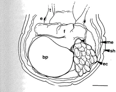
7, — Camera lucida drawing of ventral view of a female
Antisabia juliae sp. nov. with eggs capsule attached to the basal plate (paratype; scale bar, 1000 pm); bp,basalplate; e, eye; ec, eggs
psule; f, anterior part of the propodium; me, mantle edge;
sh, shell contour;t, tentacles.
nearly straight. The stomach(st, Fig. 6) is quite wide
and pouched, disposed at the middle to right side of
the posterior body. The digestive gland is greenish brown, mainly disposed dorsally, to the left part of stomach. The intestine is quite long and occupies rge part of the pallial cavity embedded in the stive gland, containing numerous brownish-gray to green oval fecal pellets.
The mantle edge is thickened without apparent differenciations (p, Fig. 1D). The pallial cavity is quite deep. The osphradium appearsas an ovalridge close to the mantle edge (0, Fig. 1D). The ctenidium is quite long, transversally disposed, with an L shape (approx. 50 short filaments; c, Fig. 1D).
_In females, egg capsules are attached at the
pos-‘o-lateral left side of the foot, at the edge of the
Sal plate (Fig. 7). Egg capsules (3-24 eggs, 500 in diameter) are thin walled pyriform bodies ith a short stalk. Young snails escape at the crawl-g stacrawl-ge (Ficrawl-g. 2C). Reproduction (many females earing egg capsules) was observed at the begin-g¢ of the rainy season (October to November)and, lesser extent, at the end of it (May). Hatching (2 to 10 young snails per capsule) was observed in
November and December.
-omparison with other related species in the Indopacific region
_Antisabia juliae sp. nov. is quite easy to
distin-guish from other Indopacific species of
Hipponicidae except from coexisting Antisabia con-ica (Schumacher, 1817) which has quite similar morphology and life habits: A. juliae has a smaller size, a much thinner basal plate, weaker sculpture on the shell and a narrower “preference” regarding the supporting shell, almost only live turbinids. Egg capsules of A. conica contain several hundred very small yellowish eggs, compared to the 3-24 big whitish eggs in the capsule of A. juliae. Both species appear evolutively very close.
Other hipponicid species are generally fixed on rocks, in crevices or on coral heads. Antisabia foli-acea (Quoy et Gaimard, 1835) is relatively flat, with many growth lamellae piled up at the surface of the shell, each lamella is finely sculptured by radial threads. Pilosabia subrufa (Lamarck, 1819), Pilosabia trigona (Gmelin, 1791) and Hipponix pilosa (Deshayes, 1831) are readily distinguished by their shaggy or squamose periostracum. Hipponix antiquatus (Linné, 1767) has a bigger, heavy shell with axial sculpture composed of very prominent rugose ribs crossed by microscopic incisedlines. Choice of supporting shell and localization
All specimens of Antisabia juliae sp. nov. of Laing Island are found on 4 species of Turbinid gastropods, 3 of which form abundant populations on the beach rock : Turbo setosus Gmelin, 1791, T. crassus Wood, 1829, T. brunneus (Röding, 1798). T. sparverius Gmelin, 1791 is less common. No other shell was found to harbour any hipponicid species until 1995 when we found rare specimens on Trochus maculatus Linnaeus, 1758 (one sample), Tectus pyramis (Born, 1778) (3 specimens on the same sample) and Notohaliotis sp. (one sample).
Bigger shells wear a significantly higher number of commensal individuals than smaller ones (N=60). There is a direct significant correlation of the num-ber of A. juliae with the size of the host shell (Table
TABLE 1. — Matrix of Spearman correlation coefficients (r,) betwe-en number of Antisabia juliae sp. nov. shells and the size and
spe-cies of the Turbo host shell (number of observations = 60) Number Number
Size males females Species Number males 0.808* 1.000 = -Numberfemales 0.814* 0.938* 1.000 -Species 0,223" 0.074? 0.0312 1.000 * Highly significant (o < 0.01)
° Not significant
Fic. 8 — a, Detail of the epiphytic community scraped off the shell of Turbo setosus (mainly cyanobacteria). Scale bar, 10 pm. b, Composition of a fecal pellet collected inside the intestine of a female Antisabia juliae nov. sp. living on the Turbo shell illustrated at A: pennate (benthic) diatoms and cyanobacteria similar to that observed on the shell. Scale bar, 10 pm. c, Detail of the epiphytic community scraped off the shell of Turbo brunneus (mainly pennate diatomsand filamentous algae). Scale bar, 10 um. d, Composition of the stomach content of a female Antisabia juliae sp. nov. living on the Turbo shell illustrated at C: pennate (benthic) diatoms, filamentous algae and
cyanobacteria similar to that observed on theshell. Scale bar, 10 um. 12 M. POULICEKetal.
A. juliae is rare on shells smaller than 20 mm in ength, whereas shells bigger than 60 to 80 mm fre-uently harbour one or two females (rarely >3) of latively large size (6-14 mm diameter) surrounded y 9:t0 6 smaller males (up to 21, 1-3 mm diameter). he number of males is also strongly correlated to he numberof females (r= 0.94, a < 0.01).
At the same size, there is no significant differ-nce in the number of specimensof A. juliae on the “more abundant species of Turbo (N=57). Thereis Iso no significant correlation betweenthe size of A. uliae and the size of the hostshell.
As already observed by Laws (1971) for intisabia conica, most specimens of A. juliae are ound on the ventral columellar region of the shell, lose to the upper part of the peristomium (87 % of
hespecimens sampled, N = 250)either on thelast
whorl or at the base of the last spiral whorl, 10 % are ound at the external upper part of the peristomium and 3 % close to the external anterior part of the hell. Several specimens are generally found close ogether with females surrounded by smaller males.
istribution around Laing Island
Antisabia juliae sp. nov. is quite abundant on g Island, where about 50% of specimens of rbo (all species together) harbour one or more
pecimens. But its distribution on the beach rock is
10t homogeneous: it is much more abundantat the orthern part of the island where more than 80% of
le turbinids wear one or more hipponicid snails. Along the western side, its occurrence remainsfair-high (45% to 60% of turbinid shells) whereas this species is less represented at the southern sites (5% 20% of turbinid shells). There is no important 0. population on the eastern (lagoonal) side. On
hebeach rock, this distribution is mainly explained
the meansizes of Turbo spp.: large specimens (60 o90 mm high) mainly occur directly on the beach k and underneath big dead corals at the northern ind western sides of the island whereas smaller pecimensshelter in crevices of boulders and under
maller rocks at the southern part of the island.
oraging
orphological features (long snout, relative mall size of the gill filaments, structure of the adula, worn teeth at the anterior edge; Figs. 1D, ‘A, 2B) together with observations of live speci-mens attest that Antisabia juliae sp. nov. does not
behave as a filtering snail like Hipponix antiqua-tus (= H. cranioides) observed by Yonge (1953). No evidence of host fecal pellet eating was ever encountered as observed in Hipponix australis (Risbec, 1935). Living specimens are frequently seen with the shell not closely affixed to the sub-strate, the snout and tentacles extruded well away through the space so produced and browsing on the host shell. Moreover, the area aroundA. juliae shells is generally relatively “clean” compared to other similar part of the host shell. When com-pared to the epiphytic community (Figs. 8A, C) found on the host shell, the composition of stom-ach content and of fecal pellets (Figs. 8B, D) shows striking similarities: benthic pennate diatoms, rhodophyta, chlorophyta and cyanobacte-ria constitutes the bulk of the pellets with a very similar composition when compared to the epi-phytic community living on the same host shell. No sand grains were observed as in Hipponix aus-tralis analysed by Knudsen (1991).
Any contact stimulus around the snout or tenta-cles immediately triggers the protrusion of the radu-la so that this species appears even able to grasp meiofaunal organisms: a partially digested. harpacti-coid copepod was even found in two cases. It seems that A. juliae feeds upon the epiphytic community, microorganisms and perhaps meiofauna in the area that can be reached by the snout.
As the basal plate secreted by the foot is only very loosely cemented to the host shell, we tried to verify the real sedentary nature of this species even if no specimen was ever observed moving or even loose on the. host shell. Several attempts. (approxi-mately 30 specimens) to let a carefully removed snail fix itself again on the same or another shell (with or without the basal plate) never succeeded: all A. juliae died within 6 to 96 hours except some small males remaining unattached for more than 6 days. It appears thus probable that this species, like other in the family Hipponicidae, has real seden-tary habits.
DISCUSSION
The hipponicid Antisabia juliae sp. nov. was first tentatively attributed to the species Antisabia conica (Schumacher) but it appears to present sev-eral distinct features that let us consider it a new species. A. juliae sp. nov. appears to have adapted to its mode oflife on live turbinids in several ways
resulting in morphological, ethological and func-tional changes.
Its preferential location on the basal fasciolar area of the host shell implies it should be progres-sively recovered by the growing Turbo shell. Nevertheless, a displacement reaction of A. juliae towards “safer” areas is not likely to occur even if the basal plate secreted by the foot is very thin and only very loosely cemented to the host shell (com-pared to the attachment of other Hipponicid species living on dead shells or on the beach rock,like Sabia foliacea for example). This can explain the prefer-ential “choice” of large (adult) supporting shells (with much slower growth) that so limits the risk of being quickly recovered.
Life on large active intertidal snails also changes the foraging behaviour of this species with correlated morphological changes: it appears to feed on the epiphytic community growing on the host shell, in the vicinity of the sedentary hip-ponicid; larger supporting shells are likely to wear a more diversified and quick growing epibiontic biocenose with a relatively high turnover. Associated with this kind of food collecting habit, this species has developed specific morphological features: long extensible snout with short mobile lateral appendages, functional grasping radula, presence of eyes, etc.
Except for these characteristics, the modeoflife appears quite similar to that of other Hipponicids with a few large females surrounded by several much smaller males. Development occurs within the egg mass inside the female shell and a few young snails escape at the crawling stage. Indirect observa-tions of the shell structure suggests a quick growth that should also be considered as an adaptation to this modeoflife.
14 M. POULICEKetal.
ACKNOWLEDGEMENTS
This study was supported by the National Fund for Scientific Research of Belgium (FNRS) as an FRFC grant n°29008/90.F. The field work was done at the King Leopold III Biological Station in Laing Island (Papua New Guinea). Manythanksare due to the directors (Prof. J. Bouillon, then Prof. M. Jangoux), the local manager, technicians and occa-sional samplersfor help and logistic facilities during our missions. We also want to thank scientific and administrative authorities of Papua New Guinea in Port Moresby and Madangfor their help and sup-port. The work of Nicole Decloux who prepared the samples for SEM is greatly acknowledged. Thanks are due to Professor Charles Jeuniaux and to an unknownreferee for their criticism and constructive suggestions to improve this paper.
REFERENCES
Knudsen, J. — 1991. Observations on Hipponix australis
(Lamarck,1819) (Mollusca, Gastropoda, Prosobranchia) from the Albany area, Western Australia. In: A. Wells, A. Walker, A.
Kirkman and A. Lethbridge (eds.): The marine flora andfauna ofAlbany, pp. 641-660. Proc. 3rd. Intern. Mar. Biol. Workshop.
Knudsen, J. — 1993. Observations on Sabia foliacea (Quoy and Gaimard, 1835) (Mollusca, Gastropoda, Prosobranchia,
Hipponicidae) from off Rottnest Island, Western Australia. In:
A, Wells, A. Walker, A. Kirkman and A. Lethbridge (eds.):
The marine flora and fauna of Rottnest Island, pp. 481-495. Proc. 5th Intern. Mar. Biol. Workshop.
Laws, H.M. — 1971. Reproductive biology and shell site preference in Hipponix conicus (Schumacher). Veliger, 13: 115-121, Risbec, J. ~ 1935. Biologie et ponte de Mollusques Gastéropodes
néocalédoniens. Bull. Soc. Zool. France, 60: 387-417.
Quoy, J.R.C. and Gaimard, J.P. — 1835. Voyage de découvertes de l’Astrolabe pendant les annees 1826-1829, Zoologie 3, J.Tastu. Paris.
Yonge, C.M. — 1953. Observations on Hipponix antiquatus (L). Proc. Calif. Acad. Sci., 4(28): 1-24.
Yonge, C.M. — 1960, Further observations on Hipponix antiquatus (with notes on North Pacific pulmonate limpets). Proc. Calif. Acad. Sci., 431): 111-119.