FACULTE DE MEDECINE VETERINAIRE
DEPARTEMENT CLINIQUE DES ANIMAUX DE COMPAGNIE ET DES EQUIDES SERVICE DE PATHOLOGIE MEDICALE DES PETITS ANIMAUX
LA FIBROSE PULMONAIRE IDIOPATHIQUE CANINE:
AMELIORATION DE LA CARACTERISATION CLINIQUE,
RECHERCHE DE BIOMARQUEURS,
ET D’AGENTS ETIOLOGIQUES
CANINE IDIOPATHIC PULMONARY FIBROSIS:
IMPROVEMENT OF THE PHENOTYPE CHARACTERIZATION,
AND SEARCH FOR BIOMARKERS
AND FOR ETIOLOGIC AGENTS
Elodie ROELS
THESE PRESENTEE EN VUE DE L’OBTENTION DU GRADE DE DOCTEUR EN SCIENCES VETERINAIRES
REMERCIEMENTS
Je tiens à adresser toute ma gratitude et mes remerciements les plus sincères à ma promotrice de thèse, le Professeur Cécile Clercx, qui a su me guider et m’encourager tout au long de ces 4 années de doctorat. Dans les couloirs de la clinique vétérinaire des petits animaux, il a été souvent entendu que j’étais votre portrait tout craché. C’est un compliment qui me va droit au cœur car j’admire votre énergie, votre enthousiasme, votre gentillesse, votre sens de l’écoute et votre capacité à nous pousser au meilleur de nous-même. Merci à vous, Cécile, de m’avoir toujours soutenue et d’avoir supporté mon caractère parfois un peu rebelle. Votre disponibilité et votre optimisme ont été pour moi des bouffées d’oxygène lors des moments plus difficiles et c’est à vous que je dois l’accomplissement de ce travail.
Je tiens également à remercier tout particulièrement ma co-promotrice, le Professeur Kathleen Mc Entee, qui a su m’aiguiller tout le long de ce parcours avec beaucoup de bienveillance et d’encouragements. Merci de m’avoir donné gout à cette magnifique discipline qu’est la cardiologie. Merci également d’avoir passé tant d’heures à faire des mesures sur un échocardiographe pas toujours conciliant !
Merci aux membres de mon comité de thèse, les Professeurs Géraldine Bolen et Fabrice Bureau, pour leur collaboration, leur aide précieuse et le temps consacré à l’évaluation de ce travail. Merci Gégé d’avoir ouvert les portes de la radio à ma horde de Westies et d’avoir toujours été disponible pour les échographier, scanner et radiographier. Merci pour ta bonne humeur à toute épreuve et pour le temps que tu as passé à lire et relire les images récoltées toute au long de ce travail. Merci Fabrice pour ta disponibilité et pour m’avoir accueillie dans ton laboratoire où je me suis directement sentie acceptée et intégrée.
Merci aux membres de mon jury de thèse, les Professeurs Tania Art, Didier Cataldo, Benoit Nemery, Paul De Vuyst et Mutien-Marie Garigliany, et le Docteur Thomas Marichal d’avoir accepté de juger ce travail. Merci au Professeur Laurent Gillet d’avoir accepté de présider ce jury de thèse.
Je souhaite également remercier de tout cœur toutes les personnes qui ont contribué de près ou de loin à l’accomplissement de ce travail et qui m’ont aidé à m’épanouir tant sur le plan professionnel que sur le plan personnel. La liste est longue et j’espère n’avoir oublié personne… !
Merci à l’équipe vétérinaire finlandaise composée de Lisa, Saila, Henna et Minna pour leur précieuse collaboration au fil des années et leurs encouragements lors de mes présentations
en congrès internationaux. Merci Saila d’avoir toujours répondu présente pour l’échange d’informations et de matériels biologiques nécessaire à la réalisation de ce travail.
Merci aux collaborateurs vétérinaires extérieurs à l’université de Liège pour leur participation dans la récolte de nouveaux cas et leur implication dans notre projet. Merci aux Docteurs Fergus Allerton, Ghita Benchekroun, Éric Bomassi, Domingo Casamian, Cécile Damoiseau, Aleksandra Domanjko, Andrea Fischer, Reinhart Hirt, Rachel Lavoué, Davide Delorenzi, Frederic Mergenthal, Nina Müller, Viktor Szatmari, Benoit Vanbrugghe, Nicole Van Israël, Audrey Vrancken et Hans Von Euler.
Merci à tous les membres du laboratoire de physiologie cellulaire et moléculaire du GIGA pour leur accueil chaleureux, leur aide technique et leurs excellents conseils. Merci Dimitri pour ta patience, ton calme et ta disponibilité. Grace à toi, j’ai appris à gérer un qRT-PCR du début à la fin et à remplir correctement une plaque 384 puits en un temps record. Merci Claire pour ton aide en cytométrie en flux même si on n’a pas eu beaucoup de chance sur ce coup-là ! Merci Coraline et Joey d’avoir été des partenaires de chants idéaux (à quand l’établissement d’une chorale ?) et merci Thibaut et Pierre pour vos petites blagues qui m’ont fait beaucoup rire. Merci Cédric d’avoir été mon fournisseur officiel de carboglace !
Merci au laboratoire d’immunologie et de vaccinologie de la FMV de m’avoir initiée à la recherche en virologie. Merci au Professeur Laurent Gillet de m’avoir si gentiment accueillie, conseillée et guidée tant dans la démarche scientifique que dans la rédaction qui s’en est suivie. Merci Mickael pour ton calme et ta rigueur dans les manips ainsi que pour ta gentillesse et ton optimisme, ce fut un plaisir de travailler avec toi. Merci également au Professeur Berhard Ehlers du Robert-Koch Institute d’avoir accepté de collaborer sur notre projet et de s’être investi à 100% dans notre travail malgré la distance entre nos institutions.
Merci au laboratoire d’histologie de la FMV de m’avoir épaulé dans la réalisation des immunohistochimies et des ELISA. Merci au Professeur Nadine Antoine d’avoir été disponible pour les lectures des immuno-marquages et pour les rédactions scientifiques associées. Merci Joëlle d’avoir toujours répondu présente pour m’assister dans les manips. Ta patience, ta bonne-humeur, ton calme et ta précision font de toi une technicienne en or avec laquelle j’ai eu la chance de pouvoir à travailler.
Merci au Professeur Frédéric Farnir pour son aide précieuse en statistiques mais également pour son réconfort et son soutien dans les moments plus difficiles. Merci d’avoir toujours trouvé les mots justes quand mon moral n’était pas au beau fixe.
Merci au Professeur Michael Day pour son aide dans la lecture des coupes histopathologiques de poumon ainsi que dans la rédaction d’abstract et d’articles scientifiques.
Merci à la team Olson (Amy et Patricia) d’avoir pris le temps de relire et de corriger entièrement ce manuscrit en un temps record. Thank you
Merci à toute l’équipe du service de médecine interne de la CVU pour m’avoir supporté pendant ces 4 années de doctorat. Merci d’avoir partagé mes joies et mes réussites tout comme mes doutes, mes angoisses et mes peines. Merci aux chefs, Fred, Dominique et Kris pour leur confiance, leur soutien et leur côté taquin. Merci Kiki d’avoir toujours accepté (presque sans broncher) d’échocardiographier mes Westies. Merci également pour tes conseils quant à mon futur et à la rédaction de ma thèse. Merci aux copines, Chachou, MiniMi, Maudinette, Elo, Morganette et Cécile d’avoir été toujours présente à mes côtés et de m’avoir fait rire aux éclats tellement de fois. Chaque moment passé avec vous a été un cadeau, que ce soit au boulot ou en dehors. Vous êtes des filles géniales et le MIPA power restera gravé à tout jamais. Merci Val, Isabelle, Charlotte et Iris pour votre écoute attentive et vos conseils. Merci Jacques et Jérôme pour votre bonne-humeur et votre sens de l’humour. Merci également à François pour qui rapidité et efficacité sont les principaux mots d’ordre (bang-bang !), atouts indispensables quand il s’agit de faire face à une personne aussi impatiente que moi ! Merci Sylvain d’avoir été tellement méticuleux et ordonné avec la tonne de prélèvements que je t’ai demandés de ranger et répertorier. Merci de t’être gelé les mains dans le -80°C autant de fois pour moi. Enfin merci aux Nadines, à Audrey, Patricia et Dominique pour leur dévouement à toute épreuve et leur gentillesse hors du commun.
Merci également à toute l’équipe du service d’imagerie de la CVU pour leur aide dans l’acquisition et l’interprétation des examens d’imagerie et pour l’accueil irréprochable qu’ils ont réservés à mes petits Westies. Mercis particuliers à Anne-Laure et Annalisa ainsi qu’à Philippe et Laurie pour vous être particulièrement investis dans le bon déroulement des opérations. Merci également au Professeur Johny Verschakelen et au Docteur Thierry Couvreur pour leurs collaborations et le partage de leur expertise dans la lecture et l’interprétation des images.
Merci à toute l’équipe du service d’anesthésie de la CVU pour avoir gérer avec succès toutes les anesthésies des Westies réalisées pendant ce travail. Merci Olivia et Véro pour votre délicatesse et votre efficacité ainsi que pour vos qualités humaines irréprochables. Merci Alexandru pour ta zen-attitude à toute épreuve.
Merci également à tous les autres membres de la CVU avec qui j’ai eu l’occasion de partager et d’échanger sur ma thèse, ma vie, le travail, la fac ou encore la météo. Ceux qui me connaissent diront en effet que j’ai la langue bien pendue !!! Merci Mikou, Oli, Martinos, Bart et Fabinou = la team des chirurgiens (DP ne soit pas jaloux) de m’avoir accompagné mainte et mainte fois à la cafèt et de m’avoir diverti avec vos sujets de conversation plus que variés (et souvent graveleux avouons-le). Merci aux fées de l’ICU, Céline et Catherine, pour leur simplicité et leur joie de vivre (et aussi pour leur talents de coiffeuse !). Merci aux filles de la compta, Catherine, Fred et Antonella pour avoir géré mes commandes, débours et autres paperasseries à la perfection. Merci à Maurizio pour ton dévouement aux beagles du chenil et tes qualités d’électricien ! Et enfin merci aux internes (en particulier Caroline et Fabienne) et aux étudiants pour leur disponibilité sans faille.
Je tenais également à remercier tous les propriétaires et éleveurs de Westies qui ont cru en notre projet et ont été d’accord d’y participer avec leurs chiens. Un merci tout particulier à Madame Véronique Desramé, présidente du club français des Westies, qui nous soutient dans nos recherches depuis plusieurs années et est toujours motivée pour organiser des évènements et rencontres afin d’améliorer la communication et les avancées sur la maladie.
Je terminerai en adressant un énorme merci à mes amies proches, Ana, Mapi, Sam, Céline, Steph L., Steph D. et Aurélie, ainsi qu’à mes parents, à mes grands-parents, à ma sœur et à mon frère pour leur soutien, leurs encouragements et leur présence dans ma vie. A mes amies, merci d’être toujours là pour moi, même si la distance et nos vies bien remplies nous empêchent de nous voir aussi souvent qu’on en aurait envie. A ma famille, merci pour la fierté que je peux lire sur vos visages à chaque fois que vous évoquez mon parcours. Merci de m’avoir guidé jusqu’ici. Vous êtes un exemple de courage, de volonté, de simplicité, de respect et d’amour.
LIST OF ABBREVIATIONS
5-HT 5-hydroxytryptamine, serotonin
5-HTR 5-hydroxytryptamine receptor
5-HTT 5-hydroxytryptamine transporter
6MWD 6-minutes walked distance
6MWT 6-minute walking test
AE Acute exacerbation
AEC Alveolar epithelial cell
AHV Asinine herpesvirus
ALP Alkaline phosphatase
Ao Aorta
AT Acceleration time of the pulmonary artery flow
AT:ET Acceleration to ejection time ratio of the pulmonary artery flow
ATS American thoracic society
ARDS Acute respiratory distress syndrome
BALF Bronchoalveolar lavage fluid
CB Chronic bronchitis
CCL2 Chemokine (CC-motif) ligand 2
CHF Congestive heart failure
CIPF Canine idiopathic pulmonary fibrosis
CPFE Combined pulmonary fibrosis and emphysema
CXCL8 Chemokine (CXC-motif) ligand 8
DAD Diffuse alveolar damage
DLCO Diffusion lung capacity for carbon monoxide
DMVD Degenerative mitral valve disease
EBP Eosinophilic bronchopneumopathy
ELF Epithelial lining fluid
ELISA Enzyme-linked immunosorbent assay
EMPF Equine multinodular pulmonary fibrosis
EMT Epithelial to mesenchymal transition
ERS European respiratory society
ET-1 Endothelin-1
EVG Elastic Von Gieson
FGF Fibroblast growth factor
FVC Forced vital capacity
GGO Ground-glass opacity
GOR Gastro-oesophageal reflux
GWAS Genome wide association study
HHV Human herpesvirus
HP Hypersensitivity pneumonitis
ILD Interstitial lung disease
IPF Idiopathic pulmonary fibrosis
MPA Main pulmonary artery
MPA/Ao Main pulmonary artery to aortic diameters ratio
MUC5B Mucin 5B
NSIP Non-specific interstitial pneumonia
NT-proBNP N-terminal pro-brain natriuretic peptide
LTBP Latent TGF- binding protein
p(A-a)O2 Alveolar to arterial oxygen gradient
PAP Pulmonary arterial pressure
pCO2 Partial pressure of carbon dioxide
PDGF Platelet-derived growth factor
PH Pulmonary hypertension
pO2 Partial pressure of oxygen
PPFE Pleuroparenchymal fibroelastosis
PR Pulmonary regurgitation
PV/PA Pulmonary vein to pulmonary artery ratio
qRT-PCR Quantitative reverse-transcription polymerase chain reaction
RPAD Index Right pulmonary artery distensibility index
Se Sensitivity
Sp Specificity
TBB Trans-bronchial biopsy
TGF- Transforming growth factor-beta
T-HRCT Thoracic high-resolution computed tomography
TR Tricuspid regurgitation
UIP Usual interstitial pneumonia
VEGF Vascular endothelial growth factor
VmaxTR Tricuspid regurgitant jet maximal velocity
TABLE OF CONTENTS
REMERCIEMENTS ... 3 LIST OF ABBREVIATIONS ... 7 TABLE OF CONTENTS ... 10 SUMMARY ... 12 RESUME ... 15 INTRODUCTION ... 191. Canine idiopathic pulmonary fibrosis: an update ... 19
1.1. Description of CIPF - History... 19
1.2. Clinical characterization of CIPF ... 20
1.2.1. Clinical presentation ... 20
1.2.2. Blood haematological and biochemical analyses ... 21
1.2.3. Cardiopulmonary function testing ... 21
1.2.4. Echocardiography ... 22
1.2.5. Diagnostic imaging ... 27
1.2.6. Bronchoscopy and bronchoalveolar lavage fluid analysis ... 28
1.2.7. Histopathological features ... 29
1.2.8. Long-term outcome and prognostic indicators ... 31
1.3. Understanding the pathogenesis ... 32
1.3.1. Surfactant protein C mutation ... 32
1.3.2. Endothelin-1 ... 33
1.3.3. Procollagen type III amino terminal propeptide ... 34
1.3.4. Proteomic analysis of bronchoalveolar lavage fluid ... 35
1.3.5. Microarray analysis of pulmonary gene expression ... 35
1.3.6. TGF-beta 1 ... 37 1.3.7. Activin A and B ... 38 1.3.8. Serotonin ... 39 1.4. Unexplored fields ... 40 1.4.1. Prevalence rate ... 40 1.4.2. Genetic background ... 40 1.4.3. Etiologic agents ... 41 1.4.4. Treatments ... 41 1.4.5. Follow-up tools ... 42
2. Pulmonary fibrosis in other animal species ... 43
2.1. Pleuropulmonary fibrosis and elastosis in donkeys ... 43
2.2. Equine multinodular pulmonary fibrosis ... 45
2.3. Pulmonary fibrosis in cats ... 47
3. Idiopathic pulmonary fibrosis in humans ... 49
3.1. Incidence and prevalence rates ... 51
3.2. Clinical presentation ... 51
3.3. Diagnostic approach ... 51
3.3.2. Blood analyses ... 52
3.3.3. Cardiopulmonary function testing ... 52
3.3.4. Diagnostic imaging ... 53
3.3.5. Bronchoscopy and bronchoalveolar lavage fluid analysis ... 54
3.3.6. Histopathological features ... 55 3.4. Risk factors ... 55 3.4.1. Cigarette smoking ... 56 3.4.2. Environmental exposures ... 56 3.4.3. Gastro-oesophageal reflux ... 56 3.4.4. Infectious agents ... 57 3.4.5. Genetic background ... 58 3.5. Disease progression ... 58 3.6. Prognostic factors ... 59 3.7. Co-morbidities ... 61
3.7.1. Pulmonary hypertension ... 61
3.7.2. Emphysema ... 62
3.7.3. Neoplasia ... 62
3.7.4. Thromboembolic disease ... 62
3.8. Overview of the pathogenesis ... 63
3.8.1. Cellular mechanisms of pulmonary fibrosis ... 64
3.8.2. Molecular mechanisms of pulmonary fibrosis ... 65
3.9. Therapeutic options ... 68
4. Other pulmonary fibrotic disorders in humans ... 70
4.1. Chronic hypersensitivity pneumonitis ... 70
4.2. Fibrosing non-specific interstitial pneumonia ... 71
OBJECTIVES ... 73
1. Case – control recruitment ... 73
2. Improvement of the phenotypic characterization ... 73
2.1. T-HRCT interpretation: sedation vs. general anaesthesia ... 73
2.2. Echocardiographic investigation of pulmonary hypertension: PV/PA ... 74
3. Investigation of biomarkers ... 75
4. Search for etiologic agents ... 76
RESULTS SECTION ... 77
1. Case – control recruitment ... 77
2. Improvement of the phenotypic characterization ... 81
2.1. T-HRCT interpretation: sedation vs. general anaesthesia ... 81
2.1.1. Title & authors ... 81
2.1.2. Abstract... 81
2.1.3. Introduction ... 82
2.1.4. Materials and Methods ... 83
2.1.5. Results ... 86
2.1.6. Discussion ... 95
2.2. Echocardiographic investigation of pulmonary hypertension: PV/PA ... 98
2.2.1. Title & authors ... 98
2.2.2. Abstract... 98
2.2.3. Introduction ... 99
2.2.4. Materials and Methods ... 100
2.2.5. Results ... 103
2.2.6. Discussion ... 110
3. Investigation of biomarkers ... 113
3.1. Serum CCL2 and CXCL8 concentrations: CIPF WHWTs vs. healthy controls ... 113
3.2. BALF CCL2 and CXCL8 concentrations: CIPF WHWTs vs. healthy controls ... 114
3.3. Serum CCL2 concentration in CIPF WHWTs: a survival prognostic marker ... 116
3.4. Lung CCL2/CCR2 and CXCL8/CXCR2 expression assessed by qRT-PCR ... 117
3.4. Lung cellular sources of CCL2 and CXCL8 assessed by immunohistochemistry... 120
3.5. CCL2, CXCL8, VEGF, and 5-HT blood concentrations in healthy dogs from 7 breeds with variable predisposition for CIPF ... 124
4. Search for etiologic agents ... 127
4.1. Panherpesvirus PCR assay (DPOL gene) ... 127
ARTICLES ... 130
DISCUSSION ... 148
LIMITATIONS, PERSPECTIVES AND CONCLUSIONS ... 156
SUMMARY
Canine idiopathic pulmonary fibrosis (CIPF) is a progressive parenchymal lung disease of unknown origin, mainly described in old-aged West Highland white terriers (WHWTs). It is characterized by exercise intolerance, cough and dyspnoea/tachypnea with a progressive deterioration until death from respiratory insufficiency. CIPF shares clinical features with human idiopathic pulmonary fibrosis (IPF), while tomodensitometric and histopathological findings do not appear to be exactly the same. Over the past 10 years, several studies have been performed to improve our knowledge about CIPF. However, this disease is still misunderstood and clinicians are dealing with several challenges including the absence of clinical or biological markers for estimating the presence, severity or progression of the disease and related co-morbidities such as pulmonary hypertension, the absence of etiologic agent, and the absence of targeted therapy. Consequently, the aims of the present project were (1) to investigate whether high-resolution computed tomography (HRCT) of the lungs obtained under sedation can be used for the diagnosis and for the follow-up of the disease, (2) to study a new echocardiographic parameter for the diagnosis of precapillary pulmonary hypertension induced by CIPF, (3) to study the potential roles of 2 chemokines of interest, CCL2 and CXCL8, as biomarkers of fibrosis and as actors in the pathogenesis of the disease, (4) to determine breed variation of basal blood concentrations of the same chemokines, vascular endothelial growth factor (VEGF), and serotonin, and (5) to search for the presence of herpesvirus as a possible etiologic agent.
To meet objective (1), lung HRCT findings found in WHWTs affected with CIPF, as well as unaffected WHWTs matched for age were described. The effect of the sedation vs. anaesthesia on the identification and gradation of the lung lesions was studied in order to determine the utility of the sedation for the diagnosis of CIPF, given that anaesthesia is a potential risk in pulmonary-diseased dogs, particularly in the presence of pulmonary hypertension. Results showed that both sedation and general anaesthesia allowed identification of ground-glass opacities (GGO), the classical HRCT features described in CIPF. This suggests that sedation may be a good alternative for the CIPF HRCT diagnosis when general anaesthesia is not recommended. GGO was also observed in some control dogs, essentially localised to the cranial lung lobes, which may correspond to methodological artefact or to sub-clinical early CIPF lesions. Differences between sedation and general anaesthesia were found in the identification of consolidation and in the gradation of the GGO and mosaic attenuation pattern extent, more probably related to the different breathing pattern of the dogs between the two examinations: spontaneous breathing under sedation vs. end-expiratory pause following hyperventilation under general anaesthesia, inducing a variable content of air within the alveoli
and influencing the interpretation of lung lesions on HRCT images. However, the differences in lesion gradation extent did not prevent the establishment of the CIPF diagnosis, given that GGO was observed in all CIPF dogs in a greater extent than in controls or in association with other CIPF features not present in controls.
Objective (2) was to study a novel echocardiographic index, the pulmonary vein to pulmonary artery ratio (PV/PA) in correlation with other non-invasive echocardiographic indices of pulmonary hypertension (PH): the maximal velocity of tricuspid regurgitation (VmaxTR), the acceleration to ejection time ratio of the pulmonary flow (AT:ET), the main pulmonary artery to aorta diameter ratio (MPA/Ao), and the right pulmonary artery distensibility index (RPAD Index). Echocardiographic measures were performed in WHWTs affected with CIPF and control WHWTs matched for age. Correlations were calculated with the partial pressure of oxygen (pO2), the distance walked in the 6-minute walking test (6MWD), and serum concentrations of NT-proBNP. A tricuspid regurgitant jet allowing the estimation of pulmonary pressure gradient was found in 50% WHWTs affected with CIPF and 12.5% controls. In CIPF WHWTs with concomitant PH (VmaxTR above 2.8 m/s) significantly lower PV/PA values were found in comparison with control WHWTs. This decrease in PV/PA was due to an increase in the pulmonary artery diameter and a decrease in pulmonary vein diameter. A trend for a decrease of AT:ET and RPAD Index and an increase of MPA/Ao was observed in CIPF WHWTs affected with PH in comparison with controls. Significant moderate correlations were found between PV/PA and all of the other echocardiographic parameters of PH, as well as with 6MWD. There was no correlation between PV/PA and serum NT-proBNP concentrations or arterial pO2 values. Those results suggest the potential benefit of the PV/PA ratio for the non-invasive diagnosis of precapillary PH in WHWTs affected with CIPF. Further investigations in a larger cohort of dogs with precapillary PH, and with a measurable tricuspid regurgitant jet, are needed to validate this finding.
In 2013, Krafft and collaborators highlighted an overexpression of chemokines CCL2 and CXCL8 in the lung parenchyma of WHWTs affected with CIPF in comparison with controls. The objective (3) of the present work was therefore to investigate serum and bronchoalveolar lavage fluid (BALF) concentrations of CCL2 and CXCL8 in CIPF and control WHWTs, and to study the lung signalling pathways of those molecules via qRT-PCR (CCL2, CXCL8, CCR2, CXCR2) and immunohistochemistry (CCL2 and CXCL8). Such tests might lead to determining the potential role of the chemokines as biomarkers of the disease and as actors in the pathogenesis. Higher serum CCL2 concentrations were found in WHWTs affected with CIPF in comparison with controls; serum CCL2 concentrations above 700 pg/mL were
difference between CIPF and control WHWTs for serum CXCL8 concentrations. For BALF concentrations, both CCL2 and CXCL8 chemokines were significantly increased in WHWTs affected with CIPF. Relative CCL2, CXCL8, and their respective receptors CCR2 and CXCR2 gene expression was not significantly different between the lungs of WHWT with CIPF and control dogs of various breeds, while an immunolabelling for CCL2 and CXCL8 was found in the lung parenchyma of CIPF WHWTs at the level of bronchial epithelial cells. Those results tend to indicate that chemokines CCL2 and CXCL8 are potentially involved in the fibroproliferative process associated with CIPF. Whether the modifications observed are a cause of, or a consequence of, the disease remain unknown.
To test our hypothesis that higher circulating basal blood concentrations of profibrotic molecules could serve as predisposing factors for CIPF development in the WHWT breed, serum concentrations of CCL2, CXCL8, vascular endothelial growth factor (VEGF) and serotonin (5-HT) were measured in healthy dogs from various breeds variably predisposed to CIPF. In addition to the WHWT breed, 6 other breeds of dogs were included: Scottish terrier and Jack Russel terrier considered as potentially predisposed for CIPF, Maltese and King Charles spaniel considered as non-predisposed breeds sharing similarities in weight and size with the WHWT breed, and Labrador Retriever and Malinois Belgian Shepherd considered as non-predisposed breeds not-matched for size and weight with the WHWT breed. Increased serum CXCL8 concentrations were found in healthy dogs from the WHWT breed in comparison with all other breeds less or not predisposed to CIPF. Serum CCL2 concentrations were significantly increased in healthy WHWTs, but also in Maltese, a non CIPF-predisposed breed, compared with King Charles spaniel and Malinois Belgian Shepherd. No relevant interbreed differences were observed for 5-HT with regard to CIPF predisposition. Breed-related differences in VEGF blood concentrations could not be investigated since most of the results obtained were below the kit detection limit.
Finally, to meet objective (5), a study was performed to search for the presence of gammaherpesvirus in the lung parenchyma of WHWTs affected with CIPF, given that an association between Epstein-Barr virus and human IPF has been proposed. A panherpesvirus nested PCR targeting the DPOL gene was applied on both lung and blood samples, but herpesvirus DPOL sequences were not identified in any of the samples tested.
RESUME
La fibrose pulmonaire idiopathique canine (FPIC) est une affection fibrosante chronique du parenchyme pulmonaire, majoritairement rencontrée chez le chien de race West Highland White Terrier (WHWT) d’âge avancé. Elle se traduit par une intolérance à l’effort, de la toux et de la dyspnée/tachypnée avec aggravation progressive jusqu’au décès de l’animal par insuffisance respiratoire. La FPIC partage de nombreuses similitudes cliniques et pathogéniques avec la fibrose idiopathique pulmonaire (FIP) décrite chez l’homme, bien que les lésions tomodensitométriques et histologiques ne soient pas strictement superposables entre les 2 espèces. Au cours de ces 10 dernières années, plusieurs études ont été menées afin d’améliorer les connaissances sur la FPIC. Toutefois, cette maladie reste encore mal comprise à l’heure actuelle et le clinicien doit faire face à plusieurs challenges incluant l’absence de marqueur clinique ou biologique permettant d’estimer la présence, la sévérité ou la progression de la maladie ainsi que la présence de comorbidités relatives dont l’hypertension pulmonaire, l’absence d’agent étiologique, et l’absence de thérapie ciblée. Le présent travail a donc été entrepris afin de répondre au moins partiellement à ces différents challenges. Les objectifs ont été (1) d’investiguer l’intérêt de l’examen tomodensitométrique à haute résolution obtenu sous sédation pour le diagnostic et le suivi de la maladie, (2) d’étudier un nouveau paramètre échocardiographique pour le diagnostic de l’hypertension pulmonaire associé à la FPIC, (3) d’élucider les rôles potentiels de 2 chémokines d’intérêt, CCL2 et CXCL8, en tant que biomarqueurs de fibrose et d’agents de pathogénèse, (4) de déterminer les variations raciales des concentrations sanguines basales en ces mêmes chémokines ainsi qu’en VEGF et en sérotonine, et (5) de rechercher la présence d’herpesvirus comme agent étiologique.
Pour répondre à l’objectif (1), les lésions tomodensitométriques pulmonaires ont été décrites chez des WHWTs atteints de FPIC, ainsi que chez des chiens sains de même race matchés pour l’âge (chiens âgés). L’impact d’une sédation versus une anesthésie sur l’identification et la gradation des lésions tomodensitométriques a été étudié, afin de déterminer l’intérêt de l’examen obtenu sous sédation pour le diagnostic de FPIC dans les cas où l’anesthésie est risquée, ce qui est particulièrement le cas chez les chiens sévèrement atteints de fibrose et d’hypertension pulmonaire concomitante. Les résultats ont montré que la sédation et l’anesthésie permettaient tous deux d’observer les images en verre dépoli classiquement décrites en cas de FPIC, suggérant l’utilisation préférentielle de la sédation pour le diagnostic de FPIC en cas d’anesthésie risquée. Des images en verre dépoli ont également été observées chez certains chiens sains, essentiellement dans les lobes pulmonaires crâniaux, suggérant un artéfact lié à la méthode ou la présence de lésions sub-cliniques de FPIC. Des différences entre
gradation des images en mosaïque et en verre dépoli. Ces différences sont attribuables à la phase respiratoire différente entre sédation (respiration spontanée) et anesthésie (pause expiratoire induite par une hyperventilation préalable) induisant une quantité variable d’air dans les alvéoles et une apparence variable des lésions pulmonaires sur les images tomodensitométriques. Cependant, ces différences ne semblent pas interférer avec le diagnostic de FPIC étant donné que les images en verre dépoli ont été retrouvées chez tous les WHWTs atteints de FIPC dans une étendue plus importante en comparaison aux contrôles et/ou en association avec d’autres caractéristiques tomodensitométriques de fibrose.
Dans le cadre de l’objectif (2), un nouveau paramètre échocardiographique (pulmonary vein to pulmonary artery ratio : PV/PA) a été étudié en corrélation à d’autres indices échocardiographiques susceptibles d’identifier de manière non-invasive l’hypertension pulmonaire : la vitesse maximale du reflux tricuspide (VmaxTR), le rapport entre l’accélération et l’éjection du flux pulmonaire (AT:ET), le rapport entre le diamètre de l’artère pulmonaire principale et le diamètre de l’aorte (MPA/Ao), et la distensibilité de l’artère pulmonaire (RPAD Index). Les mesures ont été faites chez des WHWTs atteints de FPIC et chez des WHWTs contrôles matchés pour l’âge. Des corrélations avec la pression partielle artérielle en oxygène (pO2), la distance marchée en 6 minutes (6MWD) et les concentrations sériques en NT-proBNP ont également été calculées. Un reflux tricuspide permettant d’estimer le gradient de pression dans les artères pulmonaires n’était présent que chez 50% WHWTs atteint de FPIC et 12,5% des contrôles. Des valeurs significativement plus basses en PV/PA ont été observées chez les WHWTs atteints de FPIC avec hypertension pulmonaire (VmaxTR supérieur à 2,8 m/s) en comparaison aux contrôles suite à une augmentation du diamètre de l’artère pulmonaire et à une diminution du diamètre de la veine pulmonaire. Une tendance pour une diminution des paramètres AT:ET et RPAD et pour une augmentation du paramètre MPA/Ao a été observée chez les WHWTs atteints de FPIC et d’hypertension pulmonaire. Des corrélations modérées significatives ont été observées entre PV/PA et tous les autres paramètres échocardiographies d’hypertension pulmonaire étudiés, de même qu’avec la 6MWD mais pas avec la pO2 artérielle. Il n’y avait pas de différence entre les groupes pour les concentrations sériques en NT-proBNP ni de corrélation avec le paramètre PV/PA. Ces résultats suggèrent que le paramètre PV/PA peut être utilisé comme indicateur d’hypertension pulmonaire d’origine pré-capillaire et encourage la réalisation de recherches ultérieures dans ce domaine dans une plus large population de chien pour lesquels un reflux tricuspide est présent.
Suite à la mise en évidence par Krafft et collaborateurs (2013) d’une surexpression relative des chémokines CCL2 et CXCL8 au sein du parenchyme pulmonaire des WHWTs atteints de FPIC, l’objectif (3) du présent travail a été d’investiguer les concentrations en CCL2
et CXCL8 retrouvées dans le sérum et dans le liquide de lavage bronchoalvéolaire (BALF) de WHWTs avec CIPF et de WHWTs contrôles sains, ainsi que d’étudier la signalisation de ces chémokines au sein du parenchyme pulmonaire par qRT-PCR (CCL2, CXCL8, CCR2, CXCR2) et par immunohistochimie (CCL2 et CXCL8) ; le but étant de déterminer le rôle de ces chémokines en tant que biomarqueur de la maladie et en tant qu’acteur potentiel dans la pathogénèse. Des concentrations sériques en CCL2 significativement augmentées ont été observées chez les WHWTs atteints de FPIC en comparaison aux contrôles ; les WHWTs atteints de FPIC avec des concentrations sériques supérieurs à 700 pg/mL ayant une espérance de survie significativement raccourcie. A l’inverse, aucune différence significative n’a été mise en évidence pour les concentrations sériques en CXCL8 entre les WHWTs atteint de FPIC et les WHWTs sains. Concernant les concentrations mesurées dans le BALF, les WHWTs atteints de FPIC présentaient des concentrations en CXCL8 et en CCL2 significativement augmentées en comparaison aux individus contrôles. Aucune différence significative n’a été mise en évidence par qRT-PCR pour l’expression relative des gènes codant les chémokines CCL2 et CXCL8 et leurs récepteurs associés CCR2 et CXCR2, bien qu’un immunomarquage fût présent au sein des poumons de WHWTs atteints de FPIC essentiellement à hauteur des cellules épithéliales bronchiques. Ces résultats indiquent une implication potentielle des chémokines CCL2 et CXCL8 dans le processus fibroprolifératif de la FPIC. Cependant, savoir si les modifications observées représentent une cause ou une conséquence de la maladie reste encore à déterminer.
Les concentrations sériques en CCL2, CXCL8, VEGF (vascular endothelial growth factor) et 5-HT (sérotonine) ont également été analysés dans le sérum d’autres races de chiens sains peu ou pas prédisposées à développer la maladie, afin de tester notre hypothèse selon laquelle la présence de concentrations basales élevées en molécules profibrotiques chez le WHWT pourrait expliquer la plus grande susceptibilité de cette race pour la maladie. En plus du WHWT, 6 races de chiens ont été inclues : le Scottish terrier et le Jack Russell terrier considérées comme potentiellement prédisposée à la FPIC, le Bichon Maltais et le Cavalier King Charles considérées comme non-prédisposées à la FPIC mais de taille et de poids similaires à la race WHWT, et enfin le Labrador et le Berger Malinois considérées comme non-prédisposées à la FPIC et non-matchés pour la taille et le poids avec la race WHWT. Les concentrations en CXCL8 étaient significativement plus élevées dans le sérum de WHWTs sains en comparaison à toutes les autres races de chiens sains inclues peu ou pas prédisposées à la fibrose. Les concentrations en CCL2 étaient significativement augmentées chez le WHWT et le Bichon Maltais en comparaison au Cavalier King Charles et Berger Malinois. Aucune différence significative remarquable n’a été observée pour les concentrations sériques en
objectivée étant donné que la plupart des échantillons ont donné des valeurs inférieures au seuil de détection du kit.
Enfin, pour répondre à l’objectif (5), une étude s’est portée sur la recherche de la présence potentielle de gammaherpesvirus dans le parenchyme pulmonaire de WHWTs atteints de CIPF, au vu de l’association décrite entre l’Epstein-Barr virus et l’IPF chez l’homme. Une PCR panherpes (gène DPOL) a été appliquée mais n’a pas permis d’identifier la présence de gammaherpesvirus dans le parenchyme pulmonaire de WHWTs atteints de CIPF.
INTRODUCTION
Fibrosis is defined as any pathological condition where fibrous connective tissue invades any organ, usually as a consequence of inflammatory or other injury (MeSH PubMed, year introduced 1987). In the particular case of pulmonary fibrosis, the normal lung tissue is progressively replaced by fibroblasts and collagen causing an irreversible loss of organ function resulting in death (MeSH PubMed). The present work will focus on pulmonary fibrosis in a specific dog breed, the West Highland white terrier (WHWT). In this introductory section, we will review what is already known about this disease in dogs, and then report what was discovered about pulmonary fibrosis in other animal species and in human medicine with the help of experimental murine models of the disease. General aspects of pulmonary fibrosis in each species will be addressed, in addition to more advanced literature review in link with the objectives of the present work.
1. Canine idiopathic pulmonary fibrosis: an update
Canine idiopathic pulmonary fibrosis (CIPF) is a chronic and progressive lung disease of unknown aetiology affecting mainly old-aged WHWTs. Since its first description in 1999, several studies have been conducted to improve the clinical characterisation of the disease, expand our understanding about the pathophysiology and identify biomarkers of the disease. At the present time, however, several aspects of CIPF remain unknown and will be addressed at the end of this section.
1.1. Description of CIPF - History
Corcoran and associates initially described, in 1999a, a chronic clinical pulmonary condition in a cohort of 29 WHWTs characterized by marked inspiratory crackles audible on auscultation of the thorax in association with radiographic interstitial changes. Additional case reports and case series were thereafter published allowing a better characterization of this clinical entity in relation with knowledge available from idiopathic pulmonary fibrosis (IPF) in humans (Corcoran et al., 2011; Heikkila et al., 2011; Norris et al., 2005; Webb and Armstrong, 2002).
The term ‘canine idiopathic pulmonary fibrosis’ was firstly introduced in 2005 by Johnson and collaborators and used by our team and Finland partners since 2011 to refer to this chronic pulmonary disease affecting the WHWT breed. This nomenclature was chosen because the disease affects the dog, is characterized by the presence of fibrosis on lung histopathology, and because no etiologic agents have been reported so far. This clinical syndrome has
al., 1999a; Schober and Baade, 2006), chronic idiopathic pulmonary fibrosis (Lobetti et al., 2001; Webb and Armstrong, 2002), idiopathic pulmonary fibrosis (Norris et al., 2002), or interstitial lung disease (Norris et al., 2005; Reinero and Cohn, 2007). It is important to point out that the term CIPF should be used cautiously, as it may suggest a complete resemblance with human IPF, but as we will discuss in this introduction section, CIPF is not the same disease than IPF in humans even though some clinical and pathological features are shared.
1.2. Clinical characterization of CIPF 1.2.1. Clinical presentation
CIPF affects mainly middle-aged to old WHWTs, with a mean age at diagnosis ranging from 9 to 13 years (Corcoran et al., 1999a; Heikkila et al., 2011). Symptoms may appear earlier in rare cases with an onset at 3 years of age described in the literature (Johnson et al., 2005; Schober and Baade, 2006). No sex predisposition has been reported (Heikkila-Laurila and Rajamaki, 2014). Rare cases of pulmonary fibrosis have also been described in other terrier breeds, such as the American Staffordshire terrier, the Bull terrier, the Cairn terrier and the Scottish terrier (Corcoran et al., 1999b; Johnson et al., 2005; Krafft et al., 2013; Lobetti et al., 2001; Norris et al., 2002). However, only limited data are available in those breeds and it is not known if pulmonary fibrosis is exactly the same as in the WHWT breed.
Clinical signs of CIPF in WHWTs vary among individuals, but generally develop slowly and deteriorate progressively over time (Corcoran et al., 1999a). Symptomatology is often attributed by the owners to normal aging process (Corcoran et al., 1999a). History in the majority of cases revealed exercise intolerance alone or in association with chronic cough in an otherwise alert and bright dog; more pronounced respiratory symptoms may be reported such as tachypnea, excessive panting, dyspnoea, cyanosis or syncope (Heikkila-Laurila and Rajamaki, 2014). The duration of clinical signs at diagnosis also varies between individuals and ranges from few weeks to several months (Heikkila-Laurila and Rajamaki, 2014). The major clinical feature is the presence of inspiratory crackles on thoracic auscultation. In some dogs, crackles can even be heard without stethoscope when the dog is breathing with an open mouth (Heikkila-Laurila and Rajamaki, 2014). Other common clinical examination findings include positive laryngo-tracheal reflex, tachypnea and excessive abdominal breathing (Heikkila-Laurila and Rajamaki, 2014). The presence of wheezes on thoracic auscultation, tachypnea, dyspnoea and cyanosis have also been reported (Corcoran et al., 1999a).
1.2.2. Blood haematological and biochemical analyses
Blood haematological and biochemical analyses are generally unremarkable in WHWTs affected with CIPF, but are useful in the diagnostic approach to rule out other causes responsible for the exercise intolerance (Heikkila-Laurila and Rajamaki, 2014). Interestingly, the alkaline phosphatase (ALP) concentration and platelet count are frequently increased in WHWTs affected with CIPF, but are also observed in age-matched unaffected WHWTs, suggesting that those blood characteristics are more likely related to the breed than to the underlying pathological condition (Heikkila et al., 2011). Such an increase in ALP concentration has also been reported in healthy dogs of the Scottish terrier breed (Gallagher et al., 2006; Nestor et al., 2006), and has been speculated to be due to benign subclinical hyperadrenocorticism in this breed (Zimmerman et al., 2010). Whether such phenomenon also exists in the WHWT breed has not been investigated to date.
1.2.3. Cardiopulmonary function testing
Diagnostic tools allowing the investigation of the cardiopulmonary function are limited in dogs in comparison with those available in human medicine given that conscious manoeuvres are not possible in dogs. The only two tests that have been investigated for the overall assessment of the cardiopulmonary function in WHWTs affected with CIPF are the arterial blood gas analysis and the 6-minute walking test (6MWT). Hypoxemia is a common finding in WHWTs affected with CIPF, arterial partial pressure of oxygen (pO2) comprised between 80 and 60 mmHg (mild hypoxemia) or below 60 mmHg (severe hypoxemia) being respectively noticed in 45% and 45% of affected WHWTs (Heikkila-Laurila and Rajamaki, 2014). In comparison with control WHWTs, WHWTs affected with CIPF displayed reduced arterial pO2 values and increased alveolar to arterial oxygen gradient (p(A-a)O2), while there was no difference between groups for the arterial partial pressure of carbon dioxide (pCO2) (Table 1) (Heikkila-Laurila and Rajamaki, 2014).
Table 1: Arterial blood gas analysis in WHWTs affected with CIPF and controls. (Heikkila-Laurila and Rajamaki, 2014)
It is important to note that arterial blood gas analysis modifications are not specific of CIPF and may also be observed in ventilation-perfusion mismatch due to any pulmonary parenchymal disease (e.g. pneumonia, acute respiratory distress syndrome, pulmonary thromboembolism) or in case of right-to-left shunting (Balakrishnan and King, 2014). Consequently, arterial blood gas analysis in WHWTs affected with CIPF are generally used to quantify the severity of the cardiorespiratory system dysfunction, rather than to confirm the diagnosis (Balakrishnan and King, 2014). Interestingly, despite such low oxygen levels, most of the WHWTs affected with CIPF were bright and alert, and not in respiratory distress, suggesting that the slow progression rate of the disease enables the dogs to adapt to progressive lower oxygen levels (Heikkila-Laurila and Rajamaki, 2014). This adaptation is apparently independent of an enhanced erythropoiesis as polycythaemia is not commonly observed in WHWTs affected with CIPF (Heikkila-Laurila and Rajamaki, 2014).
The 6MWT is a submaximal exercise test that measures the distance an individual is able to walk over 6 minutes (6MWD) (Lilja-Maula et al., 2014b). This test is widely used in human clinical practice to assess disease progression and response to treatment, as well as in therapeutic trials to serve as a primary end-point, as it is inexpensive, easily applicable and well-tolerated (Demir and Kucukoglu, 2015). In WHWTs affected with CIPF, the 6MWT is an easy and well-tolerated test (Lilja-Maula et al., 2014b). The 6MWD is significantly decreased in WHWTs affected with CIPF compared with controls and seems to correlate positively with arterial pO2 values, suggesting that the test can serve as a non-invasive means of monitoring lung function and exercise tolerance in WHWTs affected with CIPF (Lilja-Maula et al., 2014b). 1.2.4. Echocardiography
Echocardiography is an essential complementary examination in the diagnostic approach of CIPF, allowing the non-invasive diagnosis of pulmonary hypertension (PH) and ensuring the absence of any concomitant primary cardiac diseases. PH is frequently present in WHWTs affected with CIPF (Schober and Baade, 2006). Right-sided cardiac enlargement suggestive of PH has been described in 15 out of 29 (51.7%) WHWTs affected with CIPF in the study of Corcoran and collaborators (1999a) and in 6 out of 10 (60%) WHWTs affected with CIPF in the study of Heikkila and collaborators (2011). However, this subjective assessment of the right heart does not allow to quantify pulmonary arterial pressures (PAP), which is needed for definitive diagnosis of PH and therapeutic decisions.
Right heart catheterization (RHC) is considered as the gold standard technique for quantitative determination of PAP, but its invasive nature and the concomitant risk of complications preclude its routine use in clinical practice (Kellihan and Stepien, 2010; McGoon
and Kane, 2009). Consequently, echocardiography is classically used for the non-invasive diagnosis of PH in veterinary medicine. Doppler flow interrogations of tricuspid or pulmonary insufficiency jets are the current echocardiographic gold standard for the diagnosis of PH as they provide estimates of systolic or diastolic PAP by applying the Bernoulli equation (pressure gradient = 4 * [peak flow velocity]²) (Currie et al., 1985; Kellihan and Stepien, 2010). In absence of right ventricular outflow tract obstruction, right ventricular and pulmonary artery pressures are equivalent during systole and quantitative assessment of tricuspid regurgitation (TR) jet velocity provides an estimate of systolic PAP (Table 2) (Kellihan and Stepien, 2010). Pulmonic regurgitation (PR) occurs in diastole and the measurement of its peak velocity allows the quantitative assessment of estimated diastolic PAP; a PR velocity 2.2m/s (corresponding to a gradient of 19 mmHg) being suggestive of PH (Kellihan and Stepien, 2010).
Table 2: PH severity grading system based on peak TR velocity and associated TR gradient. (Kellihan and Stepien, 2010)
Obtaining an optimal peak TR or PR measurement may be challenging due to several factors such as poor patient compliance, poor image quality secondary to pulmonary disease/dyspnoea, or poor jet alignment with Doppler interrogation beam (Kellihan and Stepien, 2010). Furthermore, some patients do not have identifiable TR or PR (Schober and Baade, 2006), and, in case of right ventricular function failure, TR jet velocity may be reduced due to the inability of the right ventricular myocardium to generate high pressures leading to a significant underestimation of the severity of PH (Kellihan and Stepien, 2010). On the other hand, increased cardiac contractility (due to medication) or volume overload may be associated with increased TR jet velocity and can lead to an overestimation of the severity of PH (Bonagura and Twedt, 2014). To illustrate this challenge, a recent study in dogs, including healthy beagles with experimentally induced PH, compared non-invasive estimates of PAP obtained via echocardiography with invasive measurements of PAP obtained during RHC (Soydan et al., 2015). They demonstrated that using peak TR gradient as surrogate for invasive PAP measurement is prone to inaccuracy as the correlation between the 2 methods was moderate with wide confidence intervals (Soydan et al., 2015).
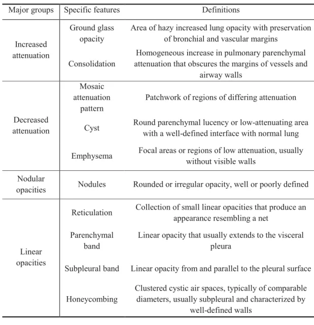
The limitations related to the use TR jet velocity for estimation of PAP greatly encourage the development of new echocardiographic measures for a non-invasive diagnostic approach of PH. The use of multiple echocardiographic imaging modalities - allowing the identification of
via Doppler flow interrogation is not possible - is highly encouraged in dogs (Kellihan and Stepien, 2010). Consequently, several authors investigated two-dimensional and Doppler echocardiographic indices that may help to predict PH. Among them, Schober and associates (2006) investigated whether systolic time intervals of the pulmonary flow (Fig. 1) may be predictive of PH in a cohort of 41 healthy WHWTs and 45 WHWTs affected with CIPF. Among the diseased WHWTs, 7 (15.5%) of them had no evidence of Doppler-derived systolic PH (tricuspid regurgitant jet maximal velocity VmaxTR < 3.1 m/s), 18 (40%) were of unknown status with regard to PAP given the absence of any measurable tricuspid regurgitant jet, and 20 (44.4%) had systolic PH (VmaxTR 3.1 m/s). A shortening of the acceleration time (AT) and a decrease of the acceleration to ejection time (AT:ET) of the pulmonary flow was identified in CIPF WHWTs with PH in comparison with healthy WHWTs. They also determined cut-off values for both measurements to predict the presence of PH (estimated systolic PAP 45 mmHg) as 0.31 for AT:ET (Se 73%, Sp 87%) and 58 ms for AT (Se 88%, Sp 80%).
Fig. 1: Normal Doppler flow pattern of pulmonary artery to demonstrate measurement of acceleration time (AT) and ejection time (ET).
(Schober and Baade, 2006)
Other indirect non-invasive echocardiographic indices that may help to predict PH have been described in dogs suffering from PH from other origins (mainly left heart disease and various respiratory disorders). These include the diameter of the main pulmonary artery in relation with the diameter of the aorta (MPA/Ao) (Serres et al., 2007), the Tei index representative of global right ventricular function (Baumwart et al., 2005; Paradies et al., 2014),
tissue Doppler imaging assessment of systolic and diastolic right ventricular function (Serres et al., 2007; Visser et al., 2014), tricuspid annular plane systolic excursion (Pariaut et al., 2012), and the right pulmonary artery distensibility index (RPAD Index) (Venco et al., 2014; Visser et al., 2016). However, none of these measures have already been studied specifically in WHWTs affected with CIPF, and their diagnostic utility for PH determination in that particular case is presently unknown.
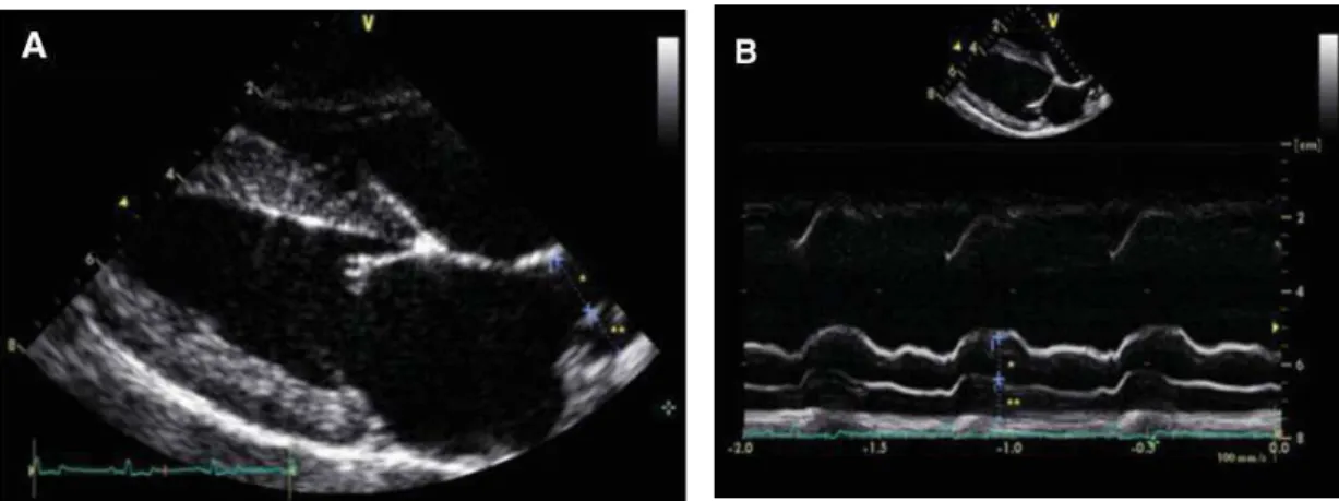
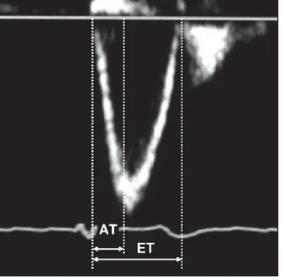
MPA/Ao is classically measured in the right parasternal basilar short axis view; values exceeding 0.98 being indicative of MPA enlargement suggestive of PH (systolic PAP 30mmHg) with sensitivity and specificity of 73% and 76%, respectively (Serres et al., 2007). The RPAD Index has been most recently studied in both heartworm infected dogs and dogs with PH from different origins. Reportedly, in heartworm infected dogs, the RPAD index, calculated as the systolic diameter minus diastolic diameter, divided by systolic diameter of the pulmonary artery imaged in M-mode (Fig. 2), can predict the presence of PH (systolic PAP 30mmHg) at a value < 35% and was strongly correlated with the PAP obtained invasively by RHC (Fig. 3) (Venco et al., 2014).
Fig. 2: M-mode echocardiography of the pulmonary artery. Systolic size is measured at the maximum diameter (usually at the T wave) and diastolic size is measured at its smallest
diameter (usually at the Q wave). (Venco et al., 2014)
Fig. 3: Correlation between RPAD Index and systolic PAP obtained by RHC. (Venco et al., 2014)
The RPAD Index has also been recently investigated by Visser and collaborators (2016) in a different echocardiographic view than previously described (Fig. 4) in dogs with PH of different origins. They demonstrated that the RPAD Index displayed the strongest correlation with TR pressure gradient (r = -0.90, P < 0.001) in comparison with AT (r = -0.69, P < 0.001), AT:ET (r = -0.68, P < 0.001) and MPA/Ao (r = 0.78, P < 0.001) and may predict a TR pressure gradient > 50 mmHg at a cut-off value of < 29.5% with a sensitivity and specificity of 84% and 95% respectively (Visser et al., 2016).
Fig. 4: Representative measurement and calculation of RPAD Index. (Visser et al., 2016)
Some authors were also interested in the use of blood biomarkers in predicting the presence of PH in dogs. N-terminal pro-brain natriuretic peptide (NT-proBNP) concentration was studied in a canine model of chronic embolic PH (Hori et al., 2012) and in dogs with
precapillary PH (Kellihan et al., 2011). Both studies yielded the same conclusions that NT-proBNP increases in case of severe PH, but lack sensitivity in cases of mild to moderate PH. 1.2.5. Diagnostic imaging
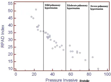
Thoracic X-rays findings described in WHWTs affected with CIPF are not specific and varied from generalised bronchointerstitial pattern (Fig. 5) to only interstitial, predominantly bronchial or patchy alveolar opacities, findings that might be observed in a diversity of other pulmonary diseases (Heikkila-Laurila and Rajamaki, 2014). The main reason for taking thoracic radiographs is to rule out other lung diseases such as neoplasia (Heikkila-Laurila and Rajamaki, 2014). An increased size of the heart shadow, especially in the dorso-ventral view resulting in a reverse-D shaped cardiac silhouette and an enlarged main pulmonary artery, may indicate the presence of PH and further encourages the realisation of a Doppler echocardiography (Heikkila-Laurila and Rajamaki, 2014).
Fig. 5: Right lateral (A) and ventro-dorsal (B) thoracic radiographs of a 10-year-old WHWT showing redundant tracheal membrane and a severe generalised bronchointerstitial pattern with main pulmonary artery enlargement (<) and pleural fissure line (v) between right lung
lobes.
(Roels and Clercx, 2015)
A B
More precise information about the lung parenchyma is obtained with the use of thoracic high-resolution computed tomography (T-HRCT), a diagnostic imaging device which become widely available in veterinary practice. T-HRCT appears to be the complementary examination of choice in the diagnostic approach of CIPF as it allows for the investigation of the lung in a better spatial resolution. The only negative point is that T-HRCT classically requires general anaesthesia, which might not be suitable in severely affected dogs due to the increased anaesthetic risk (Heikkila-Laurila and Rajamaki, 2014). The initial description of T-HRCT features of CIPF was made in 2005 in a cohort of 10 dogs (8 WHWTs and 2 Cairn terriers)
study, CIPF dogs were arbitrary classified as having mild (1 dog), moderate (7 dogs) or severe (2 dogs) disease based on clinical signs and thoracic radiographs appearance. They described patchily distributed abnormalities throughout the lung fields with ground-glass opacities (GGO) observed in all dogs, linear and reticular opacities in moderately and severely affected dogs, and bronchiectasis and honeycombing in severe cases. In 2011, two other descriptive studies were published independently using the same classification system. The first study described lesions observed in 6 WHWTs affected with CIPF and 12 controls (Heikkila et al., 2011). The dorso-caudal lung lobes were reported as predilection sites for the lesions, with GGO described in all affected dogs; parenchymal bands, consolidations and traction bronchiectasis in 4 dogs; and honeycombing in 1 dog. In the control population, 8 dogs had 1 or 2 focal lesions of unspecified origin. The second study published in 2011 cited succinctly T-HRCT changes observed in 22 WHWTs affected with CIPF. Bronchial changes were present in the half of the study population (Corcoran et al., 2011). Those 3 studies, collectively, suggest that T-HRCT findings observed in WHWTs affected with CIPF include GGO, parenchymal bands, subpleural bands, subpleural interstitial thickening, peribronchovascular interstitial thickening, consolidation, bronchial thickening, bronchiectasis and honeycombing (Corcoran et al., 2011; Heikkila et al., 2011; Johnson et al., 2005). Imaging findings in an individual dog are typically a combination of one or more of the above mentioned features with distribution of the lesions varying from patchily distributed (Johnson et al., 2005) to predominantly localised in the dorso-caudal lung lobes (Heikkila et al., 2011).
1.2.6. Bronchoscopy and bronchoalveolar lavage fluid analysis
Bronchial abnormalities have been reported in a high proportion of WHWTs affected with CIPF (Corcoran et al., 2011; Heikkila et al., 2011). However, whether those bronchial abnormalities occur secondary to the underlying interstitial lung disease or are an independent phenomenon remains to be elucidated. Age may partially contribute to abnormalities encountered given that some bronchial changes such as irregular bronchial mucosa, prominent mucosal vessels and bronchiectasis have been described in old dogs (Mercier et al., 2011). Common bronchoscopic findings detected in WHWTs affected with CIPF include tracheal collapse, bronchial mucosal irregularity, increased amount of bronchial mucus, bronchomalacia, dynamic airways collapse and bronchiectasis (Heikkila-Laurila and Rajamaki, 2014). Those bronchial changes are not specific for CIPF as they are frequently observed in other pathological respiratory conditions (Heikkila-Laurila and Rajamaki, 2014). Analysis of the bronchoalveolar lavage fluid (BALF) usually shows a moderate increase in the total cellular count due to increased numbers of macrophages, neutrophils, and mast cells (Heikkila-Laurila
and Rajamaki, 2014). The major goal to perform a bronchoalveolar lavage is therefore to exclude a parasitic or a bacterial infection from the differential diagnosis.
1.2.7. Histopathological features
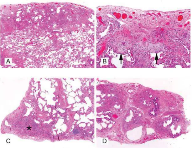
Histopathological examination of parenchymal lung tissue provides a definitive diagnosis of CIPF in the majority of cases, but lung biopsies are seldom taken due to expense and the need for invasive surgery (Heikkila and Rajamäki, 2014). Consequently, the diagnosis of CIPF is classically based on anamnestic information, findings in clinical examination and diagnostic imaging, and exclusion of other respiratory diseases. The first description of histopathological characteristics of CIPF was made in 1999a by Corcoran and collaborators in 4 WHWTs affected with CIPF. They described a multifocal to regional deposition of interstitial extracellular matrix, multifocal epithelial squamous metaplasia, and an increased number of alveolar and circulating macrophages as well as occasional lymphocytes and plasma cells. In 2005, Norris and collaborators attempted to compare histopathological characteristics of CIPF with those described in humans, such as a usual interstitial pneumonia (UIP) pattern, typical histological pattern of IPF in humans. Collagen and elastin stains were used in concert with immunohistochemical characterisation of collagen subtypes and smooth muscle actin-positive cells in 6 WHWTs affected with CIPF. Histopathological lesions observed in their study ranged from generalised deposition of septal collagen with a scant inflammatory cell infiltrate in some cases, to marked septal thickening by collagen, a more abundant cellular infiltrate of predominantly macrophages and lymphocytes and hyperplasia of alveolar-lining type II pneumocytes in more severely affected dogs. Based on results of Mason’s trichrome and immunohistochemical stains, the extracellular matrix was shown to be composed of a mixture of type-I and –III collagens with type-III collagen being the most prominent type. Elastin does not appear to be altered in CIPF. Fibroblastic foci were not observed, and smooth muscle actin immunoreactivity was limited to individual cells in the septa, a feature which differs from fibrosis in human UIP. Recent work by Syrja and collaborators (2013) further characterised CIPF histopathological lesions and compared them systematically to the human UIP and non-specific interstitial pneumonia (NSIP) histopathological patterns. All 18 WHWTs affected with CIPF included in their study were displaying a mild to moderate diffuse mature fibrosis of the alveolar walls, in addition to concentric collagen deposition around pre- and post-capillary small vessels (Fig. 6). Multifocal subpleural or peribronchiolar areas of more severe and less mature interstitial fibrosis accompanied by a diffuse presence of myofibroblasts was also observed in 60% of affected dogs. Fibroblastic foci were not observed. Hyperplastic type II pneumocytes were observed in the majority of dogs, as well as epithelial pseudostratification
accumulations were also observed in areas of severe interstitial fibrotic changes, while alveolar proteinosis (alveolar luminal homogenous dense eosinophilic material) was randomly distributed throughout the lungs in a multifocal pattern and not co-localised with areas of more severe interstitial lesions. The inflammatory changes in lungs of WHWTs affected with CIPF consisted of mild to moderate lymphoplasmacytic inflammation with few macrophages. In humans, the UIP pattern is characterised by areas of marked interstitial pulmonary fibrosis and honeycombing, distortion of the alveolar architecture in a patchy, often subpleural and paraseptal pattern (Raghu et al., 2011). On the other hand, the NSIP pattern, encountered in collagen vascular disease, hypersensitivity pneumonitis, drug-induced pneumonia and pulmonary infection, is characterised by a relative diffuse and uniform fibrosing interstitial disease with no or minimal honeycombing and rare or absent fibroblastic foci (Katzenstein et al., 2008). Consequently, Syrja and collaborators (2013) concluded that CIPF in WHWTs shares features of both human NSIP and UIP patterns, given that the diffuse interstitial lesions present in all affected dogs is in favour of NSIP, while the presence of more severely affected areas of progressive fibrosis tend to correlate with the UIP pattern (Table 3).
Fig. 6: Histopathological characteristics of CIPF in WHWTs: transition from mild diffuse fibrosis on the left, to a focus of accentuated disease, with severe interstitial fibrosis on the right. HE .Bar, 200µm. Inset: type-II pneumocytes hyperplasia and squamous metaplasia of
the alveolar epithelium. (Syrja et al., 2013)
Table 3: Comparison between the main histological criteria required for the diagnoses UIP or NSIP in man and the findings in CIPF in WHWTs.
(Syrja et al., 2013)
1.2.8. Long-term outcome and prognostic indicators
The clinical course, long-term outcomes and prognostic factors of CIPF were assessed by Lilja-Maula and collaborators (2014b) in a cohort of 15 WHWTs affected with CIPF and 10 healthy WHWTs. WHWTs affected with CIPF were prospectively followed every 3- to 6-months until death or study end-point, while the majority of the control WHWT were followed by phone calls to the owners. The causes of death were divided into CIPF-related (defined as euthanasia because of acute dyspnoea or severe progression of respiratory symptoms) or non-CIPF-related. They showed that CIPF has significant negative impact on life expectancy in WHWTs (Fig. 7). They found a survival time from onset of clinical signs in the CIPF-related death group ranging from 2 months to 4.3 years with a median of 2.7 years, indicating that CIPF in WHWTs may have a rapid or slow disease progression. No significant prognostic factors at diagnostic were identified among the chosen variables including arterial pO2, arterial pCO2, p(A-a)O2, serum endothelin-1 concentrations, severity of changes in thoracic X-rays and HRCT Hounsfield unit values. However, a slight indication of high arterial pO2 values having a protective effect on survival and high p(A-a)O2 being a risk factor were noted (statistical significance set at P=0.10 rather than P=0.05). A decline in arterial pO2 values between the first and the last measurement was noted in WHWTs that died of CIPF-related causes. However, in some dogs, temporary increases in arterial oxygenation were recorded, and some owners described a temporal improvement in clinical signs. Consequently, whether repeated measurements of arterial blood gas values over time may serve as a useful tool for evaluating disease progression requires further investigations.
Fig. 7: Kaplan-Meier survival curves for all-cause survival of WHWTs with CIPF (dotted line) and control WHWTs (solid line) from study inclusion. Censored animals (WHWTs alive
at study endpoint) are presented as circles. (Lilja-Maula et al., 2014b)
1.3. Understanding the pathogenesis
The pathogenic mechanisms behind CIPF are not yet understood. The strong predisposition of the WHWT breed to CIPF raises suspicion for a genetic background (Heikkila-Laurila and Rajamaki, 2014). However, other triggering events are probably involved in the course of the disease given that not all dogs from the WHWT breed develop the disease at an advanced age. CIPF probably results from a complex dialogue between genetic and environmental factors (Heikkila-Laurila and Rajamaki, 2014). Unfortunately, no epidemiologic studies have been performed until now and investigation of the genetic background has just started at the University of Helsinki. Nevertheless, several studies, briefly reported in the next sections, were conducted to provide clues on underlying fibrotic mechanisms while others researched the use of biomarkers to differentiate CIPF from other chronic lower respiratory diseases.
1.3.1. Surfactant protein C mutation
In humans, mutations in the gene encoding surfactant protein-C was associated with rare family cases of pulmonary fibrosis (Devine and Garcia, 2012). In 2009, Eriksson and collaborators analysed by Western blots immunoreactivity the presence of surfactant proteins-B and –C in the proteins-BALF from 3 dogs affected with CIPF (2 WHWTs and 1 Tibetan terrier) and 5 controls of various breeds including 1 WHWT. In the Tibetan terrier CIPF dog, no surfactant protein-C was found in the BALF, although immunoreactivity for surfactant protein-B was detected. In the 2 WHWTs affected with CIPF and the 5 controls both surfactant proteins-B
and –C were detected in the BALF. Amplification and sequencing of the surfactant protein-C and its exon for 2 CIPF cases, including the Tibetan terrier, and for 1 control revealed no mutations that influence the expression of surfactant protein-C. Consequently, the absence of surfactant protein-C in the BALF of the Tibetan terrier remains unexplained and requires further investigations in a larger group of dogs with breed-matched controls.
1.3.2. Endothelin-1
Endothelin-1 (ET-1), a vasoactive peptide with pro-inflammatory and pro-fibrotic properties, has been identified as a mediator of IPF in humans through its contribution in fibroblasts activation, proliferation and differentiation into myofibroblasts (Ross et al., 2010; Swigris and Brown, 2010). Higher concentrations of ET-1 have been detected in lung tissue, blood and BALF from people with IPF (Ross et al., 2010; Swigris and Brown, 2010). In 2011, Krafft and collaborators investigated whether concentrations of serum and BALF ET-1 would be higher in dogs affected with CIPF compared with healthy dogs and dogs affected with other chronic lower respiratory diseases, namely eosinophilic bronchopneumopathy (EBP) and chronic bronchitis (CB). They included 2 groups of control dogs (13 healthy WHWTs and 9 healthy experimental Beagle dogs) and 3 groups of dogs with chronic lower respiratory disease (12 dogs affected with CIPF including 11 WHWTs and 1 Scottish terrier, 10 dogs of various breeds affected with CB, and 6 dogs of various breeds affected with EBP). They found that serum ET-1 concentrations were significantly higher in dogs affected with CIPF compared with healthy Beagles, healthy WHWTs, dogs affected with EBP, and dogs affected with CB (Fig. 8A). BALF ET-1 concentrations were only measurable in dogs affected with CIPF, whereas they were below the kit detection limit in healthy WHWTs and in dogs affected with CB (Fig. 8B). Dogs in the CIPF group were all from the WHWT breed expect for one dog, a Scottish terrier, while dogs in the EBP and CB groups were from various breed but non-WHWT breeds. Therefore, differences observed between pathological groups were potentially due to a physiological breed variation. However, the absence of difference between healthy Beagles and healthy WHWTs suggested that this breed effect is null or minimal and that the differences observed between CIPF, EBP and CB are related to the disease process. Elevated ET-1 may potentially relate to the presence of PH. Indeed, in human IPF patients, higher plasma ET-1 concentrations were found to be associated with higher pulmonary arterial pressures (Ventetuolo et al., 2012). However, the study in dogs showed no difference in serum ET-1 concentrations between dogs affected with CIPF that had or did not have indirect signs of PH on echocardiography (right-sided cardiac hypertrophy, septal flattening or dilatation of pulmonary trunk), and highest serum ET-1 concentrations were found in dogs with normal
![Fig. 16: Numbers of publications listed per year with the search [Idiopathic pulmonary fibrosis] in MEDLINE (PubMed) database](https://thumb-eu.123doks.com/thumbv2/123doknet/6037173.151219/49.892.254.639.689.954/numbers-publications-idiopathic-pulmonary-fibrosis-medline-pubmed-database.webp)