REVIEW ARTICLE
The thymic education of developing T cells in self
neuroendocrine principles
V.
Geenen, F. Robert, H. Martens, O. Oe Groote*, and P. Franchimont
Institute of Pathology B-23, Laboratory of Radio-Immunology, University Hospital of Liege-Sart Tilman, B-4000 Liege, *Medgenix Diagnostics, B-6220 Fleurus, Belgium
INTRODUGTION
The thymus is now recognized as the primary lym-phoid organ involved in the differentiation of T Iym-phocytes (1). In the human species, its physiolog-ical role seems to be mainly exerted along fetal de-velopment since congenital thymic hypo- or aplasia (Iike in the Di George's syndrome) is followed by a profound impairment of T cell functions, leading to the premature death of the child. On the contrary, ablation of the human thymus after birth is not fol-lowed by significant immune deficiency. T cell on-togeny is a highly complex process which can be divided in several distinct phases. Firstly, T cell pre-cursors originating from the fetal liver and bone marrow migrate through separate waves into the thymic rudiment, most probably under the influence of specific chemoattractants (2). Secondly, within the thymus, immature T cells undergo rearrange-me nt of the genes coding for the chains of their re-ceptor for antigen (TcR), and express differentia-tion markers (CDs) at their surface. These pro-cesses are not purely automatic, but are intimately controlled by the thymic microenvironment (ep-ithelial cells, macrophages, dendritic or interdigi-tating cells, and fibrobiasts) (3). Thirdly and impor-tantly, the thymus is the site for the negative selec-tion of self reactive T cells and this mechanism is basically responsible for the induction of T cell self tolerance (self/non-self discrimination) (4-6).Finally, the integration by target pre-T cells of the various differentiative signals (adhesion molecules, growth factors, and cytokines (7)) encountered in the thy-mus leads to the positive selection of mature im-munocompetent T Iymphocytes (8).
Key-words.· Cryptocrine signalling. thymus, neuroendocrine seil antigen, antigen recognition. seil tolerance, autoimmunity, positive selection.
Correspondence' Vincent Geenen, MD., PhD., Research Associate 01
Belgian FNRS, Laboratory 01 Radio-Immunology B-23, Neuroendocrin-Immunology Uni\, University Hospital 01 Liege-Sart Tilman, B-4000 Liege, Belgium.
GELL-GELL INTERAGTIONS IN THE THYMUS
Thymic epithelial cells (TEC) constitute the major component of the environment controlling T cell dif-ferentiation (9). They have a mixed, ectodermal and endodermal, origin (10, 11). The endoderm derives from the third pharyngeal pouch, while the ecto-derm originates from the cervical and from the third branchial cleft. The majority of cortical TEC seem to derive from the endoderm, whereas the ecto-derm would contribute to the subcapsular cortical epithelium, some stellate cortical cells, the medullary epithelium and the Hassall's corpuscles. Since Le Douarin's pioneering studies (12), it is weil acknowledged that the cephalic neural crest exerts an important influence upon thymic organogene-sis, even if the exact contribution of neural crest mi-grants in the thymus is still discussed. TEC of the subcapsular cortex and medulla also share com-mon immunocytochemical characteristics, like their labelling with antisera directed against thymic pep-tides (anti-thymosin a1 (13) and anti-thymulin (14)) or with monoclonal antibody (mAb) A2B5 (wh ich reacts with complex gangliosides found in the brain and the diffuse neuroendocrine system) (15). We have also demonstrated that the same subsets of TEC contain neurohypophysial (NHP)-related pep-tides and immunoreactive (ir) interleukin-1 ß (IL-1 ß) (16, 17). Thymic nurse cells (TNC) are large ep-ithelial cells which can be isolated by 1 9 sedimen-tation from murine thymuses after enzymatic di-gestion (18), and which have been identified in situ in the thymic subcapsular and outer cortex (19,20). TNC were also shown to synthetize NHP-related peptides, and to express the same immunopheno-type as cellular elements belonging to the diffuse neuroendocrine system (21).
Even if the thymus has been considered for many years to be an intrinsic component of the endocrine system, the pattern of secretion in TEC is rather dif-ferent from the classical endocrine secretory pro-cess (22). We have also demonstrated that the c1as-sical scheme of neurosecretion established for the
hypothalamo-NHP axis could not be applied to the thymic cell-to-cell signalling. The concept of cryp-tocrine signalling (23) was recently introduced by
J.w.
Funder to describe the exchange of informa-tions in secluded microenvironments created by large epithelial cells enclosing migratory develop-ing cells. Two examples at least of cryptocrine sig-nalling exist in the human body: one in the thymus, between TEC/fNC and immature T cells (pre-T cells or thymocytes); another in the testis, between Sertoli cells and spermatids. Interestingly, Sertoli cells have already been described in 1899 as "nurse" cells es-sential for the germinal cell and the process of sper-matogenesis (24). In the thymus, the mitotic index of TNC-engulfed thymocytes is rather high and recent studies have evidenced that TEC/fNC can present antigen+self major histocompatibility complex (MHC) (25). Consequently, it seems highly proba-ble that TNC display the biochemical machinery to actively intervene in the selective process of T cells (26). The tolerogenic properties of TEC have also been illustrated in several experimental paradigms (27-29). On the basis of our observations, we have proposed that thymic NHP-related peptides could serve as functional signals in cryptocrine commu-nication between TEC/fNC and pre-T cells (30). The analogy between TNC and testicular Sertoli cells is further supported by the recent observation of the expression of an oxytocin (OT) gene in cattle Sertoli cells (31), as weil as by the immuno-Iabelling of hu-man Sertoli cells with mAbs A2B5 and 033 (direct-ed against the cyclic part of OT (32)) (unpublish(direct-ed observations). If the molecular mechanisms under-Iying the intrathymic expression of NHP-like pep-tides remain to be further characterized, separate studies have confirmed the presence in the thymus of immunoreactive and bioactive OT-like peptide (33-35). Furthermore, the expression of functional NHP peptide receptors by rat thymocytes (36) and by murine immature and differentiated T cells is a prerequisite in favor of this working hypothesis (37). Since the intrathymic concentrations of NHP-relat-ed signals are in good concordance with the high affinity Kd of NHP receptors expressed by pre-T cells, the physico-biochemical conditions are en-countered to render effective in vive a thymic cryp-tocrine signalling through NHP peptides and their cognate receptors. This is obviously not the case for circulating neurohormones OT and VP, due to their low blood concentrations. The pre-T cell NHP receptors were shown to mediate mitogenic prop-erties of NHP-related signals upon human and murine pre-T cells. Their transductory properties in-volve a T cell phosphoinositide breakdown and were similar to those described for other V1/0Tre-ceptors. This effect was specifically inhibited by a V1 antagonist in pre-T cells and by an OT antagonist in cytotoxic T cells (37). This shift in the type of T cell NHP receptor may suggest a dynamic phe-nomenon of molecular maturation parallel to the progress of T cells in their differentiative pro-gramme. As evidenced by our previous radiobind-ing studies (38), the V1 receptor expressed by pre-T cells is probably an immune-specific V1 subtype different from the recently cloned V1 a receptor (39). In the murine species, other authors have also re-ported the expression of a novel V1 subtype by splenic cytotoxic Iymphocytes (40). Of course, the TNC-derived NHP signals are not the only growth factors implicated in T cell differentiation, but their mitogenic properties evidenced on pre-T cells may provide an explanation to the high mitotic index of TNC-engulfed thymocytes. They also demonstrate an implication of thymic NHP-related peptides in the process of T cell positive selection, in accordance with previous observations (41). Molecular distur-bances of cryptocrine signalling might also inter-vene in pathologic states, either at the level of NHP signal (thymoma are the third cause of inappropriate VP secretion (42, 43)), or at the level of NHP recep-tor (with potential implication in T cell lymphoma oncogenesis). Interestingly, the intervention of TNC in leukemogenic process have al ready been demonstrated before (44). The expression of NHP peptide receptors by immune cells also opens nov-el immunomodulatory strategies through the molec-ular design of neuropeptide immune-specific re-ceptor antagonists or agonists. We recently de-scribed a significant inhibition by novel OT non-peptide antagonists (Merck Sharp & Dohme Research Laboratories, West Point) of IL-1 ß, IL-6 and TNFa productions in human whole blood cell cultures stimulated by anti-CD3 mAb (45). This in-hibition was significantly more important in whole blood cell cultures derived from human female vol-unteers, suggesting an influence of the gonadal steroid hormonal environment. The future knowl-edge of the precise NHP receptor subtypes ex-pressed by immunocompetent cell populations should help computer modelling and molecular en-gineering of speciflc immunomodulatory com-pounds based on the basal structure of these OT cyclic hexapeptide antagonists. This approach could lead to selective immunotherapy in cruciallife periods, such as the postpartum, during which an immune disequilibrium is determined byan increase of the oestrogen/progesterone ratio and by a rise in immunostimulatory lactogenic hormones prolactin and OT.
chemical information mayaiso be directed from the engulfed developing cells to the large epithelial "nursing" cell. This seems to be the case with nerve growth factor (NGF) since this was shown to be synthesized by male germ cells (46), whereas NGF receptor was shown to be expressed by Sertoli cells (47). An analogous situation may exist within thymic microenvironment as NGF receptor mRNA was also detected in chick and rat thymus extracts (48), but the precise source of the thymic NGF sig-nal remains to be further defined. Of high relevant interest with regard to the role of NGF in thymic physiology is the recent observation that this neu-rotrophic factor markedly enhances the molecular and phenotypic neuronal-like features of cultured murineTEC (49).
T GELL REGOGNlTlON OF SELF NEUROENDOGRINE FUNGTIONS
Neurohypophysial (NHP) peptide family
Through the use of several well-characterized poly-clonal and mAbs, we have defined the immu-nodominant epitopes representative of the NHP peptide family expressed by TEC{TNC of several animal species (50). The first epitope is located, at least partially, in the six amino acid cyclic part shared by OT and by vasotocin (VT), the ancestral peptide precursor of the NHP family constituted by the cyclic part of OT and the three amino acid lat-eral chain of VP. The second epitope is present in the central "constant" part of the neurophysin do-main. This part is encoded by the second exon of NHP genes and show a high degree of conserva-tion (> 90%) throughout the evoluconserva-tion of species whichever the nonapeptide associated (51). In the overall evolution of intercellular communication, the cryptocrine stage evidenced in the thymus intro-duced an obligatory step which is the recognition of the self molecular structure by developing T cells. This would logically follow the presentation by thymic MHC molecules of self epitopes represen-tative of larger proteins. The size of the MHC groove allows the binding of peptide sequences ranging from a minimum of five (52) to seventeen amino acids in length (53). If demonstrated, the presen-tation by thymic MHC molecules of the NHP self epitopes could therefore induce the central immune tolerance of hypothalamo-NHP functions. This tolerogenic effect would result from the clonal dele-tion of highly reactive T cells harboring a random-Iy rearranged TcR specific for the association MHC/self NHP epitope(s). The large involvement of OT-like peptides at different levels of the repro-ductive processes implies that they are strongly
tol-erated by the immune system. This high tolerance of OT lineage protects it from a potential autoim-mune aggression and would therefore contribute to the preservation of the species. Since VP differs from NHP self epitope by one single amino acid in its cyclic part, it is less "protected" from autoim-mune process. Indeed, some authors have previ-ously reported "idiopathic" diabetes insipidus re-sulting from autoimmune aggression directed against hypothalamic magnocellular neurones (54). In the same view, experimental breakdown of NHP immune tolerance by active immunization against VP was also shown to induce inflammatory lesions in the hypothalamo-NHP axis (55). It is also inter-esting to note that the frequence and the titers of antisera against VP are usually higher than those of anti sera developed against OT or VT.
So, the deep investigation of our original observa-tions (56) led us to shift from the classical neu-rosecretion model to the novel pathways underlying cryptocrine cell-to-cell signalling and self antigen presentation by MHC molecules. At the molecular level, the "neuropeptide" model was replaced by the working concept of NHP "self peptide" (57, 58). As discussed before, thymic NHP-related self pep-tides exert a dual role in T cell differentiation ac-cording to the type of intercellular dialogue in wh ich they are engaged (cryptocrine signalling or NHP self antigen presentation by thymic MHC).
Tachykinin (TK) peptide family
This model of a dual physiological role for thymic self peptides also applies to this family whose in-trathymic expression was investigated in collabo-ration with the Laboratory of Molecular Neurobiology at the Karolinska Institute (Stockholm). Neurokinin A (NKA) is the TK peptide encoded by preprotachykinin-A (PPT-A) gene in the rat thymus (59) and is thus the first member of the TK family encountered by developing T cells. Mitogenic activities have been described for NKA and physalaemin upon murine cultured thymocytes (IL-1-like bioactivity), while substance P (SP) and other TK-related peptides did not exert any signif-icant effect (60). These observations strongly sup-port the involvement of thymic NKA in T cell positive selection. Since NKA shares with other TKs the same C-terminal immunodominant epitope, it might represent the tolerogenic TK peptide that is ex-pressed in the thymus and is facing the develop-ing T cell system. The other TK peptide SP detect-ed in rat and human thymic extracts is associatdetect-ed with sensory nerve fibers and seems to be impli-cated in the regulation of thymic blood flow (61, 62). This does not exclude that SP exerts peripheral
proinflammatory properties, however through spe-cific immune TK receptors since potential TK au-toreactive T cells would have been deleted in the thymus.
Insulin peptide family
Insulin-like growth factors (IGFs) were also identi-fied within the human fetal thymus, both at the pep-tide and mRNA levels (63,64).
However, these studies could not really discrimi-nate between IGF-I or IGF-II, and the thymic sub-cellular localization remains to be further defined because of the discrepancies observed between the localizations of IGF mRNAs and immunoreac-tivities. The fact that thymic IGFs are implicated as accessory signals in T cell positive selection is in-directly suggested by the mitogenic properties of IGF-I on pre-T thymic lymphoma cells (65). The ac-tivation of human T Iymphocytes by anti-CD3 also leads to their expression of IGF-I and IGF-II recep-tors (66). Due to the high homology (± 60%) of proinsulin and IGFs primary amino acid sequences, the tolerance of pancreatic endocrine function could result from the intrathymic deletion of T cells harboring a rearranged TcR specific of the associ-ation thymic MHC/insulin family self epitopes. The IGF-II region on chromosome 11 p seems also to be involved in HLA-DR4-dependent diabetes sus-ceptibility by a still undefined molecular mechanism (67). So, further characterization of the precise in-sulin/IGF-derived self immunodominant epitope ex-pressed in the human thymus should lead to the identification of the primary self antigen implicated in the autoimmune cascade leading to overt insulin-dependent diabetes. In another connection, sev-eral antigenic markers are shared between TEC/TNC, and pancreatic islet ß-cells, among which A2B5 and HISL (68). This cellular phenotyp-ical relationships may suggest a common embry-onic origin in the neurectoderm or in the neural crest (69).
Other members of the thymic peptide repertoire
Several neuroendocrine peptides have been de-tected in the thymus from various species, at the peptide or the mRNA level (70-75). In two of these studies, neuropeptides were ultrastructurally de-tected in perinuclear space, vesicular structures and rough endoplasmic reticulum, but not in clas-sical secretory granules. Since an atrial natriuretic peptide (ANP) signal is found in thymic stroma and that ANP receptors are also expressed by rat thy-mocytes, a functional accessory signalling through this peptide should also be further investigated in the process of T cell development (76). An
au-tocrine/paracrine signalling through luteinizing hor-mone-releasing factor (LHRH) seems also to inter-vene in the modulation of the T cell differentiation pathways (77, 78). Parathormone-related peptide (PTH-RP), as weil as calcitonin gene-related pep-tide (CGRP) were also recently found to be includ-ed in the thymic self peptide repertoire (79, 80). Both of these polypeptides shared common amino acid sequences with the hormonal members (PTH and CT) of their corresponding family and could therefore induce T cell tolerance of PTH- and CT-mediated endocrine functions.
NEURAL AND ENDOCRINE CONTROLS OF THYMIC FUNCTION
Like all peripheral organs, the thymus receives au-tonomic sympathetic and parasympathetic inner-vation (81, 82). A number of neuropeptides have also been identified in sensory nerve fibers of the thymus (83). At the present time, it is however still difficult to establish whether these innervations ex-ert a control upon thymic stromal cells, thymic blood flow or may directly act upon differentiating T cells.
The thymic physiological role is also closely influ-enced by the endocrine environment, in particular by thyroid (84) and antehypophysial hormones (85), as weil as by gonadal steroids and glucocorticoids (86, 88). There is increasing evidence that these hormonal influences may be relayed by intrathymic factors (i.e. the trophic role of growth hormone up-on the thymus (89) is most probably mediated by local actions of thymic IGF-I). To our knowledge, the existence of a specific antehypophysial thy-motrophin has never been investigated but this question deserves further attention given the inti-mate relationships between the thymus and the pi-tuitary (90).
CONCLUSIONS
The ontogenetical and phylogenetical evolutions of the endocrine system have seen the emergence of various forms of intercellular communication, from the most primitive stages of autocrine signalling (when a cell is alone) and adhesion (wh ich follows the division of one cell) to the most complex neu-rocrine networks underlying cognitive processes in the central nervous system. Parallel to the succes-sive structural levels of the evolution of intercellu-lar communication, it appeared some hierarchy in the genomic organisation responsible for the trans-fer of biochemical information between cell popu-lations (91). In recent years, new members of
en-docrine families were identified through the use of molecular techniques. These factors were not clas-sical "hormones" vehiculated by bloodstream, but rather exerted local growth-promoting "paracrine-autocrine" activities. It also appeared that these tis-sue peptide growth factors were predominantly ex-pressed during fetal life and were playing an im-portant role in embryo development and organo-genesis. Their etiopathogenic involvement in neo-plasic processes is more and more documented, and their gene overexpression is sometimes fol-lowed by a massive release in bloodflow. A para-neoplasic syndrome may then follow, due to the usual cross-reactivity of these tissue growth-factors with the relative hormone receptors. On the other side, the immune system has evolved, the primary characteristic of which is the protection of the self molecular structure against foreign infectious in-vaders (non-self). The thymus is one crucial privi-leged site for which appears as an obligatory step in the parallel evolutions of neuroendocrine and im-mune systems: the education of T cells to discrim-inate self antigens and foreign (non-self) antigens. In the line of that highly simplified perspective, the incidental breakdown of self immune tolerance as seen in autoimmune pathologies may be consid-ered as the global tribute paid by one given species for its protection against infectious non-self antigens.
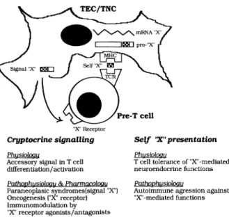
In the actual stage of our research on the neuroen-docrine self peptide repertoire expressed in the
thy-"X" Receptor
Cryptocrine signalling PhusiDloou
Aeeessory signal in T eell differen tiation/ aetivation PathpPhusiDloou & PhanruJcq!oqu Paraneoplasie syndromes(slgnal "X") Oneogenesls ("X" reeeptor) Immunomodulation by "X" receptor agonists/antagonlsts Self 'X" presentation PhusiDloou
T eell toleranee of "X"-mediated
neuroendocrine functions
PathophusiDlogu
Autoimmune agression agalnst "X" -mediated functions
Fig. 1 -Thymic education of T cells in self neuroendocrine prin-ciples.· model of the dual physiological role of thymic self pep-tides in T cell differentiation.
mus, this primary lymphoid organ appears more and more as a highly specialized school for the education of T cells in self neuroendocrine principles (Fig. 1). In the human species,that school is mainly working du ring fetal development and its highest education-al degree is the apoptosis of the autoreactive T cells (92). A failure in the educational programme would result in the emergence of T cells with autoreactivity directed against neuroendocrine self antigen(s), and this point could play an important pathogenetic role in endocrine autoimmune diseases. Of course, the appearance of autoreactive T cells is not the only etiopathogenic factor in autoimmunity (93) and the thymic T cell negative selection is not a perfect mechanism (94). Nevertheless, it is the only one for which the precise description of the underlying molecular mechanisms should allow novel diagnos-tic and preventive therapeudiagnos-tic intervention. Another important conclusion which can be drawn from our work is the fact that the thymus is not the site for the expression of all neuropeptide genes, but apparently only of the genes coding for representative mem-bers of neuroendocrine families. Although funda-mental, the central immune tolerance of neuroen-docrine functions does not of course exclude the possibility of other peripheral tolerogenic mecha-nisms, as recently demonstrated by the inhibition of the diabetogenic process in NOD mice fed with het-erologous insulin (95). This observation is another indication that the whole peripheral T cell repertoire is not shaped by self antigens alone, but that envi-ronmental factors mayaiso contribute to this funda-mental process. This latter point has already been shown for non-inherited maternal HLA antigens (96). As already shown, new immunomodulating strate-gies may be designed on the pharmacological ma-nipulation of the cryptocrine signalling (45). Innovative testing procedures for the detection of high-risk patients and preventive therapeutics of en-docrine autoimmune disorders mayaiso be con-structed on our model of the thymic repertoire of neu-roendocrine self antigens.
ACKNOWLEDGMENTS
Vincent Geenen is Research Associate 01 the National Fund 01 Scientilic Research (Belgium); Frangoise Robert is Charge de Recherches 1 ere Classe 01 INSERM (France). This work is sup-ported by the Special Funds lor Scientific Research 01 the University 01 Liege and Liege Medical School, by the Association Frangaise co nt re les Myopathies, by the Belgian Fund lor Medical Scientific Research (grants n° 3.4562.90 and 7.4611.91), by the Ministere de la Region Wallonne (DGTR), and by the European Science Foundation (Neuroimmunomodulation Network).This work was awarded by the SmithKline Beecham Prize lor Scientific Research (1989-1991) selected by the Royal Academy 01 Medicine 01 Belgium (1992).
REFERENCES
1. Miller J.FAP., Osoba D.
Current concepts of the immunological function of the thymus.
Physiol. Rev. 47: 437, 1967.
2. Bauvois B., Ezine S., Imhof BA, Denoyelle M., Thiery J.P.
A role for thymic epithelium in the selection of pre-T cells from murine bone marrow.
J. Immunol. 143: 1077, 1989. 3. Ritter MA, Crispe I.N.
The thymus.
IRL Press, In Focus Series, Oxford, 1992. 4. Davis M.M., Bjorkman P.J.
T-cell antigen receptor genes and T-cell recognition. Nature 334: 395, 1988.
5. Blackman MA, Kappier J., Marrack P. T-cell specificity and repertoire. Immunol. Rev. 101: 5, 1988. 6. von Boehmer H., Kisielow P.
Self-nonself discrimination by T cells. Science 248: 1369, 1990.
7. Tentori L., Pardoll D.M., Zuniga J.C., Hu-Li J., Paul W.E., Bluestone JA, Kruisbeek A.M.
Proliferation and production of IL-2 and B cell stim-ulatory factor 1/IL-4 in early fetal thymocytes by ac-tivation through Thy-1 and CD3.
J. Immunol. 140: 1089,1988. 8. Sprent J., Lo 0., Gao E.K., Ron Y.
T cell selection in the thymus. Immunol. Rev. 101: 5, 1988. 9. von Gaudecker B.
Functional histology of the human thymus. Anat. Embryol. (Berl.) 183: 1,1991. 10. Norris EH
The morphogenesis and histogenesis of the thymus gland in man: In which the origin of the Hassall's cor-puscles of the human thymus is discovered. Carnegie Inst. Wash. Publ. n0496, Contrib. Embryol. 27:191,1938.
11. Cordier A.C., Haumont S.M.
Development of thymus, parathyroids, and ultimo-branchial bodies in NMRI and nude mice.
Am. J. Anat. 157: 227, 1980. 12. Le Douarin N.M., Jotereau F.V.
Tracing of cells of the avian thymus through embry-onic life in interspecific chimeras.
J. Exp. Med. 142: 17,1975.
13. Haynes B.F., Shimizu K., Eisenbarth G.S.
Identification of human and rodent thymus epithelium using tetanus toxin and monoclonal antibody A2B5. J. Clin. Invest. 71: 9, 1983.
14. Savino W., Dardenne M., Papiernik M., Bach J.F. Thymic hormone-containing cells. Characterization and localization of serum thymic factor in young mouse thymus studied by monoclonal antibodies. J. Exp. Med. 156: 628, 1982.
15. Eisenbarth G.S., Walsh F.S., Nirenberg M.
Monoclonal antibody to a plasma membrane anti-gen of neurons.
Proc. Natl. Acad. Sci. USA 76: 4913, 1979.
16. Geenen V., Legros J.J., Franchimont P., Defresne M.P., Boniver J., Ivell R., Richter D.
The thymus as a neuroendocrine organ. Synthesis of vasopressin and oxytocin in human thymic ep-ithelium.
Ann. N.Y. Acad. Sci. 496: 56, 1987.
17. Robert F., Geenen V., Schoenen J., Burgeon E., Oe Groote 0., Defresne M.P., Legros J.J., Franchimont P. Colocalization of immunoreactive oxytocin, vaso-pressin and interleukin-1 in human thymic epithelial neuroendocrine cells.
Brain Behav. Immun. 5: 102, 1991. 18. Wekerle H., Ketelsen U.P.
Thymic nurse cells. la-bearing epithelium involved in T-Iymphocyte differentiation?
Nature 283: 402, 1980.
19. Defresne M.P., Goffinet G., Boniver J.
In situ characterization in freeze-fractured mouse thy-muses of Iympho-epithelial complexes ultrastruc-tu rally similar to isolated thymic nurse cells. Tissue Cell 18: 321, 1986.
20. van Ewijk W.
Cell surface topography of thymic microenviron-ments.
Lab. Invest. 59: 579, 1988.
21. Geenen V., Defresne M.P., Robert F., Legros J.J, Franchimont P., Boniver J.
The neurohormonal thymic microenvironment: im-munocytochemical evidence that thymic nurse cells are neuroendocrine cells.
Neuroendocrinology 47: 365, 1988. 22. Nabarra B., Andrianarison I.
Pattern of secretion in thymic epithelial cells: ultra-structural studies of the effect of blockade at various levels.
Cell Tissue Res. 249: 171, 1987. 23. Funder JW.
Paracrine, cryptocrine, acrocrine. Mol. Cell. Endocrinol. 70: C21, 1990. 24. Peter K.
Die Bedeutung der Nahrzelle im Hoden. Areh. Mikrosk. Anat. 55: 180, 1899. 25. Marrak P., McCormak J., Kappier J.
Presentation of antigen, foreign major histocompat-ibility complex proteins and self by thymus cortical epithelium.
Nature 338: 503, 1989. 26. Kyewsky BA
Thymic nurse cells: possible sites of T cell selection. Immunol. Today 7: 374,1986.
27. Salaün J.C., Bandeira A., Khazaai 1., Calman F., Coltey M., Coutinho A., Le Douarin N.M.
Thymic epithelium tolerizes for histocompatibility anti-gens.
Science 247: 1471,1990.
28. Good M.F., Pyke KW., Nossal G.J.V.
Functional deletion of cytotoxic T-Iymphocyte pre-cursors in chimeric thymus produced in vitra from embryonie Anlagen.
Proc. Natl. Acad. Sei. USA 80:3045,1983. 29. Webb S.R., Sprent J.
Tolerogenecity of thymic epithelium. Eur. J. Immunol. 20: 2525, 1990.
30. Geenen V., Robert F., Martens H., Benhida A., Oe Giovanni G., Oefresne M.P, Boniver J., Legros J.J., Martial J., Franchimont P.
Biosynthesis and paracrine/cryptocrine actions of self neurohypophysial-related peptides in the thymus. Mol. Cell. Endocrinol. 76: C27, 1991.
31. Ang H.L, Ungefroren H., Oe Bree F., Foo N.C., Carter 0., Burbach J.P.H., Ivell R., Murphy O. Testicular oxytocin gene expression in seminiferous tubules of cattle and transgenie mice.
Endocrinology 128: 2110, 1991.
32. Burgeon E., Chapleur M., Schoenen J., Remichius 0., Legros J.J., Geenen V, Robert F.
Monoclonal antibodies to oxytocin: production and characterization.
J. Neuroimmunol. 31: 235, 1991.
33. Argiolas A., Melis M.R., Stancampiano R., Mauri A., Gessa G.L.
Hypothalamic modulation of immunoreactive oxy-tocin in the rat thymus.
Peptides 11: 539, 1990.
34. Argiolas A., Gessa G.L., Melis M.R., Stancampiano R., Vaccari A.
Effects of neonatal and adult thyroid dysfunction on thymic oxytocin.
Neuroendocrinology 52: 556, 1990.
35. Jevremovic M., Barbijeri M., Kovacevic 0., Arambasic M., Kartaljevic G., Natalic O.J., Pazin S. Identification of neuroendocrine oxytocic activity of the human fetal thymus.
Thymus 15:181,1990.
36. Elands J., Resink A., de Kloet E.R.
Neurohypophyseal hormone receptors in the rat thy-mus, spleen, and Iymphocytes.
Endocrinology 126.' 2703, 1990.
37. Martens H, Robert F, Legros J.J., Geenen V., Franchimont P.
Expression of functional neurohypophysial peptide re-ceptors by murine immature and cytotoxic T cell lines. Prog. Neuro. Endocrin. Immunol. 5: 31, 1992. 38. Geenen V., Robert F., Fatemi M., Oefresne M.P,
Boniver J., Legros J.J., Franchimont P.
Vasopressin and oxytocin: thymic signals and re-ceptors in T cell ontogeny.
In: Yoshida S., Share L. (Eds.), Recent progress in posterior pituitary hormones.
Elsevier, New York, 1988, p. 303.
39. Morel A., O'Carroll A.M., Brownstein M.J., Lolait S.J. Molecular cloning and expression of a rat V1 a argi-nine vasopressin receptor.
Nature 356: 523, 1992. 40. Torres BA, Johnson H.M.
Arginine vasopressin (AVP) replacement of helper cell requirement in IFN-g production. Evidence for a novel AVP receptor on mouse Iymphocytes. J. Immunol. 140: 2179,1988.
41. Nikolic-Zugic J., Bevan M.J.
Role of self peptides in positively selecting the T cell repertoire.
Nature 344. 65, 1990.
42. Rosai J., Levine GD, Weber W.R., Higa E. Carcinoid tumors and oat cell carcinomas of the thy-mus.
Pathol. Annu. 95: 5, 1976.
43. Giraud F., Fabien N., Auger C, Girod C., Loire R., Monier J.C.
Human epithelial thymic tumours: heterogeneity in immunostaining of epithelial cell markers and thymic hormones.
Thymus 15: 15,1990.
44. Oefresne M.P., Rongy A.M., Boniver J.
Cellular aspects of radiation leukemogenesis in C57B1/Ka mice.
11. Thymic microenvironment and nurse cells. Leuk. Res. 1Q783, 1986.
45. Geenen V., Martens H., Robert F, Vrindts-Gevaert Y., Oe Groote 0., Bock M.G., Freidinger R.M., Franchimont P.
Immunomodulatory properties of cyclic hexapeptide oxytocin antagonists.
Endocrine Society, 74th Annual Meeting, San Antonio (TX), 1992.
46. Ayer-LeLievre C., Olson L., Ebendal T., Halbook F., Persson H.
Nerve growth factor mRNA and protein in the testis and epididimis of the mouse and rat.
Proc. Natl. Acad. Sei. USA 85: 2628, 1988.
47. Persson H., Ayer-LeLievre C., Soder 0., Villar M.J., Metsis M., Olson L., Ritzen M., Hokfelt T.
Expression of ß-nerve growth factor receptor mRNA in Sertoli cells downregulated by testosterone. Science 247: 704, 1990.
48. Enrfors P., Hallbook F., Ebendal T., Shooter E.M., Radeke M.J., Misko T.P., Persson H.
Oevelopmental and regional expression of ß-nerve growth factor mRNA in the chick and rat.
Neuron 1: 983, 1988.
49. Screpanti 1., Meco 0, Scarpa S., Morrone S, Frati L., Gulino A., Modesti A.
Neuromodulatory loop mediated by nerve growth fac-tor and interleukin 6 in thymic stroma I cell cultures. Proc. Natl. Acad. Sei. USA 89. 3209, 1992.
50. Robert F.R., Martens H., Cormann N., Benhida A., Schoenen J., Geenen V.
The recognition of hypothalamo-neurohypophysial functions by developing T cells.
Dev.lmmunol. 2: 131,1992. 51. Ivell R., Burbach J.P.H.
The molecular biology of vasopressin and oxytocin genes.
J. Neuroendocrinol. 3: 583, 1991.
52. Reddehase M.J., Rothbard J.B., Koszinowski U.H. A pentapeptide as minimal antigenic determinant for MHC class I-restricted T Iymphocytes.
Nature 337 651, 1989.
53. Rudensky A.Y., Preston-Hulburt P., Hong S.C., Barlow A., Janeway CAJr.
Sequence analysis of peptides bound to MHC class II molecules.
Nature 353.· 622, 1991.
54. Scherbaum W.A., Bottazzo G.F., Czernichow P., Wass JAH, Doniach D.
Role of autoimmunity in central diabetes insipidus. Front. Horm. Res. 13: 232,1985.
55. Cau P., Rougon-Capuzzi G.
Autoimmune alterations in the neurohypophysis of rabbits immunized against vasopressin.
Brain Res. 177: 265, 1979.
56. Geenen V, Legros J.J, Franchimont P., Baudrihaye M., Defresne M.P, Boniver J.
The neuroendocrine thymus: coexistence of oxytocin and neurophysin in the human thymus.
Science 232: 508, 1986.
57. Kourilsky P, Chaouat G, Rabourdin-Combe C., Claverie J.M.
Working principles in the immune system implied by the "peptidic self" model.
Proc. Natl. Acad. Sci. USA 84. 3400,1987. 58. Geenen V., Robert F., Ericsson A., Persson H.
Thymus gland, Neuroendocrinology.
In: Smith B., Adelman G. (Eds.), Neuroscience Year. Suppl. to the Encyclopedia of Neuroscience. Birkhaüser, Boston, 1992, p. 149.
59. Ericsson A., Geenen V., Robert F., Legros J.J., Vrindts-Gevaert Y., Franchimont P., Brene S., Persson H.
Expression of preprotachykinin-A and neuropeptide-Y messenger RNA in the thymus.
Mol. Endocrinol. 4: 1211, 1990. 60. Sader 0., Hellström P.
The tachykinins neurokinin A and physalaemin stim-ulate murine thymocyte proliferation.
Int. Arch. Appl. Immunol. 90: 91, 1989.
61. Lorton 0., Bellinger D.L., Feiten SY., Feiten DL Substance P innervation of the rat thymus. Peptides 11." 1269,1990.
62. Shigematsu K., Saavedra J.M., Kurihara M.
Specific substance P binding sites in rat thymus and spleen: in vitro autoradiographic study.
Regul. Pe pt. 16: 147,1986.
63. Han V.K.M., D'Ercole A.J., Lund P.K.
Cellular localization of somatomedin (insulin-like growth factor) messen ger RNA in the human fetus. Science 286: 193,1987.
64. Han V.K.M., Hili D.J., Strain A.J., Towie A.C., Lauder J.M., Underwood L.E, D'Ercole J.
Identification of somatomedin/insulin-like growth fac-tor immunoreactive cells in the human fetus. Pediatr. Res. 22: 245, 1987.
65. Gjerset RA, Yeargin J, Volkman S.K., Vila V., Arya J., Haas M.
Insulin-like growth factor-1 supports proliferation 01 autocrine thymic lymphoma cells with a pre-T cell phenotype.
J. Immunol. 145: 3497,1990.
66. Johnson EW., Jones LA, Kozak RW.
Expression and function 01 insulin-like growth lactor receptors on anti-CD3-activated human T Iympho-cytes.
J.lmmunol. 148:63,1992.
67. Julier C, Hyer R.N., Davies J, Merlin F., Soularue P., Briant L., Cathelineau G., Deschamps I., Rotter J.I., Froguel P, Boitard C, Bell J.I., Lathrop G.M. Insulin-IGF2 region on chromosome 11 p encodes a gene implicated in HLA-DR4-dependent diabetes susceptibility.
Nature 354. 155, 1991.
68. Dotta F., Nayak R.C., Dib SA, Di Bella E, Krisch K, Posillico J.T., Ricker A.T., Di Mario U., Eisenbarth G.S.
A novel neuroendocrine cell surface glycoprotein: identification, isolation, and initial characterizatlon. Endocrinology 122. 1263, 1988.
69. Teitelman G.
Cellular and molecular analysis of pancreatic islet cell lineage and differentiation.
Recent Prog. Horm. Res. 47.· 258, 1991. 70. Millington G, Buckingham J.C.
Thymic peptides and neuroendocrine-immune com-munication.
J. Endocrinol. 133.163,1992. 71. Fuller P.J., Verity K.
Somatostatin gene expression in the thymus gland. J. Immunol. 143.1015,1989.
72. Vollmar A., Schulz R.
Atrial natriuretic peptide is synthesized in the human thymus.
Endocrinology 126· 2277, 1990.
73. Lauriola L., Maggiano N., Larocca L.M., Ranellettl F.O., Ricci R., Piantelli M, Capelli A.
Cells immunoreactive lor neuropeptide in human thy-momas.
J. Clin. Pathol. 43. 829,1990.
74. Fukayama M, Hayashi Y, Shiozawa Y., Maeda Y Koike M.
Human chorionic gonadotrophin in the thymus. Am. J. Pathol. 136.123,1990.
Immuno-reactive and bioactive corticotropin-releas-ing factor in rat thymus.
Neuroendocrinology 55: 115, 1992. 76. Kurihara M., Katamine S., Saavedra J.M.
Atrial natriuretic peptide, ANP (99-126), receptors in rat thymocytes and spleen cells.
Biochem. Biophys. Res. Commun. 145: 789,1987. 77. Marchetti B., Guarcello V., Morale M.C., Bartoloni G.,
Farinella Z., Cordaro S., Scapagnini U.
Luteinizing hormone-releasing hormone-binding sites in the rat thymus: characteristics and biological func-tion.
Endocrinology 125: 1025, 1989.
78. Marchetti B, Guarcello V., Morale M.C., Bartoloni G., Raiti F., Palumbo G. Jr., Farinella Z., Cordaro S., Scapagnini U.
Luteinizing hormone-releasing hormone (LHRH) ag-onist restoration of age-associated decline of thymus weight, thymic LHRH receptors, and thymocyte pro-liferation capacity.
Endocrinology 125: 1037,1989.
79. Kramer S., Reynolds F.H.Jr., Castillo M., Valenzuela D.M., Thorikay M., Sorvillo J.M.
Immunological identification and distribution of parathyroid hormone-like protein polypeptide in nor-mal and nor-malignant tissues.
Endocrinology 128:1927, 1991.
80. Bulloch K., Radjocic T., Yu R., Hausman J., Lenhard L., Baird S.
The distribution and function of calcitonin gene-re-lated peptide in the mouse thymus and spleen. Prog. Neuro. Endocrin. Immunol. 4: 186,1991. 81. Bulloch K.
Neuroanatomy of lymphoid tissue: a review. In: Guillemin R., Cohn M., Melnechuk T. (Eds.), Neural modulation of immunity.
Raven Press, New York, 1985, p. 111. 82. Williams J.M, Feiten D.L.
Sympathetic innervation of murine thymus and spleen: a comparative histofluorescence study. Anal. Rec. 199: 53, 1981.
83. Kendall MD, AI-Shawaf AA Innervation of the rat thymus gland. Brain Behav. Immun. 5: 9, 1991.
84. Fabris N., Mocheggiani M., Mariotti S., Pacini F., Pinchera A.
Thyroid function modulates thymic endocrine activ-ity.
J. Clin. Endocrinol. Metab. 62: 474,1986. 85. Dardenne M, Savino W.
Neuroendocrine control of the thymic epithelium:
modulation of thymic endocrine function, cytokeratin expression and cell proliferation by hormones and neuropeptides.
Prog. Neuro. Endocrin. Immunol. 3: 18, 1990. 86. Dardenne M., Savino W., Duval D., Kaiserlian D.,
Hassid J., Bach J.F.
Thymic hormone-containing cells. VII. Adrenals and gonads control the in vivo secretion ofthymulin and its plasmatic inhibitor.
J. Immunol. 136: 1303,1986.
87. Grossman C.J, Nathan P., Taylor B.B., Sholiton L.J. Rat thymic dihydrotestosterone receptor: prepara-tion, localization and physical properties.
Steroids 34: 539, 1979. 88. Stites D.P., Siiteri P.K.
Steroids as immunosuppressants in pregnancy. Immunol. Rev. 75: 117, 1983.
89. Kelley KW., Brief S., Westly H.J., Novakofski J., Bechtel P.J., Si mon J., Walker E.R.
GH3 pituitary adenoma cells can reverse thymic ag-ing in rats.
Proc. Natl. Acad. Sei. USA 83: 5663, 1986. 90. Berczi I.
Pituitary function and immunity. CRC Press, Boca Raton, 1986.
91. Geenen V., Martens H., Robert F., Legros J.J., Defresne M.P., Boniver J., Martial J., Lefebvre P.J., Franchimont P.
Thymic cryptocrine signalling and the immune recognition of self neuroendocrine functions. Prog. Neuro. Endocrin. Immunol. 4: 135, 1991. 92. Ellis R.E., Yuan J., Horvitz H.R.
Mechanisms and functions of cell death. Annu. Rev. Cell Biol. 7:663,1991.
93. Naquet P., Ellis J., Tibensky D., Kenshole A, Singh B., Hodges R., Delovitch T.L.
T cell autoreactivity to insulin in diabetic and related nondiabetic individuals.
J. Immunol. 140: 2569,1988.
94. von Boehmer H., Kirberg J., Rocha B.
An unusual lineage of u/ß T cells that contains au-toreactive cells.
J. Exp. Med. 174: 1001,1991.
95. Zhang Z.J., Davidson L., Eisenbarth G., Weiner H.L. Suppression of diabetes in nonobese diabetic mice by oral administration of porcine insulin.
Proc. Natl. Acad. Sci. USA 88: 10252,1991. 96. Zhang L., van Rood J.J., Claas F.H.J.
The T cell repertoire is not dictated by self antigens alone.