Preservation of Honey Bee (Apis mellifera L.)
Semen
Mémoire
Marilène Paillard
Maîtrise en sciences animales
Maître ès sciences (M.Sc.)
Québec, Canada
Preservation of Honey Bee (Apis mellifera L.)
Semen
Mémoire
Marilène Paillard
Sous la direction de:
Janice L. Bailey, directrice de recherche
Pierre Giovenazzo, codirecteur de recherche
Résumé
L’abeille domestique (Apis mellifera Linnaeus) joue un rôle crucial comme pollinisateur dans l’industrie de l’agriculture. Cependant, durant les dernières décennies, une mortalité des colonies d’abeilles a été observée partout à travers le monde. La conservation du sperme d’abeille est un outil efficace pour sauvegarder la diversité génétique. Sa conservation est possible à température pièce, mais la cryoconservation serait une meilleure méthode pour la conservation à long terme. Notre objectif général est de développer une méthode de cryoconservation de la semence d’abeille. L’hypothèse no.1 était que la cryoconservation de la semence d’abeille est plus efficace à long terme que les températures au-dessus de 0 °C. Nous avons évalué l’efficacité, basé sur la viabilité des spermatozoïdes, de deux températures de conservation: -196 °C et 16 °C. Après un an de conservation, la semence congelée avait une meilleure viabilité comparée à 16°C (76% ± 5% vs 0%; p < 0,05). Cependant, le diméthyl sulfoxyde (DMSO), cryoprotectant utilisé pour congeler la semence d’abeille semble nuire à la fertilité de la reine suite à l’insémination instrumentale. L’hypothèse no. 2 était que la centrifugation de la semence post-cryoconservation élimine le DMSO et, ainsi, augmente la fertilité de la reine après l’insémination. Nos résultats démontrent que la centrifugation n’affecte pas la viabilité des spermatozoïdes (78% ± 3% vs 75% ± 4%; p > 0,05). Par la suite, la spermathèque des reines inséminées avec la semence cryoconservée a été évaluée par la migration des spermatozoïdes ainsi que la viabilité des spermatozoïdes. Il y avait beaucoup de variabilités dans nos résultats. Nous n’avons pas été en mesure de vérifier si l’ajout de la centrifugation après la conservation améliore la fertilité des reines après insémination. Toutefois, nos résultats confirment que la cryoconservation est une technique efficace pour conserver la semence d’abeille à long terme.
Abstract
Honey bees (Apis mellifera Linnaeus) are critical players in the agricultural industry for food production as they account for the vast majority of insect pollination. In the last decades, however, there have been dramatic losses of honey bee colonies worldwide. Coupled with instrumental insemination, conservation of honey bee sperm is an effective strategy to protect the species and their genetic diversity. Sperm storage is possible at room temperature, but for many mammal species, cryopreservation is the preferred method for the long-term storage of gametes. However, cryopreservation of honey bee drone semen is not optimized. Our overall objective is to develop a method of drone semen cryopreservation, therefore, two experiments were conducted. Hypothesis #1 was that cryopreservation of drone semen is more effective for long-term storage than at above-freezing temperatures. We therefore compared the efficacy based on sperm viability, of two honey bee semen preservation temperatures: frozen (-196˚C) and 16˚C. After 1 year of storage, frozen sperm viability was higher than at 16˚C (76% ± 5% vs. 0%; p < 0.05), showing that cryopreservation is necessary to conserve semen in vitro. However, the cryoprotectant used for drone sperm freezing, DMSO (dimethyl sulfoxide), is toxic to queens after instrumental insemination. Hypothesis #2, therefore, was that centrifugation of cryopreserved semen to remove DMSO prior to insemination improves queen fertility. Our results indicate that centrifuging semen does not affect sperm viability (78% ± 3% vs 75% ± 4% viable sperm; p > 0.05). After queen insemination, both spermathecae and brood production were evaluated, but the results varied greatly, possibly due to the undesirable mucus present in the semen. Therefore, we cannot yet confirm that centrifugation improves queen health after insemination. Nonetheless, our study confirms that cryopreservation of honey bee sperm is necessary and possible for long-term conservation.
Table of Contents
Résumé ... iii Abstract ... iv Table of Contents ... v List of Tables ... vii List of Figures ... viii List of abbreviations ... ix Acknowledgments ... x Preface ... xii Introduction ... 1 Chapter I: State of Knowledge ... 3 1.1 Honey bees ... 4 1.2 Caste Development ... 5 1.3 Sexual Maturation ... 7 1.3.1 Queens ... 7 1.3.2 Spermatheca ... 9 1.3.3 Drones ... 111.3.4 Sexual Maturation of Drones ... 12
1.3.5 Drone Semen ... 15 1.3.6 Spermatozoa ... 16 1.4 Mating ... 17 1.5 Drone Competition ... 21 1.6 Sperm Competition ... 21 1.7 Instrumental Insemination ... 23 1.8 Semen Preservation ... 24 1.8.1 Semen Collection ... 24
1.8.2 Dilution and Diluent ... 25
1.8.3 Temperature ... 26
1.9 Cryopreservation ... 27
1.9.1 Dilution and Diluent ... 28
1.9.2 Cryoprotectant ... 29 1.9.3 Freezing Rate ... 30 1.9.4 Thawing ... 31 1.10 Centrifugation ... 32 1.11 Problematic ... 32 1.12 Project Objectives ... 34 1.13 Hypotheses: ... 35 Chapter II ... 36 2.1 Résumé ... 38 2.2 Abstract ... 39 2.3 Introduction ... 40
2.4.2 Semen Collection ... 43 2.4.3 Semen Dilution ... 44 2.4.4 Fresh Semen at 16˚C ... 45 2.4.5 Cryopreservation at -196˚C ... 45 2.4.6 Centrifugation ... 45 2.4.7 Instrumental Insemination ... 46 2.4.8 Sperm Viability ... 46
2.4.9 Sperm Count Assessment ... 47
2.4.10 Statistical Analysis ... 47
2.5 Results ... 49
2.5.1 Preservation Temperatures ... 49
2.5.2 Sperm Centrifugation ... 49
2.5.3 Queen’s Spermatheca Assessment ... 49
2.6 Discussion ... 51
2.6.1 Cryopreservation for Long-Term Storage ... 51
2.6.2 Sperm Centrifugation to Improve Fertility ... 52
2.6.3 Fertility Parameters after Instrumental Insemination ... 52
2.6.4 Conclusion ... 54
2.7 Acknowledgments ... 55
Chapter III: General Conclusion ... 61
List of Tables
Table 2.1: Hopkin’s modified extender used to dilute semen stored at 16˚C and -196˚C (Hopkins et al. 2012). Products were dissolved in a final volume of 100 ml distilled water. ... 56 Table 2.2: Harbo’s diluent (DMSO 25% (v/v), egg yolk 25% (v/v), and Buffer 50%
(v/v) used for semen cryopreservation (Hopkins et al. 2012). ... 57 Table 2.3: Table representing results of queen I.I. with frozen-thawed semen not
centrifuged and centrifuged. Percentage of queen mortality, or sperm found in
the spermatheca after I.I. with both treatments (F (3,2) = 6.51; p = 0.14).
Comparison of sperm count (F (1,12) = 1.07; p = 0.323) and sperm viability (F
(1,12) = 0.05; p = 0.82) in the queen’s spermatheca after I.I. for both treatments. ... 58
List of Figures
Figure 1.1: Honey bee caste differentiation: Left, the larger queen, with the red spot on the thorax, surrounded by smaller workers; Right, the drone (Photo credit: Marilène Paillard). ... 5 Figure 1.2: Representation of the four developmental stages of the honey bee:
egg, larva, pupa and adult (Winston 2010). ... 6 Figure 1.3: A) Spermathecae from two different queens: Left, from a virgin queen
without sperm and right from a mated queen (Cobey et al. 2013). The presence of sperm is usually apparent in that from the mated queen. B)
Illustration of spermathecal anatomy (Koeniger et al. 2014). ... 10 Figure 1.4: Development of testes during embryogenesis (Koeniger et al. 2014). 12 Figure 1.5: Full eversion of the male’s endophallus, exposing the semen and the
mucus (photo credit: Left Marilène Paillard; Right (Cobey et al. 2013)). ... 13 Figure 1.6: The mating sign retrieved from the sting chamber of a newly mated
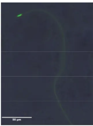
queen (Koeniger et al. 2014). The bulb, with the chitin plates, originating from the endophallus, retains secretory fluids such as the mucus and the cornual secretion from entering the queen’s lateral oviducts. ... 14 Figure 1.7: Live spermatozoa of the honey bee drone coloured in green
florescence with SYBR-14, observed in a fluorescent microscope at 400X (photo credit: Marilène Paillard). The SYBR-14 penetrates the cell membrane and binds to DNA. ... 16 Figure 1.8: A) Drone mounting the queen from behind, ready for copulation
(Koeniger et al. 2014). B) The drone is falling backwards, paralysed, after the first eversion of the endophallus (Koeniger et al. 2014). ... 19 Figure 2.1: Progression of mean sperm viability (± SEM) during 11 months, for two
temperatures (16˚C vs. -196˚C) at different evaluation days (F(3,28)= 484.27; p
< 0.0001). There was a significant difference between the two temperatures at 90 days (** p < 0.001) and 330 days (*** p < 0.0001) but not at 180 days (p > 0.05). ... 59 Figure 2.2: Mean sperm viability (± SEM) before and after centrifugation of
List of abbreviations
II: Instrumentation insemination PI: Propidium iodideAcknowledgments
First of all, I would like to thank my research supervisor Dr. Janice Bailey for welcoming me in her amazing sperm team. She has been like a second mother to me. I appreciate all the professional and personal advice she gave me during these years. It made me grow as a scientific, but also as a woman. Thank you for all the time spent in your office talking about my project or just talking about everything and nothing.
I would also like to thank my co-supervisor, Pierre Giovenazzo for introducing me to the wonderful world of beekeeping. I would have never imagined ending up working with bees and almost loving getting stung. Pierre put his full trust on me during the last few years, for this project, but also for my first research experience during my bachelor years. His optimism and his stunning enthusiasm about practically anything helped me going on and see things differently. I am grateful to continue working with his extraordinary team.
Thank you to the Ministère de l’agriculture, des pêcheries et de l’alimentation du Québec (MAPAQ) for the financial support via the program Innov’Action. Also, thank you to the Centre de recherche en reproduction, développement et santé intergénérationnelle (CRDSI) for the scholarship, helping me travel to conferences. Many thanks to the Centre de recherche en sciences animales de Deschambault and the department of animal sciences of Laval University for the material support.
I am very grateful of all the help and the support of the beekeeping team of the Centre de recherche en sciences animales de Deschambault. Thank you Michaël, Georges, Martine and Émile for believing in my crazy cryopreservation ideas. Thank you Ségolène, Olivier and Rachel for understanding what I could not, statistical analyses.
A special thank you to Andrée Rousseau, the master of drones and queen of instrumental insemination. She has been a true mentor and a true friend during these past years. I would not be here if it wasn’t for her and her contagious euphoria. She was always there to support me and listen to my tremendous whining. Working with her was and still is a breath of fresh air. Thank you Andrée for always laughing at my jokes, or simply at me, and for just being who you are.
Thank you to my lab colleagues Phanie, Maryse, Pauline, and Mathieu for the good talks and the good times we had at conferences. Thank you to Vianney Salmon for his cryopreservation wisdom. Even though he never worked with bee sperm, he always had answers to my questions. I would also like to thank Nancy Côté for always being there when I needed it the most.
Lastly, I would like to thank my family, my parents, and my friends for always believing in me. I am very grateful, even though I did not always show it, mom and dad, for all the little emails of encouragement, all the phone calls just to tell me that a bee documentary aired on television, and all the love sent from distance. These are all little thoughts that encourage me to continue and to never give up. Thank you Joanie for supporting me during these last two years. You were always there, during the good and the worst times, to cheer me up and tell me I was the best. You were the sunshine that lit my darkness. Thank you Sébastien and Yann for making exquisite drinks and staying late playing video games.
Preface
Chapter I titled “Preservation of Honey Bee (Apis mellifera L.) Semen” is a review of the pertinent research literature relating to this master’s project. Chapter II has been written as a scientific article and for submission to the scientific periodical, The Canadian Entomologist. The first author of both the review and the article is the master’s degree candidate, Marilène Paillard. The co-authors are Andrée Rousseau M.Sc., research professional at the Centre de recherche en sciences animales de Deschambault, Janice L. Bailey Ph.D., professor at the Animal Sciences Department and vice Dean of research at the faculté des sciences de l’agriculture et de l’alimentation of l’Université Laval, and Pierre Giovenazzo Ph.D., professor at the Biology Department of Laval University. These persons contributed intellectually and technically to the project and corrections and participated in writing the article.
Introduction
Honey bees (Apis mellifera L.) are critical players in the agricultural industry for food production as they account for the vast majority of insect pollinators. At least 35% of the human diet depends on these pollinators (Klein et al. 2007). An average of 1/3 of food on our plate is the result of insect pollination, mostly honey bees. Food and fibres from insect pollination was reported to be valued at approximately CAD $2 Million/year in Canada (Darrach and Page 2016) and CAD $217 Billion/year globally (Gallai et al. 2009).
Furthermore, honey bees are the source of other industries based on the production of honey, royal jelly, wax, pollen, and propolis. Honey brings the highest income for beekeepers, while the other products only represent 6% of honey bee revenues (Massicotte 2015). In 2014, there were approximately 8 777 beekeepers registered in Canada, owning 694 217 hives, and 309 beekeepers in the province of Québec (49 635 hives) (Massicotte 2015; Statistique Canada 2015). Hives, also known as colonies, are not equally distributed between beekeepers. Many beekeepers have 1-5 hives, while others possess more than a hundred, even thousands of colonies. An average-sized colony produces about 43 kg of honey annually (Massicotte 2015). In Québec, honey production was about 1,926 metric tons in 2014, which represented revenues of $13.4M (Massicotte 2015). In Canada, honey production is about 36,993 metric tons and worth $201.3M (Statistique Canada 2015). Furthermore, many beekeepers rent their hives for crop pollination at a cost of about $115.5 per colony for the cultivators (Massicotte 2015). In 2014, 44,214 colonies were rented in the province of Québec, resulting in an income of $5.1M to beekeepers.
In the last decade, however, there have been important losses of honey bee colonies worldwide, including Canada (Moritz and Erler 2016). These population declines have been attributed to various factors. The monocultures depending on honey bee pollination are highly prominent in the province of Québec, Canada, which greatly limits food diversity to the honey bees (Girard et al. 2012). Moreover, the lack of food diversity combined with high unintentional pesticide exposures make the honey bees more vulnerable to parasites (Goulson et al. 2015). The most devastating parasite of the honey bee health is Varroa destructor, a parasite mite that feeds on honey bee larvae (Allen Wardell et al. 1998; Bromenshenk et al. 2010; Currie et al. 2010; Neumann and Carreck 2010; Ratnieks and Carreck 2010). The combination of these factors (pathogens, food restrictions of homogenous crops, moving stress of hives from one crop plant to another, and pesticide exposure) increases the risk of losses of honey bees (Vanengelsdorp et al. 2009; Goulson et al. 2015). Adding to their vulnerability, many of the honey bee subspecies have been replaced by commercially available stock, which has further reduced the genetic diversity of Apis mellifera (Buchler et al. 2014).
Preservation of honey bee sperm is an effective strategy to protect genetic diversity and to facilitate genetic lineage to prevent as much colony loss as possible. The goal of this review is to summarise honey bee reproduction and provide an overview of drone semen preservation research and results since the early 1960s.
1.1 Honey bees
The honey bee, Apis mellifera Linnaeus (Hymenoptera: Apidae) originates from Europe, Africa and the Middle East, and is now widespread around the world (Seeley 1995). Honey bees have survived in various climatic conditions and evolved into many different subspecies (Louveaux et al. 1966). Honey bees are social insects of the order Hymenoptera, which includes more than 100,000 species of sawflies, wasps, ants and bees (Honeybee Genome Sequencing 2006). Social insects live in a colony where parents collaborate with their offspring, who give up their own reproduction for the reproductive success of their parents.
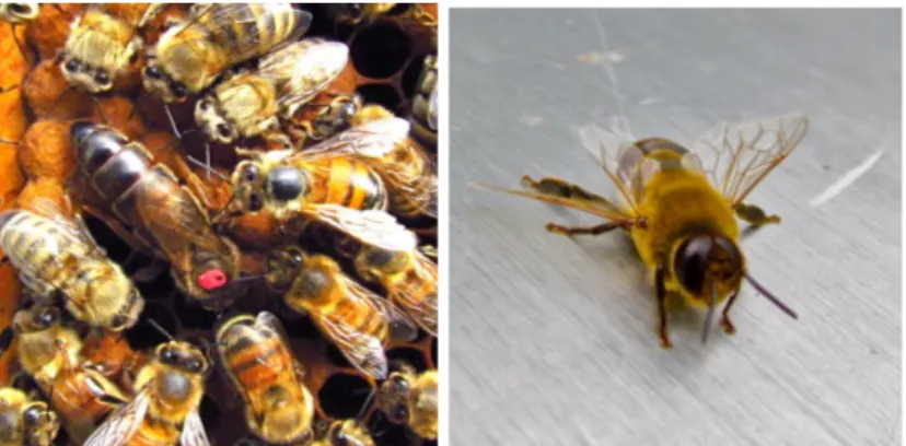
There are three castes in a honey bee colony (Figure 1.1): the queen, the female workers, and the drones (males). The queen honey bee, like other social hymenopterans, is the only fertile female and is thus responsible for maintaining the colony’s population and productivity. Her offspring are mainly sterile female workers, which are the most abundant caste in the colony. Workers are responsible for most of the tasks in the hive, such as protecting the colony, cleaning the hive, collecting pollen and nectar from flowers to produce honey, nursing the larvae, and constructing the wax combs. Depending on the worker’s age, she will have different tasks in the hive (Lindauer 1952). For example, the youngest workers will nurse the larvae while the oldest collect nectar for the colony. The principal function of the drones is to mate with a virgin queen (Gullan 2010; Winston 2010). Depending on the time of year, the colony may contain approximately 50 to 60 thousand workers and a few thousand males (Winston 2010; Koeniger et al. 2014).
Figure 1.1: Honey bee caste differentiation: Left, the larger queen, with the red spot on the thorax, surrounded by smaller workers; Right, the drone (Photo credit: Marilène Paillard).
1.2 Caste Development
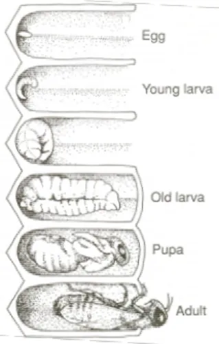
Honey bees are holometabolous insects, meaning that individuals go through multiple phases during development, including a complete metamorphosis, before emerging as an adult. Four stages occur within the same comb (Figure 1.2): egg, larva, pupa and adult (Jay 1963). The embryo develops during the egg stage. Afterwards, the larva emerges from the egg and grows rapidly. During the pupal stage, the organism is in an inactive state, and metamorphosis occurs. Finally, the adult honey bee, either worker, queen, or drone, emerges from the comb and immediately initiates its functions (Winston 2010).
Figure 1.2: Representation of the four developmental stages of the honey bee: egg, larva, pupa and adult (Winston 2010).
The duration of development before emerging into an adult differs for each caste. Queens, workers and drones take 16, 21 and 24 days to develop, respectively (Winston 2010). Female caste differentiation depends on the quality and quantity of food provided by the worker bees during their early development (Haydak 1943; Jay 1964). Female larvae can develop either into queens or workers during the first three days after the egg has been laid (Shuel and Ddxon 1960; Jay 1964). The first 48 h after egg emergence, the female larva is bipotent, meaning that she can follow one of two different developmental pathways (Winston 2010). During these two days, female larvae are fed royal jelly and it is at the third day that feeding differs and determines which developmental path is executed. Royal jelly is a creamy and viscous glandular secretion from the worker’s hypopharyngeal gland (Hebert 2010), composed of water (60-70%), sugar (10-16%), crude protein (12-15%), lipids (3-6%), traces of salts, free amino acids, and vitamins (reviewed in Buttstedt et al. 2013). Of note, royal jelly contains a protein called royalactin that helps larvae develop more ovarioles in the ovaries (Kamakura 2011). The queen larva receives high amounts of nutrient-rich royal jelly throughout all the development stages, ensuring her reproductive capacity. In contrast, workers receive less royal jelly than future queens and feeding will be switched to honey and pollen after two days of development (Weaver 1955,1957). Workers’
reproductive organs, therefore, remain undeveloped (Page and Peng 2001). This nutritionally-induced developmental pathway is driven by epigenetic changes via DNA methylation (Kucharski et al. 2008)
Like other hymenopteran insects, honey bees are haplodiploid. Females (workers and queens) developed from fertilized eggs (diploid), and drones from unfertilized eggs (haploid) (Beye et al. 2003). Thus the male offspring has 50% of the maternal genome. The sex-determination mechanism in hymenopteran insects is different from most animal species, including many insect orders, because they lack sex chromosomes (Beye et al. 2003). There is no sex-specific chromosome as in mammals, for example, where males have the XY chromosomes and females have the XX chromosomes. The haploid honey bee drones randomly receive half of the queen’s genome. It is, therefore, the quantity of one specific locus, the “complementary sex determiner” (csd) that determines the offspring sex (Mackensen 1951; Beye et al. 2003; Honeybee Genome Sequencing 2006). Eggs containing one csd locus become males, and eggs with a diploid locus become females (workers or queens). Furthermore, the fertilized egg must have two different csd loci, originating from unrelated parents, to create a female worker; otherwise the egg will develop as a diploid male (Mackensen 1951). When incestuous mating occurs, workers will usually cannibalize the diploid male larvae (Woyke 1965).
1.3 Sexual Maturation
1.3.1 Queens
Queens develop for 16 days before emerging as an adult: three days as an egg, six days as a feeding larva and seven days as a pupa (Winston 1987). She becomes sexually mature and takes her mating flight 5-6 days after emergence
two ovaries (Kaftanoglu and Peng 1982). Each queen ovary contains approximately 150 ± 180 ovarioles, contrarily to only 2-12 ovarioles in workers (Page and Peng 2001). Ovarioles are long tubes, located in the ovaries of most insects, that produce eggs (Snodrass 2010). The queen can lay up to 2 000 eggs in a single day, or approximately 200 000 eggs in one year (Winston et al. 1991).Egg production tends to decrease as the queen ages. She usually will live up to 3 years (Seeley 1978; Page and Peng 2001), during which time she will lay up to 1.0 million fertilized eggs throughout her life (Baer 2005).
Competition occurs between young queens, coming from the same hive. When they are newly emerged, sister queens fight each other until there is only one survivor. Queens use their mandibles and stinger as weapons to eliminate the opponent. The defeated queen, stung with venom, falls paralyzed and dies. The surviving queen then searches the entire hive to eliminate additional competitor sisters (Butz and Dietz 1994; Pflugfelder and Koeniger 2003). Young queens also attack developing queens who have not yet emerged from cells. The last queen remaining inherits the hive. Meanwhile, the old queen, who is the mother of the young queen, will leave the hive with half the workers to establish a new colony. This form of colony reproduction is called colony fission (swarming) (Winston et al. 1991).
The young queen remains in the hive for few days to eat and mature, following which the behaviour of workers changes and becomes more aggressive toward the queen. They bite her, presumably to “encourage” her to take her mating flight (Koeniger et al. 2014). It is important for the honey bee colony that the new queen’s development is as fast as possible. When the old queen leaves the hive with half of the colony or dies, there is a time lapse where no brood is produced. Therefore, the presence of newly mated queen must take the least possible time to repopulate and prevent colony collapse (Koeniger et al. 2014).
1.3.2 Spermatheca
The spermatheca is a female organ that stores sperm from previous copulations (Figure 1.3a). Although spermathecae are present in most insects, the honey bee queen is exceptional in being able to store semen for years while maintaining sperm viability (Verma 1973; Klowden 2013).
The mechanism by which the sperm remain fertile for such a long period in the spermatheca remains to be elucidated, however, a high protein concentration (8.5-15.3 mg/ml) is present in the spermathecal gland secretions (Klenk et al. 2004; Wegener et al. 2013) and may play a role on long-term storage of sperm (Klenk et al. 2004).Some proteins are thought to be antimicrobial in the spermatheca (Collins et al. 2006). Other proteins related to sperm energy metabolism are found in both the ejaculated semen and the stored semen but in higher concentrations in ejaculated semen (Poland et al. 2011).
Since the environment of the queen’s spermatheca is aerobic and sperm maintain their respiratory activity during storage, sperm are at risk of oxidative damage (Weirich et al. 2002; Collins et al. 2004). Therefore, antioxidants such as catalase, glutathione S-transferase and superoxide dismutase are found in the queen’s spermatheca (Weirich et al. 2002). These enzymes protect the sperm against oxidative stress by reducing the levels of reactive oxygen species (ROS) and improve sperm longevity (Weirich et al. 2002). The spermatheca also has a high
concentration of Na+ and K+ and a high pH (8.6) that encourage a low rate of
sperm metabolism (Verma 1973).Sperm are stored in an immobile state, which also minimizes their metabolic activity. Sperm motility is then activated upon contact with the spermathecal gland secretions, when the sperm are released for fertilization (Flanders 1939; Lensky and Schindler 1967).
Figure 1.3: A) Spermathecae from two different queens: Left, from a virgin queen without sperm and right from a mated queen (Cobey et al. 2013). The presence of sperm is usually apparent in that from the mated queen. B) Illustration of spermathecal anatomy (Koeniger et al. 2014).
In the honey bee queen, the spermatheca is a small sphere with a diameter of 0.9-1.3 mm with a volume of about 0.45-1.15 microliters (Verma 1974). The spermatheca is located at the rear end of the abdomen above the queen’s oviducts (Figure 1.3b) (Koeniger et al. 2014). It has two glands and is surrounded by a dense tracheal net that helps transport oxygen to the sperm inside the spermatheca (Koeniger et al. 2014). A narrow duct connects the spermatheca to the common oviduct (Bresslau 1905) and contains the Bresslau’s sperm pump, which allows the queen to control sperm release as the egg transits the oviduct (Harbo 1979c; Koeniger et al. 2014).
The queen is able to control fertilization of the egg by measuring the brood cell with her forelegs (Koeniger 1970a,b). If the queen lays an egg in a male cell, which has a bigger diameter than an average cell, she will not release sperm to fertilize the egg. In contrast, for the female castes, the queen releases sperm to fertilize the egg before laying it in a worker or a queen cell (Koeniger 1970a,b). The queen deposits the egg on the bottom of the cell, with or without sperm.
1.3.3 Drones
A drone requires 24 days of development from egg to emergence (Koeniger et al. 2014). The drone egg hatches after three days, the time required for embryo development. The larva then feeds and grows for days. Finally, the pupal stage last 15 days (Page and Peng 2001). Drones need to be nursed by workers, during their
larval stage and as young adults. The young drones stay in the warm brood area
for the first days where workers feed them. Drones then move to the honeycomb regions and gradually start feeding autonomously with the food present within the colony (Winston 1987; Koeniger et al. 2014). Drones can weigh up to 196 ± 225 mg when they emerge as adults, which is the average size of a queen, but
twice the size of a worker bee (Winston 1987). Before they become mature, young
drones aged from five to eight days, perform orientation flights to locate the Drone Congregation Area (DCA), where they will attempt to mate with a virgin queen (Koeniger et al. 2005b). The longevity of a drone is between 20 and 40 days (Ruttner 1966; Page and Peng 2001; Rueppell et al. 2005) with an average of 21.1 days (Fukuda and Ohtani 1977).
In Eastern Canada, drone-producing season is from mid-April to late August with a peak occurring during the swarming season, which is from June to July (Rousseau et al. 2015). This period is associated with abundant food sources that are essential to produce drone broods (Lee and Winston 1987; Rowland and McLellan 1987; McNally and Schneider 1994). Since sexual maturation of drones is longer compared to queens (36 days vs. 22 days, respectively), drones must be produced and raised earlier than queens to be sexually synchronized with queens (Koeniger et al. 2014). Several factors have been linked to drone production in honey bee colonies (Conner 2008): increasing photoperiod, nutritional conditions and queen age. At the end of the summer, the queen stops laying eggs in drone cells. When drone cells are not used for brood production, they are filled with honey.
1.3.4 Sexual Maturation of Drones
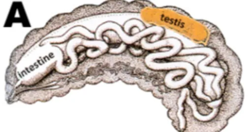
Honey bees appear to be the only insect in which spermatogenesis occurs only during their developmental stages, therefore, drones have a predetermined quantity of sperm after embryo development (Bishop 1920; Page and Peng 2001). Other insects produce spermatozoa throughout their life (Bishop 1920). Formation of the male reproductive system starts early during development. Testes are formed at the embryonic stage, before the larva emerges from the egg (Figure 1.4). Spermatogonia undergo multiple mitoses, resulting in primary spermatocytes (Hoage and Kessel 1968). Spermatogenesis is initiated on the third day of the larval stage. The primary spermatocytes undergo a reductional meiosis where two cells are produced: one secondary spermatocyte containing 16 chromosomes (haploid) and one cell containing only cytoplasm and an empty nucleus. The secondary spermatocytes then undergo a non-reductional meiosis, resulting in two spherical spermatids. During the larval stage, drones grow quickly, including the testes and spermatid proliferation, due to the high food provisions from the workers (Hrassnigg and Crailsheim 2005). Spermatid multiplication stops prior to pupation (Hoage and Kessel 1968), and spermiogenesis is initiated, which is the morphological differentiation resulting spermatozoa.
Figure 1.4: Development of testes during embryogenesis (Koeniger et al. 2014).
Approximately 2-3 days after pupal emergence, sperm start migrating from the testes to the seminal vesicles, which is the enlarged area of the vas deferens used for sperm storage, where they remain for approximately 13 days (Koeniger et al.
2014). Testes size is greatly reduced after sperm migration. Once in the seminal vesicles, the spermatozoa attach via the head to the single layer glandular cells that cover the vesicle walls. Here, it is thought that the spermatozoa go through a maturation phase where they absorb nutrients from the gland cells to become fully functional and remain so until copulation (Ruttner 1976; Koeniger et al. 2014). Little information regarding this apparent maturation is present in the literature. Additional research is required to understand bee sperm physiology during transit in the male tract.
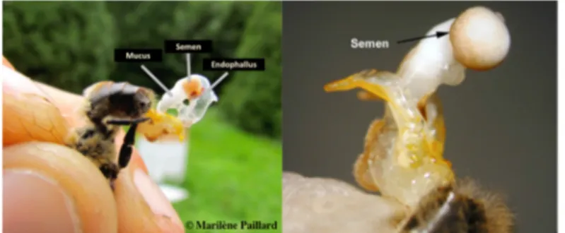
A drone is not yet sexually mature at emergence, despite having produced mature sperm. The copulatory organ, the endophallus, is fully developed and tightly stored in the abdomen (Koeniger et al. 2014). Endophallus eversion is possible on a newly emerged drone, but no mucus and semen are present for emission because the accessory gland cells are not yet active. At 5-6 days after emergence, drones may ejaculate mucus during eversion but without semen (Ruttner 1976). Drones are sexually mature twelve days port-emergence, which is six more days required for sexual maturation of the queen (Koeniger et al. 2014). At this age, drones can completely evert their endophallus and creamy coloured semen, containing spermatozoa, can be located at the posterior extremity of the ejaculate, on top of the white mucus, which is void of spermatozoa (Figure 1.5). The fluids do not mix together.
Figure 1.5: Full eversion of the male’s endophallus, exposing the semen and the mucus (photo credit: Left Marilène Paillard; Right (Cobey et al. 2013)).
Drones have three accessory sex glands: the mucus gland, the bulbus gland (Woyke and Ruttner 1958), and the cornual glands. At the end of the larval stage, the mucus gland develops from the vas deferens, the duct connecting the testes to the seminal vesicles, and produces white mucus (Koeniger et al. 2014). The cornual and bulbus glands both consist of single layers of cells (Moors and Billen 2009) and are found near the extremity of the endophallus, also known as the bulb (Koeniger et al. 1996; Moors et al. 2012). The cornual glands secrete an orange-coloured secretion that helps strengthen the attachment of the mating sign in the queen’s reproductive tract, while the endophallus, engorged with mucus, reinforces the connection between the drone and the queen during mating (Koeniger and Koeniger 2000; Colonello and Hartfelder 2005). The drone’s reproductive secretions are predominantly composed of proteins, except from the cornual glands, which produce lipids (Colonello and Hartfelder 2003; Moors and Billen 2009). The mucus glands start secreting mucus after the drone’s emergence and are filled at five to six days of age (Ruttner 1976). Each mucus gland contains approximately 50-60 µg of protein (da Cruz Landim and Dallacqua 2005). The mucus is used to produce a mating sign in the queen after a successful copulation (Koeniger 1988). The drone’s bulb is filled with mucus and other secretions (Figure 1.6). The chitin plates ensure that no secretory fluid will penetrate inside the newly mated queen’s reproductive tract, except for the semen, containing sperm and the seminal fluid, which is described later. The mating sign represents 10% of the drone’s body weight (15 mg) (Koeniger et al. 2014).
Figure 1.6: The mating sign retrieved from the sting chamber of a newly mated queen (Koeniger et al. 2014). The bulb, with the chitin plates, originating from the endophallus, retains secretory fluids such as the mucus and the cornual secretion from entering the queen’s lateral oviducts.
1.3.5 Drone Semen
Semen is the combination of sperm and seminal fluid produced by the seminal vesicles. The seminal fluid is mostly composed of more than a hundred proteins that are different from the other gland secretions. Seminal fluid proteins have many functions, such as defense against microbial attacks or oxidative stress, energy production for sperm, and metabolism of carbohydrates and lipids (Baer et al. 2012; Gorshkov et al. 2015). The concentration of proteins involved in signalling and maintaining viability differs by 16% among genetic lineages, which could influence sperm competition by reducing fertilization success of other drone sperm (Baer et al. 2012). The seminal fluid also contains sugars such as fructose, glucose, and trehalose, which serve as energy sources for the spermatozoa (Blum et al. 1962; Verma 1974).
Drones produce an average of 1.5-1.7 microliter semen with approximately 7.5 million spermatozoa/µl (Rousseau et al. 2015). Sexually mature drones have a yellowish creamy coloured semen. The semen becomes darker, thicker and the semen volume decreases with age (Woyke and Jasinski 1978; Czekonska et al. 2013; Rousseau et al. 2015). To the best of our knowledge, the reason is still unknown. Insemination with thicker semen has a risk of blocking the queen’s oviducts and increases the likelihood the queen will die (Woyke and Jasinski 1978). Contradictory reports have demonstrated that sperm viability decreases with age (Locke and Peng 1993), while others observed an increased viability with age (Czekonska et al. 2013). Rousseau (2015) reported that age has no effect on sperm motility and viability. It is still unclear whether sperm viability increases or decreases with age, however, the drone’s ability to copulate varies with age, depending on the colony strength and quality (Rhodes et al. 2011).
1.3.6 Spermatozoa
Most insects have very long spermatozoa with an elongated head. Honey bee spermatozoa measure 250-270 µm in length (Lino-Neto et al. 2000) with the head measuring 8 to 10 µm (Figure 1.7) (Peng et al. 1993). The whole cell is 0.7 µm wide (Peng et al. 1993). The head, containing the nucleus filled with DNA, is asymmetric and is composed of a bi- or tri-layered acrosomal complex (Lensky et al. 1979; Peng et al. 1993). The length of the acrosome is approximately 5 µm (Lensky et al. 1979). Unlike vertebrates, honey bee sperm lack the middle piece (Rothschild 1955). The tail is a flagellum composed of two mitochondrial derivatives, an axoneme, and two triangular shaped accessory bodies (Peng et al. 1993; Lino-Neto et al. 2000). The axoneme consists of nine single accessory microtubules, nine doublets, and a central singlet pair (9 + 9 + 2). Two mitochondrial derivatives are parallel to the axoneme, starting at the base of the nucleus and finishing at the end of the flagella (Rothschild 1955). The necessity for sperm capacitation and the acrosome reaction during the fertilization process are still unknown in the insect taxa compared to many other animal species (Peng et al. 1993).
Figure 1.7: Live spermatozoa of the honey bee drone coloured in green florescence with SYBR-14, observed in a fluorescent microscope at 400X (photo credit: Marilène Paillard). The SYBR-14 penetrates the cell membrane and binds to DNA.
1.4 Mating
Honey bees are polyandrous and this behaviour is not found in most social insects (Gencer et al. 2011). Polyandry means that the queen mates with multiple males (Koeniger and Koeniger 2000; Palmer and Oldroyd 2000). It is an important reproductive phenomenon for honey bees because queens mated with only one drone produce weaker colonies (Fuchs and Schade 1994; Moritz and Fuchs 1998; Tarpy 2003; Page et al. 2006). Consequently, workers’ genetic profiles are more
diversified and optimise their productivity (Koeniger et al. 2014). Honey bees have
an intense male-biased sex ratio estimated at 1,000 to 2,000 males per reproductive female (Page and Metcalf 1984; Koeniger et al. 2014). The sex ratio is usually 1:1 in most animal species, including insects of the order Hymenoptera such as wasps, solitary bees and many ant species (Koeniger et al. 2014).
Honey bee mating takes place in the afternoon, during flight, when weather conditions are optimal, which must be suitable for both drones and the queen to fly to DCAs (Ruttner 1956; Koeniger et al. 2014). Weather is an important mating factor in northern regions. The ideal temperature for the mating flight is 18˚C for drones and 20˚C for the queen (Koeniger et al. 2014). Furthermore, mating will only occur during sunny days to avoid strong winds and rain (Koeniger et al. 2014). It is not known how drones and young queens, residing in the hive, are able to sense these weather conditions, since the hive is thermo regulated by workers.
Drones leave their hive around 2 pm until 5 pm (Koeniger et al. 2014). Both drones and queens use the light cycle to regulate their flight (Koeniger et al. 2014). They can take several mating flights lasting up to 25 minutes each until copulation occurs (Witherel 1971; Koeniger et al. 2014). Drones can execute a maximum of about six mating flights daily (Koeniger et al. 2014). As for the queen, approximately one week after emerging, she executes short orientation flights of about five minutes (Koeniger et al. 2014). The queen then flies to a DCA about 30
to 60 minutes after drones have left the hive. Her mating flights can last between 13 (Taber 1954) and 21 minutes (Woyke 1960), and occur between 2:30 and 4 pm.
The DCA is already established when the virgin queen arrives to mate (Winston et al. 1991). The young queen mates for a short period of time (2-3 days) and, once she starts laying eggs, never mates again (Winston 1987). She can have one or multiple mating flights, sometimes up to three flights, depending on the volume of semen deposited (Taber 1954; Ruttner 1980; Tarpy and Page 2001).
Honey bee queens and drones originating from the same hive have adapted behaviour to avoid inbreeding by flying at different DCAs. Drones fly to closer DCAs, while the young queen will go to a further DCA (Koeniger et al. 2014). The energy and the flight time are very valuable to drones since they have to stay long periods of time at DCAs to locate and possibly mate with the queen. Therefore, drones fly at closer DCAs to preserve their time and energy. Queens can focus their energy on flying further, since mating with multiple males only takes few minutes (Koeniger et al. 2014).
The virgin queen heads to a DCA where more than 10,000 to 30,000 drones of different genetic lineages, originating from approximately 240 colonies, await (Gary
1963; Koeniger 1986; Loper et al. 1987; Baudry et al. 1998). During the mating
flight, drones fly in the DCA at 15 to 60 m above the ground and form a circle of 60-200 m diameter (Gary 1962,1963; Loper et al. 1987). Drones are attracted to the queen’s sex pheromones, of which the main component is 9-ODA (9-oxo-decenoic acid). They are also attracted visually, as she arrives at the DCA (Koeniger and Koeniger 2000). Once in the DCA, over 20-100 drones pursue the queen, forming a “mating comet” behind her, ready to copulate (Koeniger 1990). These drones follow her and, eventually, one will grasp the queen and mate.
The copulation between a queen and a drone takes a few seconds (Gary 1963). The flying drone mounts the queen from behind (Figure 1.8a) (Koeniger et al.
2014). The male holds the queen’s abdomen from each side with his front and middle legs. Once the male holds the queen tightly, the drone orients the tip of his abdomen in the queen’s open chamber and inserts the first half of his endophallus
(Koeniger 1986; Woyke 2008). Since the endophallus is folded in the drone’s abdomen, muscles of the abdomen contract simultaneously. A large amount of hemolymph then enters the endophallus from the abdomen (Figure 1.8b) (Koeniger et al. 2014).
Figure 1.8: A) Drone mounting the queen from behind, ready for copulation (Koeniger et al. 2014). B) The drone is falling backwards, paralysed, after the first eversion of the endophallus (Koeniger et al. 2014).
When half of the eversion is complete, the drone’s wings stop moving and its body is paralysed since almost all of the hemolymph is pressed into the endophallus (Koeniger 1986). The male swings backwards, still attached to the queen by his legs (Koeniger et al. 1979), and the queen contracts her sting chamber to complete the full eversion of the drone’s endophallus. Sperm are then transferred from the endophallus to the queen’s oviducts where the vaginal valve and sphincter hold back the sperm from exiting the reproductive tract (Koeniger et al. 2014).
Sperm-free white mucus, found at the extremity of the endophallus, pushes the semen to
migrate from the endophallus to the queen’s lateral oviducts (Woyke 2010). Sperm transfer occurs in a sterile environment, because the drone’s membranous endophallus seals the queen’s vagina, thus no communication between the exterior environment and the queen’s oviducts occurs (Koeniger et al. 2014).
Once ejaculation completed, the paralysed drone dies. After the mating, the male leaves the detached bulb from the endophallus as a “mating sign” in the queen’s sting chamber (Woyke 2010). The mating sign left by the drone increases the queen’s visibility in the DCA to attract additional drones (Koeniger 1990; Koeniger 1991). The following drone removes the previous mating sign to copulate with the queen (Koeniger 1984).This male attracting system reduces the duration of the queen’s mating flight.
The queen copulates successively (Gries and Koeniger 1996), with an average of 17 drones during one or more mating flight (Adams et al. 1977). After this mating period, the queen never copulates again. The queen then returns to the hive and the workers remove the mating sign of the last copulation (Koeniger et al. 2014). The queen receives approximately 200 million spermatozoa in her lateral oviducts. However, the spermatheca can only retain 5-7 million sperm (Woyke 1962; Koeniger 1991). Therefore, the sperm of some drones will not successfully reach
the spermatheca. The excess will be expelled out of the queen’s oviducts, by
contracting abdominal muscles, as the retained sperm migrate into the spermatheca. Sperm migration is complete approximately 40 h after mating (Woyke 1983).
Many mechanisms have been proposed to explain the migration of the sperm from the queen’s lateral oviducts into the spermatheca: oviduct contractions (Ruttner and Koeniger 1971), spermatozoa motility (Collins 2000a), the spermathecal pump (Koeniger et al. 2011), and even flagellar movement of sperm (Tofilski 2014). The queen uses the stored sperm for fertilization over her lifetime (1-3 years).
1.5 Drone Competition
The competition between males happens individually, meaning that they do not “fight” physically against each other to win the queen. It is the fittest males who can overcome obstacles and survive to copulate with the queen. Drones must be able to locate the DCA and the queen faster than other drones to copulate. Therefore, queens potentially mate with drones that have a better flight ability (Jaffe and Moritz 2010). The chance of any one drone mating with a queen is estimated to be 0.0001% (Koeniger et al. 2014).
1.6 Sperm Competition
It is still unclear whether sperm competition occurs in honey bees and how it works (Moritz 1986; Harbo 1990; Woyciechowski and Krol 1996; Shafir et al. 2009).
Sperm competition may occur after mating, within the oviducts, during sperm
storage in the spermatheca or at the time of fertilization (Woyciechowski and Krol
1996; Shafir et al. 2009; Tofilski et al. 2012). Some proteins found in the seminal fluid might displace other males’ sperm (Harshman and Prout 1994), owever, it has not yet been demonstrated in honey bees(Woyciechowski and Krol 1996). Usually, spermatozoa from the same male will not compete against each other (Parker and Pizzari 2010). Studies have demonstrated that sperm viability is reduced when mixed in seminal fluids of other males than their own seminal fluid (den Boer et al. 2010). However, these results are contradicted by another study, which found that sperm viability is not affected, and drones may possibly have sperm polymorphism
(Tofilski et al. 2012).
Because of the extreme male-male competition and the high requirements of sperm by the polyandrous queen, males have required a high level of sperm viability (Hunter and Birkhead 2002). Moreover, since the queen mates for only a
ejaculate filled with highly viable sperm, is important (Hunter and Birkhead 2002;
Simmons 2002; den Boer et al. 2008). To perpetuate their genetic lineage, drones
should maximize the number of spermatozoa produced. The number of sperm can
vary between 3 and 12 million, depending on the drone size (Berg et al. 1997; Schluns et al. 2003; Koeniger et al. 2005a) and genetic lineage (Rousseau et al. 2015).
Sperm viability plays a crucial role during fertilization, and, therefore, sperm competition (Franck et al. 2002), which is more likely to occur at this time because the queen releases 4 to 25 sperm (Harbo 1979c; Yu and Omholt 1999) from the
spermatheca (Harbo 1990). Drones producing more viable motile sperm increase
their chance to fertilize eggs. Furthermore, the length of the sperm can vary from
250 to 270 µm (Lino-Neto et al. 2000). Theoretically, the longest sperm will reach the spermatheca fastest, therefore, increasing their chances of fertilizing the egg (Gomendio and Roldan 1991; Montgomerie and Briskie 1992; Gage and Gage
1994; Morrow and Gage 2000). Studies on bumble bees (Bombus terrestris),
demonstrated that sperm length is a genetic trait that responds to selection (Koeniger et al. 2014). In polyandrous bumble bee species where queens have multiple mating, drones produce longer sperm than in monogamous bumble bee species (Brown et al. 2002; Baer et al. 2003).
Spermatozoa can potentially cooperate together by producing a pseudopodium, a foot-like projection, to more rapidly reach the spermatheca (Tofilski 2014). Sperm coordinate their flagellar beats so they can swim forward and increases their chance of fertilizing more eggs (Tofilski 2014). To our knowledge, it is unknown if the sperm cooperation happens between sperm coming from the same male, or not.
1.7 Instrumental Insemination
Instrumental insemination (I.I.) is performed in many species. Instrumental insemination is a crucial technique to control animal breeding to accelerate genetic selection. I.I. allows mating of domesticated honey bees to bypass the DCA. Many factors such as unfavourable climate and undesirable drones from other colonies can negatively affect genetic improvement through natural breeding selection. In 1927, I.I. was first successfully reported in honey bees (Watson 1927). The technique was then perfected during the 1940’s - 1950’s (reviewed in Cobey 1983; Laidlaw 1987). Instrumental insemination is currently a highly successful method for genetic control, for research, and for stock improvement (Cobey 2007), even though this technique is difficult to execute.
Instrumental insemination is difficult to introduce in the commercial industry because of the technical complexity and is thus more often used during scientific research. Therefore, it requires a minimum of expertise to perform I.I., such as advanced knowledge breeding principles and beekeeping skills to produce queens and drones (Cobey et al. 2013).
The success of I.I. depends on adequate semen quality (Collins 2000a). Males must be between 2 and 3 weeks of age, because sperm viability decreases with age and fewer sperm migrate to the spermatheca. Moreover, as mentioned earlier, semen of older males will leave residues in the queen’s lateral oviducts, thus increasing the risk of queen mortality (Woyke and Jasinski 1978). The minimum sperm viability required for queen I.I. was calculated to be 43% (Collins 2000a,2004). If sperm viability is lower, the queen will lay more drones (unfertilized eggs) and fewer workers (fertilized eggs). Therefore, the colony will die because of the lack of workers.
1.8 Semen Preservation
The development of I.I. in honey bees, has stimulated interest in semen preservation. The use of stored sperm greatly facilitates I.I., genetic improvement of the colony, and prolonging the honey bee production season. Both short and long-term semen conservation enhances selection and genetic improvement in honey bee populations (Collins 2000b). Thus, it would help developing superior honey bee stocks that are resistant to certain parasites such as varroa mites (Varroa destructor), for example (Collins 2000a). Furthermore, shipping honey bee semen instead of live drones would reduce the risk of spreading bee pathogens (Cobey 2007).
Additionally, in northern climates, such as in Canada, preserving semen could be very helpful to advance the honey production season (Collins 2000b). Since drones take more time to be sexually mature (36 days from egg to sexual maturation) and colonies only start producing them in late spring (May), I.I. can be used with semen collected the prior year to accelerate and elongate the season. Moreover, studies carried out in eastern Canada, where winters are cold and long, demonstrated that the early drones reared in May were less fertile than any other time of the beekeeping season (Rousseau et al. 2015). Therefore, by preserving semen during the winter, semen could be available sooner in spring for I.I. and have better sperm quality from drones reared late the previous summer. Successful sperm preservation is an effective strategy to preserve honey bee genetic diversity, and also to facilitate the selection of lineages tolerant to pests and diseases and prevent further colony loss (Cobey et al. 2013).
1.8.1 Semen Collection
Semen can be collected by one of two methods: (1) directly from the seminal vesicles by dissecting the drone (Mackensen 1955) or (2) by manual eversion of
the endophallus. Collecting semen directly in the seminal vesicles results in more viable sperm, however, it is less practical for I.I., since it takes more time to perform and less semen is collected compared to the manual eversion technique (Collins 2004).
To collect honey bee semen, the endophallus must be manually everted. For the first half of the eversion, the drone is held by the head so that the abdomen is facing upward and a gentle pressure is applied to the thorax with the other hand. Additional light pressures are applied to the base of the abdomen to complete eversion. The creamy coloured semen is expelled out of the endophallus, next to the mucus, and is located at the extremity of the endophallus (Mackensen 1955; Collins 2004). The eversion must be performed in sanitary conditions. It is crucial to avoid contact between semen and the drone’s body, using semen that has been mixed with fecal material, and touching the semen (Andere et al. 2011). A sanitary environment reduces semen contamination and ensures that sperm viability is not negatively affected (Andere et al. 2011).
There are many factors that must be considered for sperm preservation such as storage temperature, semen dilution, and the diluent. As early as the 1960s, experiments were conducted with different diluents and temperatures on drone semen (Taber and Blum 1960).
1.8.2 Dilution and Diluent
The diluents used for drone semen preservation are intended to imitate the spermathecal fluid, since sperm stored in the spermatheca maintain high viability for up to several years. Many laboratory diluents can maintain high sperm viability (Moritz 1984; Taylor et al. 2009). Usually, a Tris buffer composed of sugars, amino acids and antibiotics, with a pH of 8.6 is recommended for drone semen
effects on the queen, as several studies have demonstrated that diluted semen can decelerate the beginning of oviposition and fewer sperm are stored in the spermatheca (Kaftanoglu and Peng 1980,1982; Moritz 1984).
1.8.3 Temperature
Drone semen can be stored at room temperature for a few weeks, without losing sperm viability (Cobey 2007; Cobey et al. 2013). The earliest research on drone sperm preservation successfully stored semen at room temperature for four weeks, but bacterial contamination rapidly decreased sperm viability (Taber and Blum 1960). Therefore, by introducing antibiotics to the stored semen, bacterial growth was reduced and semen could be stored at room temperature for 3-4 months (Poole 1969).
Storage below 10˚C and above 32˚C rapidly decreases sperm viability (Taber and Blum 1960; Harbo and Williams 1987), however, spermatozoa can tolerate a brief, one-hour storage at 40˚C (Hopkins and Herr 2010). The optimal temperature for two days storage is 21˚C but semen can be stored between 13 and 25˚C (Harbo and Williams 1987; Locke and Peng 1993). Several studies have reported 70% to 80% sperm viability after storage at 12˚C or 25˚C for 6 weeks (Locke and Peng 1993; Collins 2000b). Semen quality was better preserved at 15˚C than at 24˚C for a storage period of 13 weeks (Poole and Taber 1970). Furthermore, 65% sperm viability was reported after 39 weeks at 12 ̊C, which is acceptable for I.I. (Collins 2000b,a). In 2015, we observed that more sperm preserved at 12-15 ̊C survived than at 25 ̊C, and our best protocol tested resulted in 47% sperm survival after 17 weeks (Paillard, unpublished data).
Although drone sperm storage is clearly achievable at above-freezing temperatures, it is inadequate for long-term storage since sperm rapidly loses viability after few weeks. Therefore, many researchers have turned to
cryopreservation as a solution for long-term sperm storage.
1.9 Cryopreservation
As for many mammalian species, cryopreservation can be a good method for long-term sperm storage. Cryopreservation is a technique that freezes and conserves live cells and tissues for long periods at very low temperatures, usually in liquid
nitrogen (-196°C). Semen cryopreservation has only been reported on few insect
species such as the silk moth Bombyx mori (Takemura et al. 2000) as well as embryo cryopreservation in Drosophila melanogaster (Gardner et al. 1990) and Spodoptera exigua (Luo et al. 2006). Frozen semen could be very useful for honey bee I.I. by having access to sperm year round.
Since the late 1970’s and early 1980’s, multiple honey bee semen cryopreservation techniques have been tested with variable but relatively poor results (Mel'Nichenko and Vavilov Yu 1976; Harbo 1977,1983; Kaftanoglu and Peng 1984). Despite good sperm viability and motility after freezing-thawing, fertility was highly reduced (Harbo 1983; Kaftanoglu and Peng 1984; Cobey et al. 2013) and queens inseminated with frozen-thawed semen did not perform as well as queens inseminated with fresh semen (Peng et al. 1992). Normally, ideal worker production by a queen is approximately 95-99%, but queens inseminated with frozen-thawed semen produced fewer than 50% of workers and the remainders were drones (unfertilized eggs) (Mel'Nichenko and Vavilov Yu 1976; Harbo 1977,1979b,a,1983; Kaftanoglu and Peng 1984; Hopkins et al. 2012).
Many aspects of the cryopreservation protocol can cause sperm damage such as the cryoprotectant used and freezing rate. Rapid freezing and thawing can damage the cell membrane, the acrosome, the nucleus, the flagellum, and split the flagellar mitochondria (Peng et al. 1992). Furthermore, damage to the sperm genome can
occur (Harbo 1981). Eggs fertilized with frozen-thawed sperm had lower viability and emerging drones from the first generation were mosaic, meaning that cells from within one individual had different genetic makeups (Harbo 1979b,1980). The proposed explanation is that the pronucleus of both the spermatozoa and the egg did not fuse properly, hence, both pronuclei developed into independent, haploid tissues (Harbo 1980).
Only recently has drone semen cryopreservation yielded promising results (Taylor et al. 2009; Hopkins and Herr 2010; Hopkins et al. 2012; Wegener and Bienefeld 2012; Wegener et al. 2012; Wegener et al. 2014a). Hopkins (Hopkins et al. 2012) successfully froze drone semen and inseminated queens to obtain a second-generation of queens that were inseminated with the same frozen-thawed semen. Two of the five initial queens inseminated with frozen semen produced a majority of workers. Six of ten first-generation queens inseminated with the same frozen semen laid eggs but only three produce a majority of workers. From one queen, 14 second-generation queens were produced. A third generation was interrupted due to seasonal conditions. These results provide “proof of concept” that honey bee sperm cryopreservation is possible.
Several key factors such as dilution/diluents, cryoprotectants, freezing rates, and thawing are important for successful cryopreservation of honey bee sperm.
1.9.1 Dilution and Diluent
Apart from the cryoprotectant, the choice and the quantity of the diluent added to the drone semen can influence cryopreservation. A higher semen dilution (> 1:3 semen: diluent) helps the spermatozoa to survive during cryopreservation and can be used to inseminate more queens (Taylor et al. 2009). However, diluting sperm increases their motility (Lensky and Schindler 1967), and sperm longevity is often
reduced after motility is activated. Indeed, sperm motility can be higher after cryopreservation than fresh semen, yet result in poor queen fertility after insemination (Kaftanoglu and Peng 1984; Wegener et al. 2014a). The dilution ratio most frequently used is 40% semen and 60% diluent, usually Kiev solution (0.3 g glucose, 0.41 g potassium chloride, 0.21 g sodium bicarbonate, and 2.43 g sodium citrate dihydrate in 100 ml distilled water with 0.05% di-hydrostreptomycin), containing 10% cryoprotectant (Kaftanoglu and Peng 1984).
Hen’s egg yolk is often used in diluents for cryopreservation, because it protects cells from cold shock (Amann and Graham 1993). The high concentration of cholesterol in egg yolk helps sperm to survive freezing (Bergeron and Manjunath 2006). However, its composition is not uniform and can vary. Egg yolk also increases the risks of contaminating the semen (Bergeron and Manjunath 2006). Replacing the egg yolk with a synthetic solution (10% w/v cholesterol solution dissolved in 95% ethanol and 20% w/v BSA solution) resulted in low fertility after insemination (Hopkins et al. 2012). Sperm motility after thawing was lower than fresh semen and queens produced mainly drones (unfertilized eggs).
1.9.2 Cryoprotectant
Sperm are stored in liquid nitrogen (-196˚C) thus; sperm need to be mixed with a cryoprotectant to protect from freezing damage. Cryoprotectants prevent ice crystal formation inside the cell by reducing the water freezing temperature (Hopkins and Herr 2010). Many cryoprotectants have been tested on drone semen, including dimethyl sulfoxide (DMSO), glycerol, methyl sulfoxide (MeSO), dimethyl formamide, 1,3-propane diol, 2,3-butane diol, and ethylene glycol (Harbo 1977,1979a; Kaftanoglu and Peng 1984; Hopkins and Herr 2010; Wegener and Bienefeld 2012; Wegener et al. 2014a). Hopkins (2010) compared many and demonstrated that sperm preserved with ethylene glycol for one hour lost motility and had lower viability compared to sperm preserved with DMSO (90% vs. 95%
respectively). He also demonstrated that glycerol killed the most sperm. Ironically, glycerol is commonly used for mammalian semen freezing (Curry 2007). Wagener (2012) found that even if short-term toxicity appeared to be low for most of the cryoprotectants tested (DMSO, 1,3-propane diol, 2,3-butane diol, ethylene glycol, and dimethyl formamide), there was a significant reduction of sperm migration into the spermatheca after I.I..
Hopkins (2010) reported that DMSO was the best cryoprotectant to preserve honey bee semen and was the least toxic compared to the other cryoprotectants. DMSO has been widely used for drone semen cryopreservation since 1976 (Harbo 1977; Wegener and Bienefeld 2012) and maintains good sperm viability (Harbo 1977; Taylor et al. 2009; Hopkins and Herr 2010; Hopkins et al. 2012). Harbo (1977) also demonstrated that 10% DMSO did not affect the quantity of sperm reaching the spermatheca. However, DMSO has been reported to be toxic for both the spermatozoa and the queen after I.I. (Harbo 1986; Wegener and Bienefeld 2012). Hopkins (2010), however, demonstrated that 15% DMSO, highly decreased sperm viability and DMSO can damages the sperm genome through breakage of the chromatids (Kapp and Eventoff 1980). Effectively, 3% of queens’ progeny following I.I. with sperm diluted with DMSO, laid eggs that did not hatch, which is a very rare phenomenon occurring in honey bees (Hitchcock 1956; Harbo 1986). Genetic damage occurs in treated spermatozoa (F0), which disrupts the meiotic division in a F1 queen and results in sterile F2 female honey bees (Harbo 1986). However, since it only affected 3% of queens were affected, Wegener & Bienefeld (2012) concluded that the impact of DMSO was less severe than previously thought.
1.9.3 Freezing Rate
It is crucial to freeze semen at an optimal rate; otherwise sperm undergo “cold shock”, which means that cells are damaged by the very rapid cooling (Robertson et al. 1990). The ideal sperm-freezing rate differs among species due to sperm
size, morphology, metabolism, and within their membrane phospholipid composition (Barbas and Mascarenhas 2009). Some species require rapid freezing, while others need slow freezing rates (Hopkins and Herr 2010). For honey bee sperm, controlled slow cooling rates yields good sperm viability and motility after thawing (Harbo 1979a; Kaftanoglu and Peng 1984; Hopkins and Herr 2010). By slowly cooling the semen, ice crystals are formed outside the sperm and dehydrates the interior of sperm cells. Thus, ice crystals do not form inside the cells (Mazur 1963).
Early attempts at bee semen cryopreservation using slow cooling of diluted semen (40% semen and 60% Kiev solution, containing 10% DMSO) at 3-4˚C/min to obtain good sperm motility after thawing, however, inseminated queens with the frozen-thawed semen produced fewer than 50% workers (Kaftanoglu and Peng 1984). More recently, a potential freezing rate for drone semen cryopreservation was established (Hopkins and Herr 2010), during which semen is initially cooled to 4˚C over 2 h. The semen is then slowly frozen at a rate of 3˚C/min using a programmable freezer until -40˚C. The semen is then directly plunged into liquid nitrogen. For semen diluted in Harbo’s diluent (Harbo 1983), egg yolk and 10% DMSO (ratio of 3:2; semen to diluent), over 90% sperm viability was reported after six days at -196˚C (Hopkins and Herr 2010). However, the fertility of the frozen-thawed semen was not assessed.
1.9.4 Thawing
Thawing protocols are variable in the literature depending on the study. Thawing temperatures vary between 25˚C and 40˚C, and usually requires less than 1 minute, mostly few seconds (Harbo 1979a; Peng et al. 1992; Taylor et al. 2009; Hopkins and Herr 2010; Wegener and Bienefeld 2012). Rapid thawing and temperatures above 40˚C is to be avoided on drone sperm (Peng et al. 1992;
rate on bee sperm biology and fertility capacity has been conducted. In some mammals, however, thawing protocol does appear to influence post-thaw parameters (Senger 1980; Nur et al. 2003).
1.10 Centrifugation
Centrifugation of cryopreserved drone semen is useful prior to I.I., to concentrate diluted sperm, homogenize sperm from multiple males and remove cryoprotectants from frozen-thawed sperm (Wegener and Bienefeld 2012; Wegener et al. 2014b). Drone sperm, however, are fragile and centrifugation can damage the cell membrane, causing lethal or sub-lethal injury (Moritz 1984; Harbo 1990; Collins 2004; Cobey 2007; Wegener et al. 2014b). Consequently, only small amounts of centrifuged sperm reach the spermatheca, resulting in poor fertility after I.I. (Kaftanoglu and Peng 1980; Fischer 1987).
More recently, however, worker brood has been successfully obtained after I.I. with centrifuged, unfrozen drone semen (Wegener et al. 2014b). There was no significant difference between the fertility of queens inseminated with centrifuged semen versus fresh semen. This study did not address the use of centrifugation of cryopreserved semen, however, the success of this technique suggests that it could be an effective strategy for post-thaw cryoprotectant removal.
1.11 Problematic
For the last decade, the bee population worldwide has been declining due to many factors such as natural habitat destruction, parasites, pesticide exposure, starvation, transport stress, poor queen health, and winter colony losses (Stokstad 2007; Vanengelsdorp et al. 2009; Moritz and Erler 2016). Colony loss ranges from 35% to 75%, depending on the country (Stokstad 2007). The combination of
multiple factors increases the harm to both honey bees and wild pollinators. The lack of food diversity, due to monoculture agriculture, added to the presence of unintentional pesticide exposure makes the honey bees more vulnerable to various pathogens (Goulson et al. 2015). The most devastating parasites to honey bees in North America include the Varroa mite, and Nosema ceranea (fungus) (Bromenshenk et al. 2010; Currie et al. 2010; Neumann and Carreck 2010;
Ratnieks and Carreck 2010). Furthermore, many indigenous honey bees have
been replaced by commercial stock (domesticated honey bees) which has reduced the genetic diversity of Apis mellifera subspecies. Finally, winter colony losses are relatively high. Whereas average winter mortality is approximately 15%, the winter loss average has been 20-25% in Canada and the province of Québec between 2006 and 2015 (Leboeuf et al. 2015).
The global honey bee colony loss has a direct impact on genetic diversity. Successful sperm cryopreservation is an effective strategy to preserve honey bee genetic diversity, and to contribute to the selection of lineages tolerant to pests and diseases and prevent further colony loss (Cobey et al. 2013).
1.12 Project Objectives
The main goal of this project is to evaluate different techniques of drone semen preservation using sperm viability and fertility after instrumental insemination as end points. Very few studies have been conducted on comparing the efficiency of long-term drone semen preservation at above freezing temperatures versus frozen storage. Moreover, although cryopreservation of semen is a well-established strategy in many species, the cryoprotectants used are toxic to the queen after instrumental insemination. We therefore to evaluated the efficacy of semen centrifugation after preservation to eliminate the cryoprotectant prior to I.I.
Specific objectives:
Determine which semen preservation temperature (16˚C vs. -196˚C) is better for 1 year storage.
Determine if semen centrifugation to eliminate the cryopretectant after freezing-thawing will improve fertility after instrumental insemination.
1.13 Hypotheses:
1. Cryopreservation is more effective than fresh preservation at 16˚C for storage of honey bee sperm for periods up to one year.
2. Post-thaw centrifugation to remove cryoprotectants from the sperm will improve fertility after I.I.
Preservation of Honey Bee (Apis mellifera L.) Semen
This manuscript has been submitted to periodical The Canadian Entomologist for
publication. The first author is Marilène Paillard. Co-authors are Andrée Rousseau M.Sc. (Centre de recherche en sciences animales de Deschambault), Janice L. Bailey Ph.D. (Centre de recherche en reproduction, développement et santé
intergénérationnelle, Université Laval) and Pierre Giovenazzo (Département de