Université de Montréal
Investigating the localization mechanism of Bsg25D
mRNA in Drosophila melanogaster
par
Sinduja Krishnarajah Velupillai
Département de Biochimie, Université de Montréal (IRCM) Faculté de Médecine
Mémoire présenté à la Faculté de Médecine en vue de l’obtention du grade de Maitrise
en Biochimie (M.Sc.) option Génétique moléculaire
Avril, 2019
Université de Montréal
Faculté des études supérieures et postdoctorales
Ce mémoire intitulé:
Investigating the localization mechanism of Bsg25D mRNA in Drosophila melanogaster
Présentée par :
Sinduja Krishnarajah Velupillai
a été évalué par un jury composé des personnes suivantes :
Dr. Vincent Archambault, président-rapporteur Dr. Eric Lécuyer, directeur de recherche
Résumé
Le transport subcellulaire et la traduction localisée des molécules d'ARNm semble être un processus très répandu et important pour contrôler la distribution asymétrique des protéines dans les cellules. L’ARNm, Bsg25D, connu pour se localiser aux centrosomes et aux microtubules astraux dans les embryons de drosophile au cours des premiers événements d'embryogenèse, a été sélectionné pour déterminer le rôle et l'importance du ciblage de l'ARNm à l'appareil mitotique lors de la division cellulaire. La localisation de Bsg25D aux centrosomes dans les embryons de drosophile est conservée entre espèces telles que D. melanogaster, D. simulans et D. yakuba. Bsg25D encode une protéine qui est étroitement liée à la Ninein (Nin) et à la Ninein-like protein (Nlp), deux protéines associées aux centrosomes présentes dans les cellules mammifères. L’analyse structure-fonction démontre que la région codante et la région 3’UTR de Bsg25D sont nécessaires pour son ciblage. Ceci suggère qu’un élément de régulation en cis, qui favorise sa localisation se situe dans la région codante + 3’UTR.
Mots-clés : Localisation des ARNm, Centrosomes, Hybridation in situ de fluorescence, Embryogenèse.
Abstract
The subcellular transport and localized translation of mRNA molecules is emerging as a highly prevalent and important process for controlling asymmetric protein distribution in cells. A candidate mRNA, Bsg25D, known to localize to centrosomes and astral microtubules in Drosophila embryos during early events of embryogenesis, was selected to determine the role and importance of mRNA targeting to the mitotic apparatus during cell division. The localization of Bsg25D to centrosomes in Drosophila embryos is conserved between species such as D. melanogaster, D. simulans and D. yakuba. Bsg25D encodes a protein closely related to centrosome-associated proteins Ninein (Nin) and Ninein-like protein (Nlp) in mammalian cells. Structure function analysis revealed that the coding and 3’UTR of Bsg25D are necessary for its targeting pattern, suggesting that a cis-regulatory motif that drives its localization, is in the coding + 3’ UTR region.
Keywords: mRNA localization, Centrosomes, Fluorescent in situ hybridization, Embryogenesis.
Table des matières
Résumé
i
Abstract
ii
Table des matières
iii
Liste des tableaux
v
Liste des figures
vi
Liste des abréviations
vii
Remerciements
x
1 – Introduction
12
2 – mRNA Localization
13
2.1 – General Importance and functions of mRNA localization 13
2.2 – Why localize mRNAs? 15
2.3 – Mechanisms of mRNA localization 17
2.4 – Localization Signals within the RNAs 18
2.5 – Translational regulation 20
3 – The Cell Cycle and Mitosis
22
3.1 – The Cell Cycle 23
3.2 – Cell Division 23
3.3 – Mitotic Apparatus 24
4 – Overview of the Centrosome
24
4.1 – Centrioles 26
4.2 – Ninein and Ninein-like Proteins 28
5 – Pathways of Control
29
5.1 – Aneuploidy and Chromosomal Instability 30
6 – mRNA localization and the Mitotic Apparatus
31
6.1 – Candidate mRNA 35
7 – An Overview of the Model Organism: Drosophila melanogaster 39
7.1 – Life Cycle 39
7.2 – Early Development 41
7.3 – Delta 2-3 (Δ2-3) 42
7.4 – Gal4-UAS Driver 42
7.5 – Site-Specific transgenesis 44
9 - Materials and Methods
47
9.1 Fly Stocks and Genetics 47
9.2 P-element mutagenesis 47
9.3 Nanos Gal4-VP16 and Matα4 48
9.4 Embryo Collection, Harvesting and Fixation 48
9.5 Fluorescent in Situ Hybridization 49
9.6 Western blotting and Immunofluorescence microscopy 50
9.7 Immunofluorescence microscopy 50
9.8 DNA constructs/Expression vectors 51
9.9 Cell Culture 53
10 – Results
54
10.1 Bsg25D mRNA localization properties in early Drosophila embryogenesis 54 10.2 Bsg25D is an ortholog of human Ninein and Ninein-like protein 60
10.3 Bsg25D Protein Localization 61
10.4 Mutagenesis of Bsg25D through P-element Excision 65 10.5 Bsg25D Transgenic Flies via microinjection of pUAST-attB GFP-Bsg constructs
68
11 – Discussion
72
11.1 Bsg25D mRNA localization is dynamic and diverse 72 11.2 Bsg25D is an orthologue of human Ninein and Ninein-like protein 72 11.3 Characterization of mutants generated through P-element excision 73
11.4 Future Perspectives 75
12 – Conclusion
77Liste des tableaux
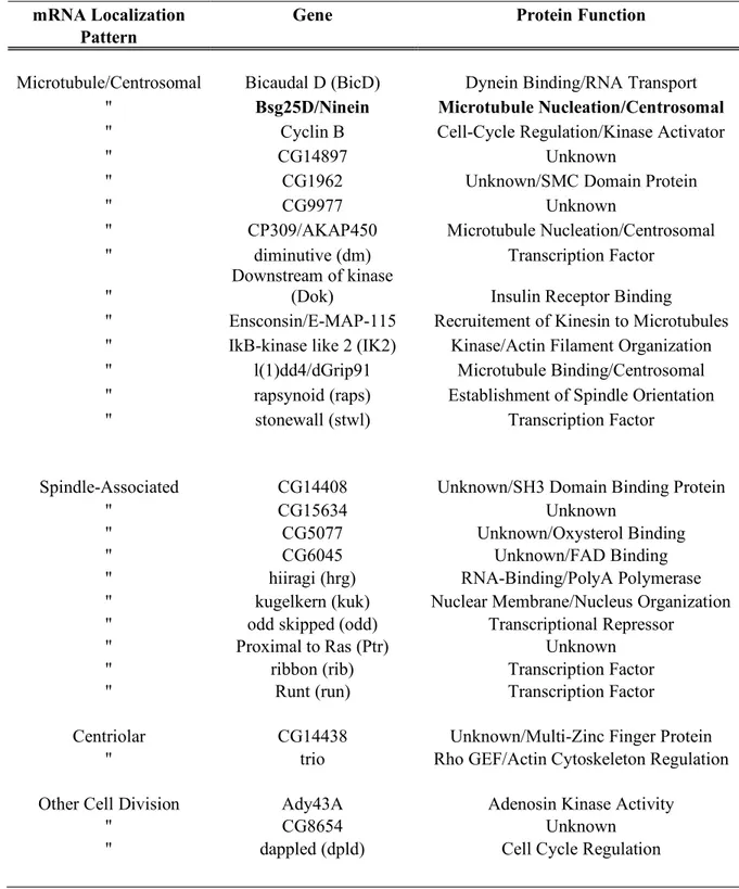
Table 1: mRNA localized to components of the cell division apparatus in Drosophila
Liste des figures
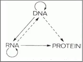
Figure 1 : Central dogma of molecular biology 12
Figure 2 : Examples of Localized mRNA 16
Figure 3 : Overview of the Cell Cycle and Mitosis 22
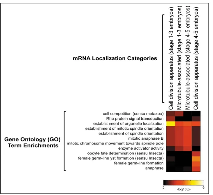
Figure 4 : Centrosomes, Centrioles and Kinetochores 25 Figure 5 : Functional enrichment of mRNAs localized to the cell division
apparatus
37
Figure 6 : Preliminary finding of Bsg25D mRNA 38
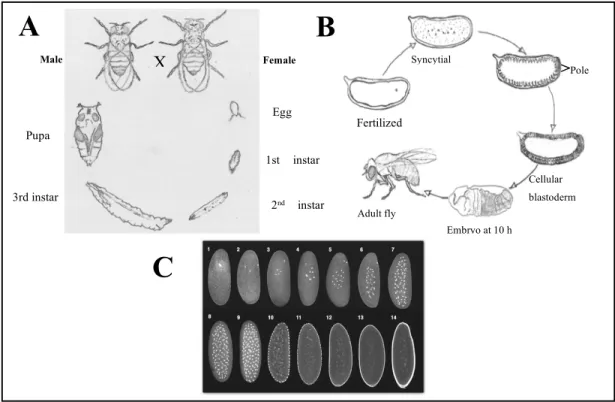
Figure 7 : Life Cycle, Development and Embryogenesis of Drosophila melanogaster
40
Figure 8 : Schematic diagram of GAL4 – UAS Driver 43
Figure 9 : Site-Specific Transgenesis Using the Phi-C31 System 45 Figure 10 : Diverse Localization of Bsg25D in Drosophila Embryos 55
Figure 11 : Bsg25D Localization in S2 cells 56
Figure 12 : Evolutionary conservation of Bsg25D within different Drosophila species
57
Figure 13 : Bsg25D an ortholog of Ninein/Ninein-like proteins 59
Figure 14 : Characterization of Bsg25D antibodies 63
Figure 15 : Fly cross for generating Bsg25D loss-of-function mutants 67 Figure 16: Bsg25D targeting is mediated by both coding and 3’ UTR regions 70
Liste des abréviations
3’UTR 3’ Untranslated Region
A/P Anterior/Posterior
Ash1 Achaete-Scute Homologue-1
Bub Budding uninhibited by benomyl
CIN Chromosomal Instability
C-terminus Carboxyl terminus
D. melanogaster Drosophila melanogaster
D/V Dorsal/Ventral
DGRP84 Drosophila Gamma-Tubulin Ring Protein 84
DGRP91 Drosophila Gamma-Tubulin Ring Protein 91
DNA Deoxyribonucleic acid
DAPI 4’,6-Diamidino-2-phenylindole
EGFR Epidermal growth factor receptor
G1 Gap 1
G2 Gap 2
Grk Gurken
GFP Green fluorescent protein
Hsp83 Heat shock protein 83kD
Hb Hunchback
IF Immunofluorescence
MTOC Microtubule organizing centers
MTs Microtubules
N-terminus Amino terminus
NLP Ninein-like protein
ORF Open reading frame
PCM Pericentriolar material
P53 Tumour protein 53
RBP RNA binding protein
RNA Ribonucleic acid
S Synthesis
SAC Spindle Assembly Checkpoint
SPB Spindle pole body
S. cerevisiae Saccharomyces cerevisiae
TGFα Transforming growth factor alpha
Tgf-β Transforming growth factor beta
Top torpedo
UTR Untranslated regions
Vg1 Vestigial
wt Wild type
γ- tubulin Gamma tubulin
Remerciements
I would like to thank all the wonderful people who have helped me get to where I am today. This has undeniably been a journey, one with a whirlwind of change throughout the entire process. I am fortunate to have the unconditional love and support of my family and friends.
First and foremost, thank you Eric for being my supervisor and mentor. From the very first day we met, you have shown sincere dedication to the project and our team. I am grateful to have worked with a great scientist. You taught me everything there is to know about RNA biology and more importantly how to cope with graduate studies. I have also learned a lot about life and hard work just shadowing you over the years. The lab was a great place to be and still is because of the wonderful people. Thank you everyone for all your help over the years and a big thank you to the IRCM community for your love and support along the way. என் அன்பான ெபற்ேறார்க் நன் ெசால்ல ம் ேறன்! You both have always ensured that education is a priority in our lives, and similarly I will continue to inspire many young minds for years to come. Maman et papa, je voudrais vous remercier pour vos conseils, votre soutien et votre amour inconditionnel. Vous m’avez aidé d'une manière que vous ne comprendrez jamais! Sendhuri, Kaviena and Anujan: thanks for putting up with me and helping me get to this point.
Ariyan and Ashviya - You are my world and my life. You each have given me strength in ways I cannot find words to explain. You gave me a purpose in life. Finishing this degree has been one of the most challenging and yet fulfilling experiences. When faced with adversity, never doubt your strength. If you really want it, just go for it! Anything is possible! The best for last: Nitish. Thank you for never giving up on me. Thank you for giving me hope and always showing me how much I am loved. Love you forever and always.
1
–Introduction
The central dogma of molecular biology was first described by Francis Crick (Crick, 1958). He explained the flow of genetic information from DNA being transcribed into RNA (transcription) and subsequently being translated from RNA to protein (translation) (Crick, 1970). As seen in Figure 1, DNA undergoes replication, forming two identical strands of DNA. This idea of the central dogma suggests that genes encode proteins and proteins alone are responsible for the expression of the genetic material inscribed in our DNA. At the time, only the lines that are solid were known interactions, and the dotted lines were predicted interactions. There are several mechanisms that have since come to light that involve regulation at the RNA level. The addition of 5’ cap or 3’ Poly-A tail, the splicing of introns and exons and alternate splicing, are a few examples of such mechanisms. These short segments of mRNA are subsequently translated into protein molecules of different shapes and sizes to serve as structural supports, chemical catalysts, and molecular motors, in order to enable cells to communicate with each other and regulate gene expression (Alberts, 2002; Ding et al., 1993).
Figure 1. Central dogma of molecular biology
All living cells follow this fundamental process coined the central dogma of molecular biology. Figure was adapted from (Crick, 1970).
Following transcription, RNAs undergo several maturation steps. RNA maturation is controlled at various points including 5’-end capping and 3’-end cleavage/polyadenylation (Neve et al., 2017; Ramanathan et al., 2016), splicing of introns (Shi, 2017), RNA nuclear export through the nuclear pore (Sakuma and D'Angelo, 2017), cytoplasmic transport (Singh et al., 2015), translation and eventually degradation (Corbett, 2018). These steps are further regulated by RNA binding proteins (RBPs), which often control the trafficking, stability and translation of the mRNA (Gebauer et al., 2012; Glisovic et al., 2008; Keene, 2007).
2 - mRNA Localization
2.1 – General Importance and Functions of mRNA localization
Localized transcripts are found in numerous organisms ranging from unicellular fungi to animals and plants, and in a diverse array of cell types including oocytes, fibroblasts, glia, neurons and epithelial cells (Bashirullah et al., 1998; Chartrand et al., 2001; Jansen, 2001; Kloc et al., 2002; Lipshitz and Smibert, 2000; Palacios and St Johnston, 2001; Seydoux and Schedl, 2001). Asymmetric mRNA localization is an important mechanism by which cells achieve polarity and generate asymmetric protein distributions that are important for development both functionally and morphologically. Subcellular localization of mRNA is a post-transcriptional mechanism that allows for delicate regulation of when and where proteins are made and function, as well as confining target proteins to specific subcellular compartments (Bashirullah et al., 1998). There are highly regulated cis and trans-acting elements that mediate specific targeting of mRNAs spatially and temporally. Early discoveries and different studies of mRNA localization were restricted to a few transcripts in a small number of model organisms. Today, there is looming evidence that mRNA localization is a very widespread process in eukaryotes (Bashirullah et al., 1998; Blower et al., 2007; Cody et al., 2013; Diehn et al., 2000; Jansen, 2001; Lecuyer et al., 2007; Marc et al., 2002; Martin and Ephrussi, 2009; van Heesch et al., 2014; Wilk et al., 2016). Though the
process of mRNA localization was thought to be a very rare phenomenon, a previous study analyzing 3370 Drosophila genes has shown that more than 70% of mRNAs that are expressed are found to be localized subcellularly (Lecuyer et al., 2007). Several other such large genome wide studies have shown the importance of RNA localization subcellularly (Medioni et al., 2012; Mili et al., 2008; Rapoport, 2007; van Heesch et al., 2014; Wickner and Schekman, 2005; Wilk et al., 2016).
Among the earliest evidence of localized mRNAs was the finding by Jeffery and colleagues (1983) in the eggs of ascidians (sac-like marine invertebrate filter feeders), who showed that β-actin mRNA localizes to the myoplasm of ascidian eggs and participates in ooplasmic segregation (Jeffery, 1986). Our growing knowledge of mRNA localization in different model systems is derived from several different genetic, cytological and biochemical assays performed in various model systems. In budding yeast Saccharomyces cerevisiae, Ash1 mRNA localizes to the bud tip during anaphase of the cell cycle, leading to an asymmetric distribution of Ash1 to daughter cells, an important process for mating type switching (Chartrand et al., 2002; Long et al., 1997; Takizawa et al., 1997). Several well-characterized mRNAs are found to localize in early embryos and oocytes, where they work to regulate the development of the germline and control the formation of axis plan. Embryonic patterning is established during oogenesis in Xenopus and Drosophila through the localization of maternally provided mRNAs that are polarized (Bashirullah et al., 1998; Deshler et al., 1998; Ephrussi et al., 1991). Asymmetric subcellular localization of mRNAs is not limited to the germline. It is also observed in somatic cells like neurons where it is shown to play an important role in memory and learning (Amtul and Rahman, 2016). These studies collectively have provided us with the foundation to understand the biological and functional importance of mRNA localization.
2.2 - Why localize mRNAs?
Transcript localization can potentially serve many important biological functions (see Figure 2 for summary). mRNA localization is a mechanism used to regulate expression of proteins in a temporal and spatial manner. Firstly, mRNA localization is an effective means to concentrate proteins synthesized at a specific site since each template can serve many rounds of translation. Local protein synthesis can also be regulated temporally in response to stimuli, which is efficient since synthesizing proteins that are not necessary is energetically costly. This allows cells to rapidly respond to local requirement making it possible to regulate gene expression (St Johnston, 2005). Each gene can be transcribed and translated at different efficiencies depending on the cells’ needs. Bicoid mRNA, for example, localizes to the anterior end of a Drosophila embryo. There, it encodes Bicoid protein and induces cells to adopt anterior cell fates (Driever and Nusslein-Volhard, 1988). Normal bicoid gradient is necessary for establishing an anterior/posterior (AP) axis and proper development of the head and thorax (Nusslein-Volhard et al., 1987). Secondly, transcript localization can provide a way to prevent proteins from being targeted to the wrong compartment, where they could exert harmful or toxic effects. Drosophila nanos mRNA localizes to the posterior of the early embryo where its corresponding protein product induces posterior cell fates. When nanos transcripts are mislocalized to the anterior end of the embryo, it induces cells to adopt posterior fates thereby causing severe abnormalities, such as development of a second abdomen in place of the head and thorax (Gavis and Lehmann, 1992; Smith et al., 1992). This example illustrates how the presence of a protein in the wrong place at the wrong time can have harsh implications on a developing organism. This is particularly important in large or highly polarized cells. Thirdly, in certain cases, the localized mRNAs code for proteins that have their own targeting signals which allows these proteins to be sorted to organelles and subcellular domains such as the mitochondria, mitotic microtubules and endoplasmic reticulum (Pelham, 1990; Rapoport, 2007; Rothman and Orci, 1992; Wickner and Schekman,
2005). However, many of these proteins do not contain this sorting signal and therefore may solely rely on the subcellular localization of their transcripts.
Figure 2: Examples of Localized mRNA
Various examples of RNA localization playing an important role in asymmetric cell division, synaptic plasticity, morphogen gradient formation and cell migration. (A) In Xenopus oocytes, Vg1 mRNA localizes to the vegetal pole. (B) In budding yeast, S. cerevisiae, Ash1 mRNA localizes to the bud tip. (C) In Drosophila embryos, bicoid mRNA localizes to the anterior pole of the developing embryo. (D) In neurons, CamKIIα mRNA localizes to the dendrites of the axon. (E) In fibroblasts, β-actin localizes to the lamellipodia.
2.3 Mechanisms of mRNA localization
There are several different mechanisms by which RNAs get localized, including active transport, localized synthesis, diffusion and localized entrapment, localized degradation, and polarised nuclear export (Lipshitz and Smibert, 2000). A combination of these processes may take place to localize a transcript. Active transport along the cytoskeletal filaments is considered the major localization mechanism in most cells (Jansen, 2001; Kloc et al., 2002; Palacios and St Johnston, 2001; Tekotte and Davis, 2002). The transcripts can be moved from the site of synthesis to their destination within the cytoplasm on tracks made by microtubules or microfilaments since the cytoskeleton is composed of actin and microtubule networks that are important in short-distance and long-distance transport, respectively. They are important in the transport of RNA cargos (Kloc et al., 2002) which are transported along the cytoskeleton by molecular motors such as kinesin, kinesin-like and dynein as well as ribonucleoprotein (RNP) complexes that are necessary to associate with these molecular motors (Kloc et al., 2002).
Local synthesis, though rare, is a simple way to target mRNA to a specific area in the cell. Transcripts can also diffuse from where they are made until they become anchored at a specific site, thereby creating a gradient. Drosophila Nanos is a well-characterized example of diffusion and entrapment, which is perhaps the easiest form of localization. Nanos localizes to the posterior pole of an embryo during late oogenesis. There, it translationally represses hunchback (hb) RNA and promotes abdominal development (Tautz, 1988). hb RNA is initially maternally expressed and evenly distributed in the embryo (Tautz, 1987; Tautz, 1989). Nanos then inhibits maternally derived hb from being translated at the posterior end thereby forcing it to congregate in the anterior end (Tautz, 1987; Tautz, 1989). Although nanos mRNA is known to localize to the posterior of the Drosophila embryo, only 4% of the total nanos mRNA concentrates to the posterior pole while the rest of the nanos mRNA is found throughout the embryo (Bergsten and Gavis, 1999) and the anchoring is established
through actin filaments present at the posterior pole (Forrest and Gavis, 2003). This entrapment or anchoring allows for a stable association of mRNA with parts of the cellular architecture, which in this case is required for translation (Gavis and Lehmann, 1992). Embryos that are mutant for nanos fail to develop an abdomen (Lehmann and Nusslein-Volhard, 1991). Therefore, nanos is able to establish a concentration gradient of both hb and itself from anterior to posterior pole of a mature oocyte through its inhibition of Hb translation (Pelegri and Lehmann, 1994). Germline localization of nanos appears to be an evolutionarily conserved mechanism even in primordial germ cells of zebrafish (Gavis et al., 2008).
Localized degradation is a process by which transcripts that were widely distributed throughout the cytoplasm are degraded everywhere but at the site of localization, where it is said to be protected. Many RNAs in Drosophila, like Heat Shock protein 83kD (Hsp83) localize to precursors of germ cells called pole cells at the posterior of the embryo (Palacios, 2007). Hsp83 mRNA is uniformly distributed in a mature oocyte where upon fertilization, Hsp83 is degraded everywhere except at the posterior pole plasm where it is localized (Kelley, 1993). Each nuclear division results in a decrease in the level of total RNAs with the exception of the pole plasm where they are protected (Ding et al., 1993). This degradation-protection mechanism accounts for the removal of Hsp83 during the first two hours of embryogenesis when 95% of Hsp83 is degraded (Bashirullah et al., 1999). In mutants that do not have a pole plasm, Hsp83 RNA is degraded throughout (Bashirullah et al., 1999).
2.4 Localization Signals within the RNAs
Conserved mechanisms of mRNA localization and localized translation exist in all branches of eukaryotes in order to regulate gene expression, by localizing transcripts to a specific region and activating translation of those localized mRNA (Du et al., 2007;
Kraut-Cohen and Gerst, 2010; Micklem et al., 2000). This in turn results in an asymmetric distribution of RNAs and proteins. Cis-regulatory motifs, often called “zipcodes”, are localization elements usually found in the untranslated regions of transcripts where they are not constrained to have protein coding sequences (Holt and Bullock, 2009). They mediate interactions between mRNAs and their binding partners to set up mRNA targeting pathways (Andreassi and Riccio, 2009; Jambhekar and Derisi, 2007). Zipcodes are recognized by transacting factors that specifically bind to them (Czaplinski and Singer, 2006). RNA binding proteins (RBPs) are the main players involved in recognizing, binding and mediating the translocation of the transcript to its appropriate destination. In some cases, the interaction is mediated by several RBPs forming complexes such as in the example of Vg1 mRNA in Xenopus laevis. Vg1 is a signalling molecule that belongs to transforming growth factor-β (tgf-beta) superfamily and is important for mesoderm induction during embryogenesis. Several RBPs are required to promote Vg1 mRNA localization to the vegetal pole in Xenopus oocytes (King et al., 2005; Rand and Yisraeli, 2001).
In some cases, a single localization element will efficiently localize the transcript (Serano and Cohen, 1995). This, however, is not usually the case. Multiple localization elements and sometimes, several copies of the same element, are required for proper mRNA localization. For many localized mRNAs characterized to date, cis-regulatory sequences are found within the 3’UTR. However, the coding region and 5’UTR can also harbour localization signals (Gonzalez et al., 1999). For example, in budding yeast S. Cerevisiae, Ash1 behaves as a transcriptional repressor of HO endonuclease and is crucial for asymmetric distribution of Ash1 (Bobola et al., 1996; Sil and Herskowitz, 1996). Ash1 localizes to the bud tip thereby restricting Ash1 protein expression in daughter cell to promote opposite mating types between mother and daughter (Long et al., 2001; Takizawa et al., 1997). Functional cis-acting elements are found in both 3’UTR and the coding region of Ash1 mRNA: one that spans the end of the coding sequence and the 3’UTR while three others are in the coding sequence (Chartrand et al., 1999; Gonzalez et al., 1999). It was demonstrated
that the 3’UTR contains a stem-loop structure that is necessary for the localization of this RNA (Chartrand et al., 1999; Gonzalez et al., 1999). Gurken (grk) mRNA encodes a protein called transforming growth factor alpha-like protein (TGFα-like protein). Gurken is important for establishing the Anterior/Posterior (A/P) and Dorsal/Ventral (D/V) axes of the oocyte and embryo. Localization signals for grk RNA are found in the 5’ and 3’ UTRs. While the zipcode in the 5’UTR is important for grk mRNA localization within the oocyte in early stage egg chambers, the localization signal in the coding sequence is required for mid to late stage egg chamber localization (Neuman-Silberberg and Schupbach, 1993; Thio et al., 2000). The 3’UTR localization signal is necessary to confine the message to the dorsal-anterior end (Neuman-Silberberg and Schupbach, 1993; Thio et al., 2000). Defects in grk lead to consequences in the polarity of the oocyte and the embryo where the embryos become ventralized through an expansion of ventral structures (Kelley, 1993; Manseau and Schupbach, 1989; Schupbach, 1987; Schupbach and Wieschaus, 1991; Wieschaus, 1978).
2.5 Translational regulation
Translational regulation is a means by which mRNAs on route to their destination are repressed from undergoing translation until it is necessary. This prevents the encoded proteins from being ectopically expressed during transport. However, the RNAs need to be actively translated once they reach their destination and therefore the repression is alleviated through de-repression. The overall mechanism is an interplay between repression and activation of translation governed by RNA localization. In certain instances, like Ash1 transcripts in budding yeast, this translational repression is essential to properly localize the RNA to the bud tip (Gu et al., 2004; Irie et al., 2002). Once the RNA reaches the bud tip, its translation is necessary for anchorage. However, it is not clear how this transition from repression to activation is achieved.
Our understanding of the relationship between RNA localization and translation has been gleaned from studies done in Drosophila focusing on four maternal transcripts critical for axis determination: bicoid, gurken, nanos, and oskar during oogenesis and embryogenesis. Cis- and trans-acting factors are necessary not only for proper localization of mRNA but for translational control of these mRNAs as well. Bicoid transcript localizes to the anterior of the oocyte and is translationally regulated through polyadenylation, a post-transcriptional modification made to the 3’end of the transcript upon exiting the nucleus (Salles et al., 1994). Nanos and oskar localize to the posterior end of the oocyte, and are controlled translationally via transcript localization (Kloc et al., 2002; Lipshitz and Smibert, 2000; St Johnston, 2005).
Translational regulation is not limited to the embryo and oogenesis. Studies have shown that mRNAs localized to dendrites of neurons have to be translationally regulated to achieve synaptic plasticity (Steward and Schuman, 2001), important in memory and learning (Farris et al., 2014; Kejiou and Palazzo, 2017).
3 - The Cell Cycle and Mitosis
Figure 3: Overview of the Cell Cycle and Mitosis
The cell cycle allows for the doubling of genetic material (A) and division of cells into two daughter cells. The cycle has two main phases: Interphase, where the DNA is replicated (S) and the cell grows (G1 and G2); and Mitosis where the cell divides into two identical cells (M) (Image adapted from Morgan, D., 2007. The Cell Cycle). (B) Mitosis consists of five phases: prophase, prometaphase, metaphase, anaphase and telophase. The mitotic spindle, made up of microtubules, pulls apart the chromosomes during mitosis. Once mitosis is complete, the cytoplasm is divided into two through a process called cytokinesis (Image adapted from Alberts et al., 2002. Molecular Biology of the Cell).
3.1 The Cell Cycle
Cells undergo a series of events collectively known as the cell cycle. These steps allow cells to grow, copy their genetic material, prepare for cell division and finally divide. The cell
cycle is divided into two phases: interphase and mitosis (see Figure 3). Cells spend most of their time in interphase, consisting of three subphases - G1, S and G2 phases. The DNA is replicated during the S phase (synthesis phase), which is also when the centrosomes undergo duplication (Morgan, 2007). The G1 and G2 phases (Gap 1 and Gap 2, respectively) separate DNA synthesis and mitosis. Mitosis or the M phase of the cell cycle is comprised of mitosis and cytokinesis (Morgan, 2007). The M phase is the shortest phase of the cell cycle, lasting about 30 minutes, during which sister chromosomes segregate into two daughter cells each with a complete set of chromosomes (Morgan, 2007). Cytokinesis occurs at the end of mitosis where the cytoplasm is cleaved to form two genetically identical cells (Alberts, 2002; Schafer, 1998).
Several hundred proteins coordinate the assembly and activity of the mitotic spindle and centromeres during the cell cycle (Vitre and Cleveland, 2012). Targeting RNA molecules to structures of the mitotic apparatus has long been suggested as a way to control cell cycle regulators through localized translation and to control asymmetric inheritance of genetic material during cell division (Suprenant, 1993; Vitre and Cleveland, 2012).
3.2 Cell Division
Mitosis is divided into 5 different subphases. During prophase, the chromosomes becomes very compact, condensing into visible chromosomes and the centrosomes separate while the mitotic checkpoint proteins, such as BUB1 and BUBR1 are recruited to the kinetochores (Elowe, 2011). At prometaphase, the nuclear envelope begins to break down and the kinetochore microtubules start to form and connect to the centrosomes through the kinetochores (see Figure 3). During metaphase, the chromosomes align along the equatorial plate with each chromatid attached to a kinetochore microtubule, at the centre of the cell
called the metaphase plate. The chromatids begin to separate during anaphase as they move toward the spindle poles, while tethered at the centromere. During telophase, the chromosomes reach the spindle pole and the nuclear envelope begins to re-form while chromatin decondenses. Cell division is completed with the division of the cytoplasm, during cytokinesis. Although mitosis is the shortest process during the cell cycle, there are many intricate steps and feedback mechanisms that help maintain the integrity of the genome (Alberts, B. et al, 2004).
3.3 Mitotic Apparatus
The mitotic apparatus consists of 1 spindle and 2 centrosomes (Flemming, 1882) (Wilson, 1902). For the purpose of this thesis, we will concentrate on the centrosome as a key player along with microtubules (MTs).
4 - Overview of the Centrosome
The centrosome was first discovered by Boveri and Van Beneden, in the late 19th century when it was coined the term “centrosome” as the division centre of the cell during mitosis (Boveri, 2008; Hamoir, 1992). In animal cells, the centrosome is the major microtubule-organizing centre (MTOC), responsible for nucleating, anchoring and releasing MTs (see Figure 4). Centrosomes are involved in several processes including cell motility, cell adhesion and polarity and cell division (Doxsey et al., 2005).
The centrosome, also called the spindle pole body in fungi, consists of two centrioles embedded within an electron-dense cloud called the pericentriolar matrix (PCM). The centriole provides structural stability in vertebrate cells. Each centriole is made up of a total of nine MT triplets, spanning 0.2 µm in width and on average 0.5 µm in length (Bornens, 2002; Paintrand et al., 1992), though this can vary depending on the species and cell type (Bornens, 2002; Paintrand et al., 1992). For example, in D. melanogaster the microtubules can be arranged as doublet or triplet MT (Bettencourt-Dias and Glover, 2007). Centrioles are
polarized along the proximo-distal axis (Bettencourt-Dias and Glover, 2007; Nigg and Stearns, 2011).
Figure 4: Centrosomes, Centrioles and Kinetochores
(A) Schematic diagram showing main players in cell division: microtubules, centrosomes, kinetochores, chromosomes and motor proteins. The chromosomes are aligned at the metaphase plate, ready for division. The microtubules are attached by kinetochores and will draw apart the sister chromatids to their respective spindle poles. (B) A phase-contrast micrograph depicting a similar image to (A) the schematic. (E.D. Salmon and R.R. Segall, J. Cell Biol. 86:355–365, 1980. © The Rockefeller University Press) (Both Images were taken from Alberts B, Johnson A, Lewis J, et al. Molecular Biology of the Cell. 4th edition. New York.)
4.1 Centrioles
Centrioles are stable structures and their loss has been shown to lead to PCM breakdown (Basto, 2006; Bettencourt-Dias et al., 2005; Janke et al., 2005). It is believed that post-translational modifications, such as polyglutamylation of the centriolar tubulin confers its stability (Bobinnec et al., 1998; Janke et al., 2005). Tektins and ribbon proteins are other structural components of the centriole, which might also provide structural stability (Hinchcliffe and Linck, 1998; Steffen and Linck, 1988).
To form a bipolar mitotic spindle the centriole duplicates using the mother or mature centriole as a template to give rise to the daughter. The centrosome duplicates during the S phase of the cell cycle, matures during G2 phase by accumulating PCM material, then the duplicated centrosomes separate at the beginning of M phase (Alvey, 1985; Kochanski and Borisy, 1990; Kuriyama and Borisy, 1981a; Robbins et al., 1968; Sluder and Rieder, 1985; Vorobjev and Chentsov Yu, 1982). The mother centriole has subdistal and distal appendages that dock cytoplasmic MTs. If depleted using a laser, centrioles can reform de novo (La Terra et al., 2005; Marshall et al., 2001; Riparbelli and Callaini, 2003).
Proteomic studies revealed that there are more than 200 centrosome-associated proteins, many that have yet to be characterized (Andersen et al., 2003). The PCM contains proteins such as pericentrin and AKAP450, a family of coiled-coil domain proteins that anchor MTs in the cytoplasm during interphase and mitosis (Gillingham and Munro, 2000). Gamma (ᵞ) tubulin is found on the walls of the centrioles and has been shown to increase in concentration prior to mitosis (Martin et al., 1997). γ- tubulin is found in a complex called γ-TuSC or γ-tubulin small complex. In D. melanogaster, each complex has two tubulin molecules and a molecule of DGRP84 and DGRP91. These four proteins together are known as the γ-tubulin ring complex (γ-TuRC), which holds the subcomplexes of γ-TuSC. The removal of these molecules in D. melanogaster has been shown to give rise to spindles that are abnormal (Colombie et al., 2006).
MTs are anchored onto subdistal appendages of centrioles. Ninein is part of the subdistal appendage of the mother centriole, which interacts with the centriole at its C-terminus while connecting to the γ-TuRC through its N-terminus (Mogensen et al., 2000). Several cells depend on centrioles and spindle pole body (SPB) for accurate cell division in early embryonic systems. However, many other cell types such as higher plants cells and oocytes divide even in the absence of centrioles. Centrioles have been implicated in the regulation of cytokinesis and G1-S transition. The removal of the centrosome has been shown to disrupt cytokinesis (Hinchcliffe et al., 2001), even though most cells form a furrow. SPB also helps concentrate molecules that are important for mitosis exit. In some species, centrioles assure mitotic fidelity and contribute to spindle orientation (Khodjakov and Rieder, 2001). Interestingly, in D. melanogaster, the removal of centrosomes is not sufficient to halt mitosis, as cells still transition from G1 to S in their absence (Basto et al., 2008). However, if the proteins associated with the centrosome are also removed along with the centrosome, then the cells are halted at G1 of the cell cycle. This could simply be a mechanism that has evolved to prevent cells from becoming aneuploid or from developing defects that can lead to disease. The increase in centrosome number leads to multipolar spindles, as well as tumorigenesis (Basto et al., 2008; Levine et al., 2017; Timonen and Therman, 1950). Indeed, chromosome instability is a common feature of tumour cells, characterized by an elevated rate of gain or loss of whole chromosomes.
Even though the centrioles do not seem to be a universal requirement in cell division and are absent during female meiosis, they seem to be a requirement for the assembly of cilia and are important in male meiotic divisions. Centrioles serve as basal bodies when they are attached to the membrane, and they form cilia and flagella, which are involved in sensory perception, propagation of morphogenetic signals and motility. In most cells, cilia are found in their immotile form as primary cilia, while in other cells, such as sperm and gut cells, they exist in their motile form (Alberts, 2002). Both forms are equipped with sensory functions. A study done by Jonassen et al. in 2008 suggests that cilia might be important in determining
the axis of cell division and the positioning of the centrosome in kidney cells, associating two separate functions of the centriole in a process (Jonassen et al., 2008).
The proper control of centrosome and centriole number is crucial for the development of a healthy organism. Since centrosomes facilitate the organization of the spindle poles during mitosis, any errors in this process can lead to cancer and cause genomic instability (Nigg, 2002) as a consequence of aberrant cell division (Sluder and Nordberg, 2004). The increase in centrosome number could be a source of chromosomal instability thereby leading to cancer. There is an increasing number of studies being performed in the 21st century many years after the discovery of the centrosome and its link to several human diseases such as ciliopathies and diseases involved in brain development. The knockdown of p53, a tumour suppressor, leads to the increase in centrosome number in mouse fibroblasts and skin tumours (Fukasawa, 2007), but this link between centrosome abnormalities and cancer is not limited to mice. In fact, it is common in several prevalent human cancers such as breast cancer, prostate cancer, lung cancer, colon cancer and brain cancer (Lingle et al., 2002; Pihan et al., 1998).
4.2 Ninein and Ninein-like proteins
The minus-end of the microtubules are nucleated and anchored from the pericentriolar material (PCM). Elongation occurs by the addition of tubulin subunits at the distal plus-end of microtubules. Ninein is a centrosomal protein important for microtubules minus-end anchorage and plays a role in docking γ-tubulin containing complexes (Delgehyr et al., 2005; Stillwell et al., 2004). Ninein has been shown to localize to the ends of the subdistal appendages of the mother centriole as well as to the minus-ends of both centrioles (Mogensen et al., 2000; Ou et al., 2002). The centrosomal protein, Ninein-like protein (Nlp) plays a major role in centrosome maturation via γ-TuRC recruitment during interphase. Nlp has also been shown to be an important substrate for many mitotic kinases such as Aurora B, Plk1, Cdc2, Nek2 (Bornens, 2002; Casenghi et al., 2005; Casenghi et al., 2003; Rapley et al., 2005; Zhao
et al., 2010). During the cell cycle, Nlp is associated with the maturing centrosome leading up to the G2/M transition where it dissociates via phosphorylation. This is a very important step for the maturation process and mitotic spindle formation. When Nlp is overexpressed, it forms large aggregates that subsequently induce aberrations in mitotic spindle formation and the depletion of Nlp gives rise to lagging chromosomes and multinucleated phenotypes (Casenghi et al., 2003; Jin et al., 2009; Zhao et al., 2010). Understandably, since both Ninein and Nlp play a crucial role during microtubule anchoring, γ-tubulin docking and centrosomal maturation, the perturbation of these proteins during cell division can induce mitotic aberrations, spindle checkpoint defects, chromosomal missegregation and failure of cytokinesis. This can induce chromosomal instability potentially leading to tumorigenesis (Thompson et al., 2010; Thompson and Compton, 2008).
5 - Pathways of Control
The accurate transmission of genetic information from one cell to its daughters is crucial for the survival of an organism. Proper segregation of chromosomes during mitosis is therefore necessary to maintain the genomic integrity of a cell (Hartwell and Weinert, 1989; O'Farrell et al., 2004). There are several checkpoints within the cell cycle that help maintain the fidelity of each step allowing the overall organism to remain healthy.
During the cell cycle, the cell must make decisions on whether or not to proceed based on the faithfulness of each process (Ciccia and Elledge, 2010; Litwin et al., 2018). These checkpoints are highly conserved and as soon as they encounter a problem, the cycle is halted allowing the error to be corrected prior to proceeding to the next step. Since each step must be highly regulated, when these checkpoints are turned on for an excessive period of time, it may give rise to an abnormal number of chromosomes, thereby inducing apoptosis or leading to a disease state (Hanahan and Weinberg, 2011).
5.1 Aneuploidy and Chromosomal Instability
When the integrity of the genome is threatened, the cell has many feedback mechanisms to try and protect the genome while preventing the accumulation of mutations to protect future generations. However, genes that are involved in these checkpoints are also susceptible to mutations giving rise to various human diseases (Suprenant, 1993; Vitre and Cleveland, 2012). Most cancers are a result of mutations in one or more genes associated in the checkpoint pathway(s) (Lengauer et al., 1997). A few examples of defects leading to chromosomal instability include failed mitotic checkpoint signalling, defects in chromosome cohesion or attachment and assembly of multipolar mitotic spindles.
Checkpoints that guard the cell from errors can often be defective as is the case for many aneuploid cancers including types of leukemia, breast, colorectal, ovarian and lung cancers (Weaver and Cleveland, 2009). In these diseases the signalling of the Spindle Assembly Checkpoint (SAC) is inadequate as a result of an altered expression or mutations of components of the mitotic checkpoints. A rare genetic disorder called mosaic variegated aneuploidy has also been associated with mutations in BubRI (or BublB), a component of the mitotic checkpoint (Schmid et al., 2014). This disease causes growth retardation, microcephaly, and childhood cancer, which often results in death at a young age (Hanks et al., 2004; Matsuura et al., 2006). Defects in sister chromatid cohesion might also promote aneuploidy, as many studies have demonstrated that overexpression of securin or separase regulators important for the control of chromatid cohesion gives rise to aneuploidy and cell transformations (Pei and Melmed, 1997; Yu et al., 2003; Zhang et al., 2008). Merotelic attachment results from one kinetochore attaching improperly to microtubules extending from both spindle poles, leading to improper segregation of chromosomes (Thompson and Compton, 2008). Even though in most cases, the cell corrects this error prior to moving onto anaphase, it can promote aneuploidy. If aneuploidy is left undetected in the cell, it leads to lagging anaphase chromosomes at the spindle midzone, which subsequently do not segregate into daughter cells (Zhang et al., 2008). Chromosomal instability (CIN) can be caused by
missegregation of a whole chromosome. However, it is not limited to this, as structural rearrangements such as translocation, deletions and inversions can also give rise to CIN. CIN has been associated with poor patient prognosis in tumors (Choi et al., 2009; Gao et al., 2007; Heilig et al., 2010) and resistance to chemotherapeutic agents, thereby leading to tumor evolution (Kuukasjarvi et al., 1997; McClelland et al., 2009; Swanton et al., 2009). As such, it is important to supress CIN to target tumor cell adaptability as a means of therapeutically targeting the tumor.
6 - mRNA localization and the Mitotic Apparatus
The components of the mitotic apparatus were first successfully isolated as a single unit from dividing sea urchin eggs (Mazia and Dan, 1952). These components were physically associated with each other and were called the mitotic apparatus (MA). In later years, spindles were isolated from sea urchin eggs and it was determined that they were mainly composed of microtubules with chromosomes attached to them (Robbins et al., 1968).
A very early observation of mRNA associating with the cytoskeleton was documented when several polysomes were present in the cytoskeleton even after the cells were treated with detergents (Kuriyama and Borisy, 1981b). They demonstrated that the cytoskeleton retains almost all the active polyribosomes and they were attached to the cytoskeleton via mRNA (Kuriyama and Borisy, 1981b). Interestingly they ruled out microtubules (MT) of playing any part in this process, since polysomes were retained in the cytoplasm even when the MT were depleted by using detergents that created an environment unfavourable for stabilizing MT. However, it was later shown that under specific stabilization conditions it was possible to also preserve MTs even after detergent extraction (Kuriyama and Borisy, 1981a).
Microtubules are approximately 25 nm in diameter. The basic unit of a microtubule is the tubulin dimer composed of alpha and beta subunits. Microtubule arrays are not fixed and can be polymerized or depolymerized through GTP hydrolysis and have a rapid turnover
due to their dynamic instability (Kuriyama and Kanatani, 1981). They act as transport vehicles and roadways for mRNA to travel directionally (Suprenant, 1993).
When Salmon and Segall (1980) isolated spindles, they made observations using both a light microscope as well as an electron microscope (Robbins et al., 1968). Under the light microscope, they observed globular material, approximately 0.5 um diameter aligned in a circular fashion around astral fibres on the central spindle. They were able to distinguish features such as the centrosome and centriole at opposite poles, chromosomes and MT. A closer look using high magnitude electron microscopy showed particles that were spread along the central spindle MT, adhering to a thin filament, in large clumps. These particles corresponded with the globular material seen with the light microscope. At the time, they were not able to identify the thin filament and particles. They did however postulate that the particles were ribosomes or other ribonuclear proteins.
Early evidence of ribosomes associating or interacting with MTs came from morphological evidence gained through electron microscopy (EM). High voltage EM observations suggested that polyribosomes interact with microtubules. Heuser and Kirschner (1980) documented a very interesting observation they discovered through freeze-dried cell samples (Heuser and Kirschner, 1980). They reported grape-like clusters of ribosomes surrounding microtubules (Heuser and Kirschner, 1980). This data was further confirmed when Ris (1985) observed the presence of several polyribosomes, with the help of a short 2-3 nm filament, highly crosslinked to microtubules, intermediate filaments and actin filaments in cultured mammalian cells (Ris, 1985).
RNAs are not only found associated with the mitotic apparatus. Other organelles and structures have also been shown to contain RNAs. One such example is the association of RNA in muscle. Early evidence from histochemical studies of RNA distribution in muscle (Clavert et al., 1949) have later been confirmed using immunohistochemical studies with anti-ribosomal antibodies (Horne and Hesketh, 1990) to show that ribosomes are present
along the myofibrils. In some studies, the associated mRNAs have been found to be present in polysomes and are therefore being actively translated (Bag and Pramanik, 1987; Bird, 1986).
Work by Peterson and Berns (1978) using psoralens, a nucleic acid-binding drug that is light activated, demonstrated that RNA present at the centriolar region is responsible for forming spindle in dividing PTK2 cells (Peterson and Berns, 1978). This was not the first time RNA was suspected to be present in the centriolar region. Prior studies using staining agents, such as acridine orange (Randall, 1965) and ethidium bromide (McGill et al., 1976) had also suggested the presence of nucleic acid in the centriolar region, as well as in basal bodies. Other studies in the 1970s suggested a similar notion, indicating high levels of RNA were found at microtubule organising centers (Bielek, 1978; Rieder, 1979). This RNA is believed to be important in nucleating microtubules. This was concluded through a study done by Heidermann et al., (1977) where basal bodies isolated from Chlamydomonas and Tetrahymena were treated with enzymes and their ability to nucleate aster formation in Xenopus laevis was examined (Heidemann et al., 1977). Through their work, they showed that centrioles have RNA that is required and is important for aster formation.
More recently, Blower et al., (2007) performed a global study of mRNAs bound to microtubules during metaphase in X. laevis egg extracts and human cell extracts revealing conserved groups of mRNA enrichment on MTs (Blower et al., 2007). Only a select group of mRNAs were associated with MT-bound polyribosomes suggesting that mRNA that are both translationally active and inactive are present on the mitotic spindles. Blower and his colleagues proposed that mRNA subcellular trafficking to the microtubules is a mechanism for enhancing protein localization and for dividing translationally inactive transcripts to daughter cells during cell division. The evidence from this article and others mentioned above set the basis for the presence of mRNA and its association with components of the mitotic apparatus.
RNA localization was thought to be a rare phenomenon due to poor detection methods leading to poor resolution. As such, in an attempt to improve the ability to visualize the mRNA localization patterns in developing Drosophila embryos, Dr. Lecuyer and his colleagues refined their protocol to add a few crucial steps to ameliorate signal detection. The improved in situ hybridization technique was subsequently used to uncover a whole new plethora of mRNAs that localize to various parts of the cells, each potentially having different functional roles in the cell. During a genome-wide global analysis of RNA localization patterns in early Drosophila embryogenesis, Lecuyer et al. (2007) discovered that more than 70 % of RNAs are localized, a phenomenon that was otherwise thought to occur rarely (Lecuyer et al., 2007). The high-resolution fluorescent in situ hybridization protocol was instrumental in uncovering these patterns of mRNA trafficking and their importance during embryogenesis (Lecuyer et al., 2007).
Approximately 4000 genes in the Drosophila genome were analyzed and 71% of the genes that were expressed encode subcellular localized mRNAs. This was a ground breaking discovery since it was previously estimated that less than 1% of mRNAs were localized (Tomancak et al., 2002). Lecuyer et al., also uncovered several striking new patterns of localization as well as diverse subcellular locations (e.g. membranes, cytoskeleton, mitotic apparatus, chromatin, nuclei, etc.), indicating that there is more to mRNA localization than originally conceived. These patterns have been documented into a web resource (http://fly-fish.ccbr.utoronto.ca). The number of different patterns also suggests that there are probably several different mechanisms associated with the observed localization patterns. Furthermore, there was a strong correlation between the transcript distribution and protein localization/function, which suggests that mRNA localization plays a vital role in organizing cellular protein networks (Lecuyer et al., 2007).
6.1 Candidate mRNA
Among the list of mRNAs found to have a specific localization pattern in Drosophila embryos, an interesting group of mRNAs localize to parts of the cell division apparatus, including centrosomes, astral MTs, and mitotic spindles (Table 1). This raises the possibility that mRNA targeting, and localized translation might play a role in the regulation of mitosis. A subset of about 30 mRNAs was found to localize to the mitotic apparatus and they were found to be functionally enriched for transcripts encoding regulators of cytoskeleton organization and cell division-related processes (see Figure 8). Although it has long been thought that localized translation of mRNAs may occur along the mitotic apparatus as a means of targeting regulators of mitosis or asymmetric cell division, this has not been investigated thoroughly.
To elucidate the function and the mechanism of mitotic mRNA localization, we selected a candidate mRNA with a dynamic and diverse localization pattern that we are interested in characterizing. The candidate mRNA, Blastoderm-specific gene 25D (Bsg25D), localizes to centrosomes and astral microtubules in Drosophila embryos during early events of embryogenesis (see Figure 5).
Ultimately, we want to test whether mRNA targeting to the mitotic apparatus is important for the regulation of cell division.
Table 1: mRNA localized to components of the cell division apparatus in Drosophila (adapted from Lecuyer et al., 2007)
mRNA Localization Gene Protein Function
Pattern
Microtubule/Centrosomal Bicaudal D (BicD) Dynein Binding/RNA Transport
'' Bsg25D/Ninein Microtubule Nucleation/Centrosomal
'' Cyclin B Cell-Cycle Regulation/Kinase Activator
'' CG14897 Unknown
'' CG1962 Unknown/SMC Domain Protein
'' CG9977 Unknown
'' CP309/AKAP450 Microtubule Nucleation/Centrosomal
'' diminutive (dm) Transcription Factor
''
Downstream of kinase
(Dok) Insulin Receptor Binding
'' Ensconsin/E-MAP-115 Recruitement of Kinesin to Microtubules
'' IkB-kinase like 2 (IK2) Kinase/Actin Filament Organization
'' l(1)dd4/dGrip91 Microtubule Binding/Centrosomal
'' rapsynoid (raps) Establishment of Spindle Orientation
'' stonewall (stwl) Transcription Factor
Spindle-Associated CG14408 Unknown/SH3 Domain Binding Protein
'' CG15634 Unknown
'' CG5077 Unknown/Oxysterol Binding
'' CG6045 Unknown/FAD Binding
'' hiiragi (hrg) RNA-Binding/PolyA Polymerase
'' kugelkern (kuk) Nuclear Membrane/Nucleus Organization
'' odd skipped (odd) Transcriptional Repressor
'' Proximal to Ras (Ptr) Unknown
'' ribbon (rib) Transcription Factor
'' Runt (run) Transcription Factor
Centriolar CG14438 Unknown/Multi-Zinc Finger Protein
'' trio Rho GEF/Actin Cytoskeleton Regulation
Other Cell Division Ady43A Adenosin Kinase Activity
'' CG8654 Unknown
Figure 5: Functional enrichment of mRNAs localized to the cell division apparatus GO term enrichments exhibited by mRNAs classified within the cell division apparatus and microtubule-associated localization categories in stage 1-5 Drosophila embryos. The “hot metal” color scale reflects statistical significance (-log 10 of the p-value) of the GO term enrichments (Figure adapted from Lecuyer et al., 2007).
Figure 6: Preliminary finding of Bsg25D mRNA
Bsg25D exhibits targeting to centrosome and astral microtubules during Drosophila embryogenesis. The Green is Bsg25D mRNA while the Red stain is DAPI (Adapted from Lecuyer et al., 2007).
7 - An Overview of the Model Organism: Drosophila melanogaster
Drosophila melanogaster, or the common fruit fly, was initially introduced in the context of evolutionary biology but has since developed into a very powerful tool in biological research, particularly in genetics and developmental biology (Arias, 2008; Hallem et al., 2004; Kohler, 1994). Several attributes have made D. melanogaster a good model system. These flies are easily maintained at standard laboratory conditions (25º C). They are inexpensive and allow easy observation and manipulation at most developmental stages. They produce large numbers of offspring. They are small and have a rapid generation time of 10 days from an embryo to an adult fly. Most importantly, we now have well-established techniques that facilitate genetic manipulation of these flies (Brookes, 2001; Sturtevant, 1961).
7.1 Life Cycle
The life cycle of Drosophila, from egg fertilization to adult life, takes about 10 days at 25°C. Female flies produce the most number of eggs between the fourth and seventh day after they emerge (Lodish et al., 2000). They can lay several hundred eggs in their lifetime. A few hours after copulation, the female starts laying fertilized eggs. The egg is surrounded by a thin inner lining called the vitelline membrane and an outer lining called a chorion (Lodish et al., 2000). After a day of embryonic development at 25º C, the egg hatches (see Figure 7 A-B-C). Over the next several days, the larvae undergo two molts from the first instar larvae to the second and third instar larva. During this time, they primarily feed and grow. The larva then molts into puparium. As the pupa develops the pupal casing becomes increasingly darker (Lodish et al., 2000). The final stage of pupation is metamorphosis into an adult fly and eclosion (Lodish et al., 2000).
Figure 7: Life Cycle, Development and Embryogenesis of Drosophila
melanogaster
The life cycle of Drosophila, from egg fertilization to adult life, takes about 10 days at 25°C. (A) Adult male and female mate and produce an egg, which undergoes a multitude of changes from larva (4 days) to pupa (4 days) and finally leading to eclosion of the adult fly. (Image adapted from Carolina Biological Supply
Company). (B) Fertilized egg undergoes several changes as it develops into an adult fly: 24h after fertilization, embryogenesis, formation of a syncytium, cellularization, blastoderm formation, gastrulation and the emergence of an adult fly. (Image adapted from Life: The Science of Biology, Purves et al., 1998) (C) Drosophila
embryogenesis: the zygote undergoes many rounds of mitotic divisions during embryogenesis. Looking closely at fluorescently labelled nuclei through confocal microscopy, by the end of 8 consecutive nuclear divisions each lasting about 8 minutes, a total of 256 nuclei are present in the centre (yolk) of the egg. The pole cells form after the ninth division, when ~5 nuclei migrate to the posterior pole and become enclosed. The remaining nuclei migrate to the periphery and become enclosed after 13 divisions, thereby forming the blastoderm. (Image adapted from
Gilbert SF. Developmental Biology. 7th edition. Sunderland (MA): Sinauer Associates; 2003.) Egg 1st instar 2nd instar 3rd instar Pupa X Male Female
A
C
B
Fertilized Syncytial Cellular blastoderm Embryo at 10 h Adult fly >Pole7.2 Early Development
During fertilization, the male and female pronuclei appose next to each other eliciting several rapid and synchronous zygotic divisions (Zalokar and Erk, 1976). Interestingly cytokinesis does not occur in the first 13 rounds of division upon fertilization (see Figure 5 C). Instead, a multinucleated cell is formed, sharing a single large cytoplasm (syncytium). A regular D. melanogaster embryo is an ovoid shape approximately 500 um by 180 um and the first nuclear divisions take place in the yolk located in the centre of the embryo (Greenspan, 1997).
The nuclei then migrate to the periphery of the embryo forming the syncytial blastoderm (Foe and Alberts, 1983) while a few dozen nuclei remain in the centre forming the yolk nuclei (vitellophages). A couple nuclei are also incorporated into the posterior pole plasm to form the polar buds (Campos-Ortega, 1997; Mahowald, 1963). These pole cells form the primordial germ cells, which give rise to gametes. The embryo undergoes cellularization whereby the nuclei migrate to the periphery, and cell membranes are laid down between each nucleus. The embryo undergoes a series of asynchronous cell division creating the cellular blastoderm. Studies have shown early stages of cell division in a developing embryo are attributable to the maternal contribution of information (RNA and proteins) present in the egg prior to fertilization. Zygotic transcription only begins thereafter during the 14th division where the embryo relies on the production of its own transcripts and
proteins to properly develop into an adult fly. Zygotic gene mutations therefore may not produce an apparent phenotype until the maternal gene product has been consumed. Therefore, in order to study a gene’s function in a fly embryo, it is important to generate mothers that are mutant for the gene of interest thereby giving rise to eggs that are deficient in specific maternal gene products.
7.3 Delta 2-3(Δ2-3)
An example of a transposase used for P-element mutagenesis is the P (Delta) 2-3 element (Robertson et al., 1988). The name of the line is derived from the location of the intron between exon 2 and 3, which is spliced solely in germ cells (Laski et al., 1986). Normally, P-element transposition does not occur in somatic cells as it is repressed at the level of RNA processing (Robertson et al., 1988). In somatic cells, however, a protein binds to exon 2, thereby preventing the intron from being spliced (Chain et al., 1991; Siebel and Rio, 1990; Tseng et al., 1991). The intron can be removed artificially, which creates a transposase that is capable of mobilizing P-elements in all tissues, somatic and germline cells (Laski et al., 1986). When the intron is not spliced, the resulting truncated protein behaves as a repressor of P-element mobility (Gloor et al., 1993; Handler et al., 1993; Misra et al., 1993; Rio et al., 1986).
7.4 Gal4-UAS Driver
The GAL4 system is a technique used to specifically drive transgene expression in a tissue-specific and cell-specific manner in Drosophila and other model organisms (Brand and Perrimon, 1993). The eukaryotic transcriptional machinery is highly conserved across different species, thereby allowing GAL4 to activate transcription in other species (Kakidani and Ptashne, 1988; Ma et al., 1988; Webster N, 1988) The GAL4 system allows for the study of the effects of misexpressing a gene of interest on development, by selectively expressing the transgene in cells where GAL4 is expressed. Gal4 is a eukaryotic transcription factor that is responsible for activating genes involved in galactose metabolism in yeast (Hashimoto et al., 1983). The upstream activation sequence (UAS) controls the expression of the transgene.
Figure 8 depicts a cross between a transgenic line expressing the GAL4 driver (A) in a known spatiotemporal pattern and the second line (B) which contains the transgene downstream of UAS sequences. The transgene is expressed in progeny that have cells or
tissues expressing the GAL4 protein. The GAL4 system allows one to express a gene of interest ectopically which allows for inducing cell fate change (Davis et al., 1987), inducing altered cell fates in neighbouring cells, and altering the cell’s physiology (Southall et al., 2008). This method of ectopic expression can be used to test whether a gene functions autonomously or non-autonomously (Brand and Perrimon, 1993), whether the gene is sufficient for cell identity and whether the cell or tissue responds to changes in signalling pathways (Huang and Rubin, 2000; Zhu et al., 2005). It has been proposed that nearly 60% of Drosophila genes have no loss-of-function phenotype (Miklos and Rubin, 1996). Therefore, to study the functional importance of a gene of interest in these cases, expressing this gene ectopically might be the only way.
Figure 8: Schematic diagram of GAL4–UAS Driver
The expression line containing the GAL4 sequence is crossed to transgenic flies that contain the target gene under the control of UAS sequences. GAL4 is produced in the progeny and the expression of the transgene can be activated in vivo following the binding of the UAS sites (Figure adapted from Phelps and Brand, 1998).
7.5 Site-specific transgenesis
The ability to introduce and control the expression of transgenes in a model organism is a powerful tool for understanding how genes function.
The introduction of P-elements to induce mutations became an attractive technique to study gene function. Mutagenesis through P-element transposition, however, occurs in an unpredictable manner making it challenging to selectively target a gene of interest. They integrate several times throughout the genome and therefore it is almost impossible to generate integration at the same site with two different transgenes (Bischof et al., 2007; Brand and Perrimon, 1993). Also, it has been shown that in Drosophila the expression of transgenes is affected by positional effects especially when the transgene inserts into a heterochromatic region (Bischof et al., 2007; Brand and Perrimon, 1993). Finally, the frequency of integration of P-elements is relatively low in fertile adults.
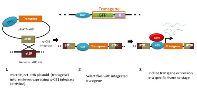
The PhiC31 integrase system was developed for D. melanogaster by Groth et al. in 2004 to tweak the system previously shown to function in other model organisms such as Xenopus laevis embryos (Allen and Weeks, 2005; Groth et al., 2004), the mosquito Aedes aegypti (Nimmo et al., 2006) and even mammalian cells (Groth et al., 2000). The PhiC31 integrase is a bacteriophage (PhiC31) which mediates sequence-specific recombination between attB (donor) and attP (recipient) sites (see Figure 9). They have a stretch of 3 nucleotides in their central region that is common, which facilitates crossover events (Thorpe et al., 2000).
Therefore, the integrase catalyzes the insertion of the transgene in a sequence specific manner and integration is unidirectional. There are a hundred known attP lines that have been created with the attP insertion sites mapped. These lines are widely available to the fly community.